DOI: 10.1590/0004-282X20160123
ARTICLE
Working memory and left medial temporal
cortical thickness
Memória de trabalho e espessura cortical temporal medial esquerda
Giuseppe Pastura1, Tadeu Takao Almodovar Kubo2, Maria Angélica Regalla3, Cíntia Machado Mesquita3,
Gabriel Coutinho4, Emerson Leandro Gasparetto5, Otávio Figueiredo6, Paulo Mattos7, Alexandra Prüfer de
Queiroz Campos Araújo1
Attention deicit/hyperactivity disorder (ADHD) is a frequent condition in childhood afecting around 5% of school-age children1
. Early cross-sectional studies indicated 4% to 5% reduction in total cerebral and cerebellar volumes in children and adolescents with ADHD, compared to typically developing children (TDC)2
. Later indings suggested that the
decrease is mainly due to reduced grey and white matter volumes and other regional abnormalities in the prefron-tal cortex, especially the orbitofronprefron-tal and dorsolateral pre-frontal cortices, basal ganglia, and cerebellum3. Attention deicit/hyperactivity disorder is a neurodevelopmental disor-der with a remission rate of approximately 60% during late
1Universidade Federal do Rio de Janeiro, Faculdade de Medicina, Departamento de Pediatria, Rio de Janeiro RJ, Brasil;
2Clínica de Diagnóstico por Imagem (CDPI), Rio de Janeiro RJ, Brasil;
3Universidade Federal do Rio de Janeiro, Instituto de Psiquiatria, Rio de Janeiro RJ, Brasil;
4Centro de Neuropsicologia Aplicada/Instituto D`Or, Rio de Janeiro RJ, Brasil;
5Universidade Federal do Rio de Janeiro, Faculdade de Medicina, Departamento de Radiologia, Rio de Janeiro RJ, Brasil;
6Universidade Federal do Rio de Janeiro, Instituto COPPEAD, Rio de Janeiro RJ, Brasil;
7Universidade Federal do Rio de Janeiro, Faculdade de Medicina, Departamento de Psiquiatria, Rio de Janeiro RJ, Brasil.
Correspondence: Giuseppe Pastura; Departamento de Pediatria da Faculdade de Medicina da Universidade Federal do Rio de Janeiro; Rua Bruno Lobo, 50; Cidade Universitária; 21941-590 Rio de Janeiro RJ, Brasil; E-mail: [email protected]
Conflict of interest: There is no conflict of interest to declare.
Received 30 April 2016; Received in final form 02 June 2016; Accepted 13 June 2016.
ABSTRACT
Objective: To perform a pilot study to investigate the association between working memory and cortical thickness in a sample of attention deficit/hyperactivity disorder (ADHD) children. Methods: Seventeen children aged 7-10 years diagnosed with ADHD and 16 healthy children underwent a magnetic resonance scan for cortical thickness measurements. Data was correlated with working memory performance using the Backwards Digit Span subtest of the Wechsler Intelligence Scale for Children. Results: Working memory impairment, evidenced by lower scores on the Backwards Digit Span, was observed in patients with ADHD compared to healthy controls. There was a direct correlation between working memory and cortical thickness of the left medial temporal lobe (Spearman’s correlation coefficient: 0.499; p < 0.005). Conclusions: Our data suggests, for the first time, a correlation between working memory, evaluated by the Backwards Digit Span, and left medial temporal cortical thickness.
Keywords: attention deficit disorder with hyperactivity; child; memory.
RESUMO
Objetivo: Realizar estudo piloto para investigar a associação entre memória de trabalho e espessura cortical em crianças com transtorno de défict de atenção e hiperatividade (TDAH). Métodos: Dezessete crianças com TDAH, entre 7 e 10 anos, e dezesseis crianças saudáveis foram submetidas a ressonância magnética para aferição de espessura cortical. Os dados foram correlacionados com desempenho da memória de trabalho usando a ordem inversa do subteste Dígitos da Escala de Inteligência Wechsler para Crianças. Resultados: Prejuízos na memória de trabalho, evidenciado pela menor pontuação na ordem inversa do subteste Dígitos, foram observados em pacientes com TDAH, em comparação com crianças saudáveis. Observou-se correlação direta entre memória de trabalho e espessura do lobo temporal médio à esquerda (coeficiente de correlação de Spearman: 0,499; p < 0,005). Conclusões: Nossos dados sugerem, pela primeira vez, uma correlação entre memória de trabalho, avaliada através da pontuação na ordem inversa do subteste Dígitos, e espessura do córtex temporal medial à esquerda.
adolescence, meaning that symptoms subdue in a majority of cases, but not all. It has been shown that ADHD symptoms are correlated to the rate of cortical thinning in the medial and dorsolateral prefrontal cortex4.
Although not necessary for a clinical diagnosis, neu-ropsychological tests provide a better understanding of the cognitive proile of ADHD patients in clinical practice as well as contributing to a better understanding of the cognitive deicits of the disorder. Working memory (WM) deicits are well described in a myriad of disorders includ-ing ADHD5 and appear to be associated with a worse
out-come, even when there is no comorbid learning disorder. he WM provides short-term storage and processing of sen-sory information. It has a critical role in guiding everyday behavior, underlying the ability to perform complex tasks such as learning, comprehension, reasoning, and planning6.
It is noteworthy that behaviors associated with WM deicits might be the main complaints that lead ADHD individuals to seek treatment in specialized centers7.
he Digit Span subtest from the Wechsler Intelligence Scale for Children (WISC-III)8
is the most commonly used test in clinical practice to assess working memory, although some authors9
have questioned its sensitivity when milder deicits are present10.
he Digit Span test consists of progressively lengthier for-ward and backfor-ward repetitions of numbers8,11. he forward
condition is considered a measure of the phonological loop whereas the backwards condition is considered a measure of central executive (i.e., working memory) given that it de-mands both storage and manipulation in order to retain and repeat the number in reverse order12.
Our group has previously demonstrated the importance of this test in discriminating between ADHD children and children with complaints of low academic performance re-ferred for neuropsychological evaluation13.
Since patients with ADHD have reduced cortical thick-ness14
, the aim of the present study is to investigate the corre-lation between WM and cortical thickness in ADHD children. To our knowledge, there is no previous correlation between those variables.
METHODS
After the institutional review board approval and par-ents’ informed consent signature, 17 children of both gen-ders, aged between seven and 10 years, were selected from the ADHD outpatient clinic of the Children’s Hospital of the Federal University of Rio de Janeiro. All of them were drug-naïve and were diagnosed using DSM-IV criteria15.
The ADHD module of the Kiddie-Schedule for Affective Disorders and Schizophrenia was used in order to confirm the diagnosis16,17. The DSM-5 criteria18
had not yet been published when the study took place. As the new changes
have occurred in relation to age of onset of symptoms (up to 12 years old) and a lower cutoff criterion for adults, our sample was not affected.
Sixteen gender- and age-matched TDC were select-ed from the elementary school of the same university. he K-SADS questionnaire was administered in order to exclude an ADHD diagnosis. Both groups underwent neuropsycho-logical evaluation, including intelligence quotient (IQ) mea-surement and the Digit Span test from WISC-III.
A 3.0 Tesla scanner (Magnetom Verio, Siemens, Germany) with a 12 channel head coil was used to ob-tain MRI data. The imaging protocol images 3D gradient echo T1-sagittal plane, T2-weighted coronal plane, 3D FLAIR images in the sagittal plane and diffusion tensor (DTI) orthogonal directions in 30 gradients. Images were transferred to a workstation (CENTOS 4.9, Linux) with 8 GB of RAM memory and two Quad-Core Intel Xeon pro-cessors (2 x 3.2 GHz). FreeSurfer version 5.0.0 was used to perform cortical reconstruction (http://surfer.nmr. mgh.harvard.edu). The procedures included motion cor-rection; removal of non-brain tissue using a hybrid wa-tershed/surface deformation procedure; automated Talairach transformation; segmentation of subcortical white matter and deep gray matter structures, includ-ing the thalamus, hippocampus, amygdala, caudate, putamen, and ventricles; intensity normalization; tes-sellation of the gray matter/white matter boundary; au-tomated topology correction; skull stripping and surface deformation and inflation of the cerebrum18
. FreeSurfer software provided correction for motion in all images, reducing interference from movement during acquisi-tion. Besides, an experienced neuroradiologist (ELG) and medical physicist (TTAK) accompanied all examinations and motion artifacts were excluded. Cortical thickness maps were calculated for each subject. The mean corti-cal thickness in regions-of-interest in the patient group and control group were computed and statistically com-pared (p < 0.01) by a single-binary application included in the FreeSurfer distribution, Qdec, based on a General Linear Model. Correction for multiple comparisons was made by Qdec using Monte-Carlo simulation (p = 0.05). Procedures for the accuracy of cortical thickness mea-surements were validated with histological analysis19,20.
Age was included as a covariate21.
he Monte Carlo method provided correction for multiple comparisons and four brain cortical regions were appraised: left superior, medial and inferior temporal cortices, and left inferi-or parietal cinferi-ortex. he non-parametric Mann-Whitney test was performed to analyze the diference between ADHD and TDC with regard to the measure of cortical thickness and the results of the Digit Span subtests – digit forwards and digit backwards. he Benjamini-Hochberg correction was used to calculate the false discovery rate for each of the p-values (Table 1).
RESULTS
he TDC and ADHD children were comparable in terms of age, gender, and intelligent quotient (IQ), as shown in Table 2.
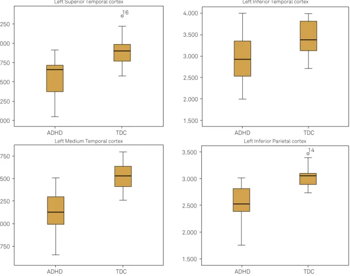
he diference between TDC and ADHD children was signiicant for the four cortical regions mentioned above. Nevertheless, Digit Span scores were not statistically signii-cant diferent between the groups of children.
Box-plots depicting diferences in the groups’ distribu-tions according to cortical thickness of the left superior, me-dial and inferior temporal cortices, and left inferior parietal regions were represented in Figure 1.
Considering that no statistically signiicant diference between Digit Span scores of TDC and ADHD children was detected, scatter-plots showing correlations between corti-cal thickness and these scores were built taking both groups into account together (Figure 2).
Table 3 shows the correlation between the cortical thickness of each brain area selected and the values ob-tained in the Digit Span subtests (Spearman’s correlation coeicient). Here, we observed a direct association between the scores on the Backwards Digit Span and thickness of the left medial temporal cortex (Spearman’s correlation coeicient = 0.499; signiicant at the 0.01 level; 2-tailed). To a lesser extent, we observed the same association with the left inferior temporal cortex (Spearman’s correlation coeicient = 0.388; signiicant at the 0.05 level; 2-tailed).
DISCUSSION
Our findings have shown a direct relationship between cortical thickness of the left medial temporal cortex and working memory, evaluated through the Backwards Digit Span test. The correlation of WM with the left inferior tem-poral cortical thickness was also observed, but to a lesser extent. No correlation was observed between the Forward Digit Span and cortical thickness in these brain regions.
his last result is in accordance with previous studies that discuss the validity of using both Digit Span conditions – for-wards and backfor-wards – separately, since they involve difer-ent neuropsychological circuits22
and only the reverse condi-tion addresses working memory13.
Traditionally, the frontal lobes are recognized as respon-sible for the control of complex cognitive processes such
Table 1. Values of cortical thickness and Digit Span scores in children with attention deficit/hyperactivity disorder (ADHD) and typically developing children (TDC).
Region ADHD TDC Mann-Whitney U p-value BH corrected (p-value)
Left superior temporal cortex* 2.5 2.9 30.5 0.000 0.000
Left medial temporal cortex* 3.1 3.5 22.0 0.000 0.000
Left inferior temporal cortex* 2.9 3.4 65.0 0.010 0.015
Left inferior parietal cortex* 2.5 3.0 21.0 0.000 0.000
Forward Digit Span 7.2 7.1 134.5 0.958 0.956
Backwards Digit Span 3.6 4.2 84.0 0.063 0.061
* in mm; BH: Benjamini-Hochberg correction for multiple comparisons.
Table 2. Comparison between the variables of age, gender and intelligence quotient (IQ) in children with attention deficit/ hyperactivity disorder (ADHD) and typically developing children (TDC).
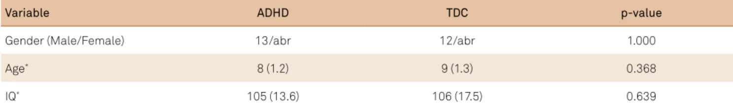
Variable ADHD TDC p-value
Gender (Male/Female) 13/abr 12/abr 1.000
Age* 8 (1.2) 9 (1.3) 0.368
IQ* 105 (13.6) 106 (17.5) 0.639
as decision-making, planning and sustained attention23. However, more recently, there has been strong evidence in-dicating the contribution of the medial temporal lobe to
WM24,25,26,27. In a less robust manner, the inferior temporal
lobe’s role in WM has also been demonstrated28.
According to the classic definition, WM relies on three interconnected subsystems: the phonological loop, responsible for the initial processing and storage of verbal information, the visual sketchpad, responsible for the ini-tial processing of nonverbal information; and the episodic buffer, responsible for the connection of the information between the former systems29
. The frontotemporal path-ways play an important role in integrating these three sub-systems of WM30.
he role of the medial temporal lobe in WM is not fully elucidated, as there are studies demonstrating that this re-gion inluences WM only when the task depends more on long-term memory processes31.
Our sample size should be considered a limitation to the study and the results may not be generalized. A larger sample
Left Superior Temporal cortex Left Inferior Temporal cortex
ADHD TDC
ADHD TDC
Left Medium Temporal cortex Left Inferior Parietal cortex
ADHD TDC
ADHD TDC
3.250
3.000
2.750
2.500
2.250
2.000
4.000
3.500
3.000
2.500
2.000
1.500
3.750
3.500
3.250
3.000
2.750
3.500
3.000
2.500
2.000
1.500 16
14
ADHD: Attention deficit/hyperactivity disorder; TDC: typically developing children.
Figure 1. Box-plots depicting differences in distributions between the groups according to cortical thickness of left superior, medial and inferior temporal cortices, and left inferior parietal regions.
Table 3. Values of Spearman’s correlation coefficient between cortical thickness of each brain area selected and the values obtained in the Digit Span test.
Sig.: significant; BH: Benjamini-Hochberg; *Correlation is significant at the 0.10 level (2-tailed); **Correlation is significant at the 0.05 level (2-tailed); ***Correlation is significant at the 0.01 level (2-tailed).
Region Forward
digit span
Backwards digit span Left superior temporal cortex
Spearman’s coefficient -0.196 0.322*
Sig. (2-tailed) 0.274 0.067
BH corrected p-value 0.419 0.089
Left medial temporal cortex
Spearman’s coefficient -0.190 0.499**
Sig. (2-tailed) 0.288 0.003
BH corrected p-value 0.419 0.012
Left inferior temporal cortex
Spearman’s coefficient -0.181 0.388*
Sig. (2-tailed) 0.314 0.026
BH corrected p-value 0.419 0.052
Left inferior parietal cortex
Spearman’s coefficient -0.026 0.136
Sig. (2-tailed) 0.886 0.451
Id ADHD TDC 12.0
10.0
8.0
6.0
12.0
10.0
8.0
6.0
12.0
10.0
8.0
6.0
12.0
10.0
8.0
6.0
7.0
6.0
4.0
2.0 5.0
3.0
7.0
6.0
4.0
2.0 5.0
3.0
7.0
6.0
4.0
2.0 5.0
3.0
7.0
6.0
4.0
2.0 5.0
3.0
2.000 2.250 2.500 2.750 3.000 3.250 1.500 2.000 2.500 3.000 3.500 4.000
2.750 3.000 3.250 3.500 3.750 1.500 2.000 2.500 3.000 3.500
2.000 2.250 2.500 2.750 3.000 3.250 1.500 2.000 2.500 3.000 3.500 4.000
2.750 3.000 3.250 3.500 3.750 1.500 2.000 2.500 3.000 3.500
Id ADHD TDC
Id ADHD TDC
Id ADHD TDC
Id ADHD TDC
Id ADHD TDC
Id ADHD TDC
Id ADHD TDC
Left Superior Temporal cortex Left Inferior Temporal cortex
Forwards Digit Span
Left Medium Temporal cortex Left Inferior Parietal cortex
Left Superior Temporal cortex Left Inferior Temporal cortex
Backwards Digit Span
Left Medium Temporal cortex Left Inferior Parietal cortex
might show a statistically signiicant diference between the Backwards Digit Span scores of TDC and ADHD children. Although many steps were taken to minimize movement bias during examinations, head motion in ADHD patients can be considered a problem and a limitation of this study.
his pilot study was not able to conirm that work-ing memory problems can diferentiate ADHD from TDC. Nevertheless, our results suggest, for the irst time, a direct correlation between the Backwards Digit Span and left me-dial temporal cortical thickness.
References
1. Polanczyk GV, Willcutt EG, Salum GA, Kieling C, Rohde LA. ADHD prevalence estimates across three decades: an updated systematic review and meta-regression analysis. Int J Epidemiol. 2014;43(2):434-42. doi:10.1093/ije/dyt261
2. Castellanos FX, Lee PP, Sharp W, Jeffries NO, Greenstein DK, Clasen LS et al. Developmental trajectories of brain volume abnormalities in children and adolescents with attention deficit/hyperactivity disorder. JAMA. 2002;288(14):1740-8. doi: 10.1001/jama.288.14.1740
3. Frodl T, Skokauskas N. Meta-analysis of structural MRI studies in children and adults with attention deficit hyperactivity disorder indicates treatment effects. Acta Psychiatr Scand. 2012;125(2):114-26. doi:10.1111/j.1600-0447.2011.01786.x
4. Shaw P, Malek M, Watson B, Greenstein D, Rossi P, Sharp W. Trajectories of cerebral cortical development in childhood and adolescence and adult attention-deficit/hyperactivity disorder. Biol Psychiatry. 2013;74(8):599-606. doi:10.1016/j.biopsych.2013.04.007
5. Kaufman AS, Lichtenberger EO. Essentials of WAIS-III Assessment. New York: John Wiley & Sons; 1999.
6. Baddeley AD. Working memory, thought, and action. New York: Oxford University Press; 2007.
7. Cockcroft K. Working memory functioning in children with attention-deficit/hyperactivity disorder (ADHD): a comparison between subtypes and normal controls. J Child Adolesc Ment Health. 2011;23(2):107-18. doi:10.2989/17280583.2011.63454
8. Wechsler DW. WISC III: escala de Inteligencia Wechsler para crianças: manual. 3a ed. São Paulo: Casa do Psicologo; 2002.
9. Groth-Marnat G, Baker S. Digit Span as a measure of everyday attention: a study of ecological validity. Percept Mot Skills. 2003;97(3 Pt 2):1209-18. doi:10.2466/PMS.97.8.1209-1218
10. Martinussen R, Hayden J, Hogg-Johnson S, Tannock R. A meta-analysis of working memory impairments in children with
attention-deficit/hyperactivity disorder. J Am Acad Child Adolesc Psychiatry. 2005;44(4):377-84. doi:10.1097/01.chi.0000153228.72591.73
11. Figueiredo VLM. Adaptação e padronização Brasileira da escala de inteligência Wechsler para crianças, terceira edição – WISC-III. São Paulo: Casa do Psicólogo; 2002.
12. Rosenthal EN, Riccio CA, Gsanger KM, Jarratt KP. Digit Span components as predictors of attention problems and executive functioning in children. Arch Clin Neuropsychol. 2006;21(2):131-9. doi:10.1016/j.acn.2005.08.004
13. Coutinho G, Mattos P, Malloy-Diniz LF. Neuropsychological differences between attention deficit hyperactivity disorder and control children and adolescents referred for academic impairment. Rev Bras Psiquiatr. 2009;31(2):141-4. doi:10.1590/S1516-44462009000200011
14. Shaw P, Eckstrand K, Sharp W, Blumenthal J, Lerch JP, Greenstein D et al. Attention-deficit/hyperactivity disorder is characterized by a delay in cortical maturation. Proc Natl Acad Sci USA. 2007;104(49):19649-54. doi:10.1073/pnas.0707741104
15. American Psychiatric Association. Diagnostic and statistical manual of mental disorders, 4th edition (DSM-IV). Washington, DC: American Psychiatric Association; 1994.
16. Kaufman J, Birmaher B, Brent D, Rao U, Ryan N. The Schedule for affective disorders and schizophrenia for school-age children. Pittsburgh: University of Pittsburgh Medical Center; 1996.
17. Brasil HHA, Bordin IA. Convergent validity of K-SADS-PL by comparison with CBCL in a Portuguese speaking outpatient population. BMC Psychiatry. 2010;10(1):83. doi:10.1186/1471-244X-10-83
18. American Psychiatric Association. Diagnostic and statistical manual of mental disorders, 5th edition (DSM-V). Washington, DC: American Psychiatric Association; 2013.
19. Desikan RS, Ségonne F, Fischl B, Quinn BT, Dickerson BC, Blacker D et al. An automated labeling system for subdividing the human cerebral cortex on MRI scans into gyral based regions of interest. Neuroimage. 2006;31(3):968-80. doi:10.1016/j.neuroimage.2006.01.02
20. Kuperberg GR, Broome MR, McGuire PK, David AS, Eddy M, Ozawa F et al. Regionally localized thinning of the cerebral cortex in schizophrenia. Arch Gen Psychiatry. 2003;60(9):878-88. doi:10.1001/archpsyc.60.9.878
21. Shaw P, Lerch J, Greenstein D, Sharp W, Clasen L, Evans A et al. Longitudinal mapping of cortical thickness and clinical outcome in children and adolescents with attention-deficit/hyperactivity disorder. Arch Gen Psychiatry. 2006;63(5):540-9. doi:10.1001/archpsyc.63.5.540
22. Salat DH, Buckner RL, Snyder AZ, Greve DN, Desikan RS, Busa E et al. Thinning of the cerebral cortex in aging. Cereb Cortex. 2004;14(7):721-30. doi:10.1093/cercor/bhh032
23. Ramsay MC, Reynolds CR. Separate digits tests: a brief history, literature review, and a reexamination of the factor structure of the Test of Memory and Learning (TOMAL). Neuropsychol Rev.1995;5(3):151-71. doi:10.1007/BF02214760
24. Gilbert SJ, Burgess PW. Executive function. Curr Biol. 2008;18(3):R110-4. doi:10.1016/j.cub.2007.12.014
25. Axmacher N, Schmitz DP, Wagner T, Elger CE, Fell J. Interactions between medial temporal lobe, prefrontal cortex, and inferior temporal regions during visual working memory: a combined intracranial EEG and functional magnetic resonance imaging study. J Neurosci. 2008;28(29):7304-12. doi:10.1523/JNEUROSCI.1778-08.2008
26. Axmacher N, Elger CE, Fell J. Working memory-related hippocampal deactivation interferes with long-term memory formation. J Neurosci. 2009;29(4):1052-60. doi:10.1523/JNEUROSCI.5277-08.2009
27. Cashdollar N, Duncan JS, Duzel E. Challenging the classical distinction between long-term and short-term memory: reconsidering the role of the hippocampus. Future Neurol. 2011;6(3):351-62. doi:10.2217/fnl.11.12
28. Stretton J, Winston G, Sidhu M, Centeno M, Vollmar C, Bonelli S et al. Neural correlates of working memory in Temporal Lobe Epilepsy: an fMRI study. Neuroimage. 2012;60(3):1696-703. doi:10.1016/j.neuroimage.2012.01.126
29. Ranganath C, Cohen MX, Dam C, D’Esposito M. Inferior temporal, prefrontal, and hippocampal contributions to visual working memory maintenance and associative memory retrieval. J Neurosci. 2004;24(16):3917-25. doi:10.1523/JNEUROSCI.5053-03.2004
30. Baddeley A. Working memory and language: an overview. J Commun Disord. 2003;36(3):189-208. doi:10.1016/S0021-9924(03)00019-4