ABSTRACT
Diabetes mellitus, a disease that has been reaching epidemic proportions, is an important risk factor to the development of cardiovascular complica-tion. Diabetes causes changes within the cardiac structure and function, even in the absence of atherosclerotic disease. The left ventricular diastolic dysfunction (VE) represents the earliest pre-clinical manifestation of dia-betic cardiomyopathy, preceding the systolic dysfunction and being able to evolve to symptomatic heart failure. The doppler echocardiography has emerged as an important noninvasive diagnostic tool, providing reliable data in the stages of diastolic function, as well as for systolic function. With the advent of recent echocardiographic techniques, such as tissue Doppler and color M-mode, the accuracy in identifying the moderate diastolic dys-function, the pseudonormal pattern, has significantly improved. Due to cardiometabolic repercussions of DM, a detailed evaluation of cardiovas-cular function in diabetic patients is important, and some alterations may be seen even in patients with gestational diabetes. (Arq Bras Endocrinol Metab 2007;51/2:168-175)
Keywords:Diabetes; Echocardiography; Diastole; Doppler; Cardiomyopathy
RESUMO
Disfunção Diastólica do Ventrículo Esquerdo no Diabetes: Uma Atualização.
O Diabetes Mellitus (DM), doença que vem atingindo proporções epidêmi-cas, é um importante fator de risco para o desenvolvimento de compli-cação cardiovascular. O DM leva a alterações cardíacas estruturais e fun-cionais, mesmo na ausência de doença aterosclerótica. A disfunção diastólica do ventrículo esquerdo (VE) representa a manifestação pré-clíni-ca mais precoce da pré-clíni-cardiomiopatia diabétipré-clíni-ca, precedendo a disfunção sistólica e podendo progredir para insuficiência cardíaca sintomática. O Doppler ecocardiograma tem se mostrado uma importante ferramenta diagnóstica não-invasiva, fornecendo dados confiáveis dos estágios da função diastólica do VE, assim como da função sistólica. Com o advento de recentes técnicas de ecocardiografia, como o Doppler tecidual e o color M-mode, a acurácia em identificar a disfunção diastólica moderada, padrão pseudonormal, aumentou significativamente. Frente às reper-cussões cardiometabólicas do DM, é importante uma avaliação detalhada da função cardiovascular dos pacientes diabéticos, sendo que algumas alterações podem ser vistas até mesmo em pacientes com o diabetes gestacional. (Arq Bras Endocrinol Metab 2007;51/2:168-175)
Descritores:Diabetes; Ecocardiograma; Diástole; Doppler; Cardiomiopatia
atualização
CLÁUDIAMARIAV. FREIRE ANALUIZAM.T. MOURA MÁRCIA DEMELO BARBOSA LUCASJOSÉ DE C. MACHADO ANELISE IMPELIZIERE NOGUEIRA ANTÔNIORIBEIRO-OLIVEIRAJR.
Laboratório de Pesquisas em Endocrinologia, Departamento de Clínica Médica, Faculdade de Medicina da Universidade Federal de Minas Gerais (CMVF, ALMTM LJCM, AIN & AR-OJr), e Ecocenter — Serviço de Ecocardiografia do Hospital
Socor (CMVF & MMB), Belo Horizonte, MG.
D
IABETES MELLITUS is one of the most commondiseases in the world and is acquiring epidemic proportions. Its prevalence is growing in developed and developing countries. More than 5% of adults have this disease, with prevalence arising from 1% in the youth to 13% in those older than 60 years. Recently the American Diabetes Association (ADA) and the World Health Organization (WHO) sharpened the criteria for diagnosing DM, contributing to the increase in the number of diagnosis in earlier ages. Because of the increasing frequency of diabetes in the past 30 years, the importance of cardiovascular disease attributable to diabetes will continue to increase, even if the incidence in the non-diabetic population contin-ues to diminish (1).
In 1997 and 2003, an International Expert Committee recommended changes to the diagnostic criteria for diabetes (2,3). So, precise statistical data regarding the prevalence of new categories of impaired glucose metabolism, such as impaired fasting glucose (IFG) and impaired glucose tolerance (IGT) might have changed in different populations with the appli-cation of the most recent diagnostic criteria for dia-betes and pre-diadia-betes. The American Diadia-betes Asso-ciation data show that 20,8 million people have dia-betes and other 54 million people have pre-diadia-betes, IFG or IGT (4). In two Brazilian population samples, they found a prevalence around 7,5% of IFG at that time (5,6).
The epidemic of diabetes represents a major burden to health care systems around the world. Both type 1 and type 2 diabetes are increasing in children and adolescents. However, more alarming is the increase in type 2 diabetes in the youth related to obe-sity and physical inactivity as was shown in clinical series and some population data from the USA and Canada (7,8).
Cardiovascular complications are known to be the main cause of death and morbidity in diabetic patients, as over 75% of all diabetic patients die from cardiovascular events (9). There is an increased rate of ischemic heart disease and cardiomyopathy, which may lead to congestive heart failure in the absence of coro-nary atherosclerosis. Heart failure is a common and seri-ous co-morbidity of diabetes. The Framingham study (10) demonstrated an increased risk in heart failure in patients with diabetes and that it has a greater impact on the incidence of congestive heart failure, especially in women. It has been shown a 2-fold higher incidence of heart failure in men with diabetes and a 5-fold increase in women. Additional trials — Studies of Left Ventricu-lar Dysfunction (SOLVD), the Heart Outcomes
Pre-vention Evaluation study (HOPE), the Cardiovascular Health Study (CHS) and nationwide case-control study — also identified diabetes as a major risk factor for the development of heart failure (11-13). Recently a study of health maintenance organization in nearly 10,000 type 2 diabetic patients, 12% had heart failure at entry and about 3.3% of type 2 diabetic subjects developed heart failure each year (14).
Hypertension and coronary artery disease, known co-morbidities of diabetes, are established causes of heart failure. The most prominent risk factor for heart failure in diabetic patients is prior history of coronary artery disease. Furthermore, heart failure is more frequent in diabetic than in non-diabetic patients with myocardial ischemic injury (11). Diabetes has been considered of such importance to the develop-ment of heart failure that it has been incorporated as an independent risk factor to it in the American Col-lege of Cardiology/American Heart Association (15). Accumulating data from experimental, patho-logical, epidemiological and clinical studies have shown that diabetes causes changes within the cardiac structure and function, in the absence of coronary ath-erosclerosis, hypertension or any other known cardiac disease. However, the coexistence of myocardial ischemia, hypertension, and a specific diabetic car-diomyopathy seems to be independent but contributes to the biochemical, anatomic, and functional alter-ations in cardiac cells and tissues that impair cardiac function. Factors that can cause microvascular abnor-malities, endothelial dysfunction, derangement of myocardial metabolism and autonomic neuropathy, such as hyperglycemia, hypertriglyceridemia and hypertension are postulated as etiological factors (13,16-18).
The most important mechanisms of diabetic cardiomyopathy are metabolic disturbances (increased free fatty acids, carnitine deficiency, changes in calcium homeostasis), myocardial fibrosis (increases in angiotensin II, IGF-I, and inflammatory cytokines), small vessel disease (microangiopathy, impaired coro-nary flow reserve, and endothelial dysfunction), car-diac autonomic neuropathy (denervation and alter-ations in myocardial catecholamine levels), and insulin resistance (hyperinsulinemia and reduced insulin sensi-tivity) (19).
dys-function represents the earliest preclinical manifesta-tion of diabetic cardiomyopathy, preceding the systolic dysfunction, and that it can progress to symptomatic heart failure (21,22).
DIASTOLIC HEART FAILURE
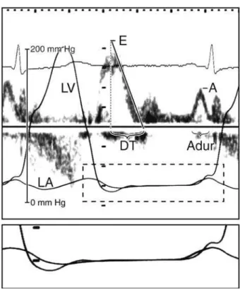
Diastole is the portion of the cardiac cycle that begins with aortic closure and ends with mitral closure. In diastolic dysfunction, the abnormality in LV relaxation and/or compliance alters the onset, rate, and extent of LV pressure decline and filling during diastole. These changes create an abnormal relation between left ven-tricular pressure and volume so that higher filling pres-sures are needed to maintain normal LV end-diastolic volume and cardiac output. This may result in higher filling pressures at rest; however, it more frequently produces elevated filling pressures during exercise, resulting in exertional dyspnea and fatigue. For the noninvasive assessment of diastolic function, we may rely on Doppler studies of mitral and pulmonary veins inflow patterns. Diastole can be divided into four stages, for descriptive purposes (23).
1st stage: Isovolumic relaxation time – the time
taken from aortic valve closure to mitral valve opening. This phase is attributed mainly to myocardial relax-ation and has been shown to be an energy-requiring process. During this interval, the intraventricular pres-sure falls at a rapid rate, while ventricular volume remains constant.
2nd stage: Rapid filling phase (E wave of mitral
inflow and D wave in pulmonary vein flow) – when left ventricular pressure falls below atrial pressure, the mitral valve opens and the rapid filling begins resulting in a rapid increase in ventricular volume. It represents an interaction of an active relaxation (suction) and passive viscoelastic properties of the myocardium (compliance). 3rd stage: Passive filling (diastasis) – in this
phase, left atrial and ventricular pressures are almost equal, so filling is the result of pulmonary venous flow. It is related to left ventricular compliance.
4th stage: Atrial contraction (A wave of mitral
inflow and atrial reversal wave in pulmonary vein flow) – this is an active process and contributes with approx-imately 15% of left ventricular filling in normal sub-jects. The subsequent rise in left ventricular pressure leads to mitral valve closure. This phase is most affect-ed by left ventricular stiffness (figure 1).
Heart failure is defined as a pathophysiological state in which an abnormality of cardiac function is responsible for failure of the heart to pump blood at a
rate commensurate with metabolic requirements or to do so only from an elevated filling pressure. It is usu-ally, but not always, caused by a defect in myocardial contraction. However, in some patients with heart fail-ure a similar clinical syndrome is present but there is no detectable abnormality in myocardial contraction function (24). Thus, heart failure, a clinical syndrome, may occur in the presence of either a normal or abnor-mal left ventricular ejection fraction (EF). It is widely accepted that the pathophysiology of heart failure in patients with decreased ejection fraction involves a predominant (though not isolated) decrease in systolic function justifying the term “systolic heart failure”. In contrast, the underlying pathophysiology of patients with heart failure with normal left ventricle systolic function — normal ejection fraction — involves a pre-dominant (not isolated) abnormality in diastolic func-tion, the “diastolic heart failure”. Further stratification of congestive heart failure (CHF) subjects into those with systolic dysfunction and those with predominant-ly diastolic dysfunction has been suggested as impor-tant for clinical decisions, once the therapeutic and prognostic differences between these 2 subsets of patients and, in most disease states, diastolic dysfunc-tion precedes the onset of systolic dysfuncdysfunc-tion (25).
Figure 1.Mitral inflow pattern and left ventricular pressure curves.
Cardiac heart failure is a major public health problem in developed countries, and a high propor-tion of patients admitted due to CHF have normal left ventricular systolic function. Diastolic heart failure is a clinical syndrome characterized by the symptoms and signs of heart failure, a preserved systolic function and abnormal diastolic function. In this situation the left ventricle is unable to accept an adequate volume of blood during diastole, at normal diastolic pressures and at volumes sufficient to maintain an appropriate stroke volume. These abnormalities are caused by a decrease in ventricular relaxation and/or an increase in ventricular stiffness (26).
This growing recognition that CHF may be caused by a common predominant abnormality, not in systolic, but in diastolic function, and that it causes significant morbidity and mortality, called for further definition of the diagnostic criteria. In the United States of America it is estimated that diastolic heart failure accounts for more than 25% of the total cost of CHF (27). The prevalence of asymptomatic diastolic dysfunction was estimated in 27% in the general pop-ulation, reaching higher prevalence with aging. Of the patients hospitalized for heart failure, 35% to 40% pre-sent with diastolic heart failure and, in the community setting, this number increases from 45% to 55% (28).
Unfortunately, the differentiation between sys-tolic and diassys-tolic heart failure cannot be made at bed-side, based on history, physical examination, ECG, or chest radiograph alone. A careful history will detect symptoms of CHF, although about 20% of patients with left ventricular systolic dysfunction do not report symptoms (table 1). Therefore, the diagnosis of heart failure, especially in diabetic patients who have many associated co-morbidities, may require further testing in order to make an appropriate diagnosis. Although electrocardiogram and chest X-ray may be helpful in
demonstrating hypertrophy or left ventricular enlarge-ment, echocardiography is important to visualize the heart for any structural/functional changes, and is the recommended test if CHF is suspected (29).
Much effort was made to propose satisfactory diagnostic criteria for diastolic heart failure. A simple method suggested by Gandhi et al. (30) addressed the requirement for the presence of an EF ≥50% within 72 hours of the heart failure event. Under most circum-stances, measurement of EF within 72 hours is suffi-cient to meet diagnostic criteria for diastolic heart fail-ure. According to Zile et al. (31), the diagnosis of diastolic heart failure can be made without the mea-surement of diastolic function if these two criteria are present together: 1) symptoms and signs of heart fail-ure and, and 2) EF > 50%. Therefore, the objective measurement of diastolic function serves to confirm rather than establish the diagnosis.
Cardiac catheterization with simultaneous pres-sure and volume meapres-surements is the “gold standard” for assessing left ventricular function. However, it is invasive and cannot be performed in the majority of patients with suspected diastolic dysfunction (32). Valuable knowledge has been acquired with the advent of Doppler echocardiography. Because diastole is a complex sequence of interrelated events, and the factors contributing to diastole are highly sensitive to changes in loading conditions, heart rate, and contrac-tility, no currently available method can completely assess diastolic function. During the last two decades, Doppler echocardiography has emerged as an impor-tant and easy method to perform noninvasive diagno-sis, providing reliable data on diastolic performance.
Based on Doppler transmitral flow, a grading system for diastolic dysfunction has been proposed. It has been demonstrated that mitral inflow shows a dis-ease progression over time within the myocardium.
Table1.Prevalence (%) of signs and symptoms in systolic and diastolic heart failure.
Symptoms and Signs Diastolic Heart Failure Systolic Heart Failure (EF > 50%) (EF < 50%)
Dyspnea on exertion 85% 96%
Paroxysmal nocturnal dyspnea 55% 50%
Rales 72% 70%
S3 45% 55%
S4 45% 56%
Edema 30% 40%
Cardiomegaly 90% 96%
Pulmonary Hypertension 75% 80%
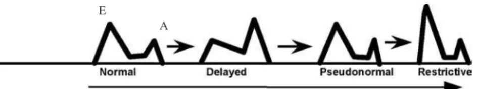
Three patterns mainly based on the E/A ratio have been proposed. The first abnormal filling pattern is the delayed relaxation that results in a reversed E/A ratio (E/A < 1), being the earliest stage of heart disease. The second pattern represents abnormalities in both relaxation and compliance and is known as pseudo-normalization because of an apparently normal E/A ratio (E/A > 1). This pattern implies an increase in left atrial pressure and represents a moderate compromise in diastolic function. The third abnormal filling pat-tern is termed restrictive filling found in patients with severe compromise of left ventricular compliance and elevated ventricular filling pressures, reflecting an advanced stage of disease (33) (figure 2).
These patterns can evolve from one to another in a single patient, with changes in disease evolution, treatment, loading conditions, and heart rate. There-fore, non-invasive assessment of relaxation or diastolic compliance should be interpreted with caution. Fur-ther echocardiographic examination must be used when diastolic function is indeterminate: the analysis of pulmonary venous flow pattern, analysis of mitral flow during the Valsalva maneuver, and new tech-niques of Doppler tissue imaging and color M-mode assessment of flow propagation velocity that are rela-tively load independent (34-36).
There is evidence that brain natriuretic peptide (BNP) levels are increased in patients with systolic and diastolic heart failure, although in diastolic heart fail-ure it is not as established as for systolic heart failfail-ure. Such biochemical abnormalities could provide further evidence of an underlying disease process, and it may play a role in diagnosis of diastolic heart failure (37,38).
Radionuclide angiography and magnetic reso-nance imaging offer opportunities for studying dias-tolic function; however, they are not performed in routine clinical practice.
No large randomized clinical trial results are yet available to guide management of patients with dias-tolic heart failure. There are some ongoing clinical
tri-als to evaluate this issue. The American College of Cardiology/American Heart Association/European Society of Cardiology guidelines reflect this paucity of data (15,39). However, they recommend: control of symptoms by reducing ventricular filling pressures without reducing cardiac output; addition of diuretics and nitrates for symptomatic patients; control of arte-rial hypertension; probable benefit of adding calcium channel blockers, angiotensin-converting enzymes inhibitors and angiotensin II receptor blockers beyond the treatment of hypertension; and control of tachy-cardia. It’s reasonable to infer that improved glucose control may improve diastolic heart failure in diabetic patients, although the effect of different diabetic treat-ments may not reverse the diastolic dysfunction. How-ever, in a large cohort of diabetic patients, it was shown that a better glycemic control reduces the risk of heart failure (40-43).
DIABETES AND LEFT VENTRICULAR DIASTOLIC DYSFUNCTION
Many conditions besides aging are associated with and are likely to contribute to diastolic dysfunction and diastolic heart failure such as hypertension, coronary artery disease, atrial fibrillation, and diabetes. Diabetes has such an important influence on the development of CHF that it has been incorporated as a risk factor in the American College of Cardiology/American Heart Association guidelines (15).
One of the factors that are associated with the development of diabetic cardiomyopathy is hyper-glycemia. Increasing evidence suggests that altered substrate supply and utilization by cardiac myocytes could be the primary injury in the pathogenesis of this specific heart muscle disease. However, even in type 2 diabetic patients without cardiac involvement, uncon-trolled hyperglycemia is described to provoke diastolic left ventricular dysfunction (44,45). Alteration in left ventricular diastolic function seems to be related to
Figure 2.Schematic drawing of mitral inflow patterns during different stages of diastolic dysfunction. E= early inflow wave; A= atrial contraction, A wave.
E
concentrations of fasting plasma glucose and glycated hemoglobin even below the threshold of diabetes (46). Furthermore, each 1% increase in HbA1c value
has been associated with an 8% increase in the risk of heart failure (12,43), and glycosylated hemoglobin > 8 has also been associated with diastolic dysfunction (47), although the glycemic control may not reverse the diastolic dysfunction (48-50).
Other changes closely associated with abnor-malities in diastolic function in diabetic patients are the impairment of gene expression to what has been called the fetal gene program, leading to myocardial impairment of calcium handling and altered regulation of genes for αand β-myosin heavy chains (29,51).
Of note, impairment of diastolic performance is non-specific and frequently observed in many diseases such as hypertension, hypertrophic cardiomyopathy and coronary artery disease, while systolic function remains intact. However, alterations in diastolic func-tion have been observed in diabetic patients without any co-morbidities and before cardiovascular tradi-tional complications. Investigations using cardiac catheterization showed alterations in left ventricular diastolic filling pressures in diabetic patients without any significant coronary artery disease or systolic dys-function (52,53). Raev et al. (21) showed alterations in diastolic function in young type 1 diabetic patients without cardiovascular disease and suggested that these alterations could be the earliest signs of the dia-betic cardiomyopathy. Their findings were quite plau-sible because diastolic abnormalities generally occur 8 years after the onset of type 1 diabetes, and systolic dysfunction establishment has been described even later in the disease evolution (48).
With the advent of recent echocardiographic techniques such as tissue Doppler imaging and color M-mode, the ability to accurately detect diastolic dysfunc-tion has significantly improved. Boyer et al. detected altered left ventricular filling in 46% in asymptomatic normotensive type 2 diabetic patients when screened by conventional Doppler, whilst newer techniques showed diastolic dysfunction in 75% of patients (54). A more recent study in patients with type 2 diabetes free of any detectable cardiovascular disease found that 47% of the subjects had diastolic dysfunction, of which 30% had the first stage dysfunction — impaired relaxation, and 17% had second stage dysfunction — pseudonormal filling, a more advanced abnormality of left ventricular relaxation and compliance, which otherwise would be classified as having a normal diastolic physiology (55).
These new techniques, especially tissue Doppler image and color M-mode, have provided information
to overcome some technical limitations concerning traditional Doppler echocardiographic studies of dias-tolic function. Until recently, the existence of the pseudonormal left ventricular filling pattern, a second stage of diastolic dysfunction, was not evaluated in all the earlier studies. Therefore it is possible that many patients with diabetic diastolic dysfunction with a pseudonormal pattern would not have missed this diagnosis if these new techniques had been available by the time the studies were done. Furthermore, this may account for the discrepancies previously related to the prevalence of diastolic dysfunction, especially in a young diabetic population.
The problem of diabetes and metabolic syn-drome appearing in young ages should prompt early interventions because by the time type 2 diabetes is diagnosed, more than 30–50% of patients will already have some evidence of vascular disease (56,57). We now know that diabetes may be doing silent and con-tinuous harm to heart, even in those with no known manifested cardiac complications, as shown by some degree of diastolic dysfunction early shown. But we do not know however, how early these damages to cardiac function occur.
We considered whether a temporary condition of diabetes such as gestational diabetes mellitus (GDM) could have any impact in cardiac function. GDM may be considered as an early expression of insulin metabolic syndrome and a harbinger to type 2 diabetes (58), providing an excellent model for the study of precursors of diabetic cardiomyopathy in a young, apparently healthy population. We found that these patients had different left ventricular diastolic filling profile when compared to normal pregnant patients, and we also showed that some of these changes persisted post partum (49).
All of these backgrounds emphasize the need of a closer follow-up and early intervention for cardiovas-cular risk factors and metabolic glucose control in patients with the diagnosis of diabetes, GDM and metabolic syndrome. However, there is no available data on the literature supporting an early investigation in clinical practice with Doppler echocardiography and its new techniques in the approach of these patients at risk for cardiovascular complications.
diastolic alterations, so as these techniques can be rou-tinely recommended as screening tests for cardiovascu-lar impairment in this population.
ACKNOWLEDGEMENTS
We are indebted to Nabila Salahudian for kindly reviewing the English of this manuscript.
REFERENCES
1. Howard BV, Rodriguez BL, Bennett PH, Harris MI, Hamman R, Kuller RH, et al. Prevention conference VI: diabetes and car-diovascular disease: writing group I: epidemiology. Circula-tion 2002;105(18):e132-7.
2. The Expert Committee on the diagnosis and classification of Diabetes Mellitus: report of the expert committee on the diag-nosis and classification of diabetes mellitus. Diabetes Care 1997;20:1183-97.
3. The Expert Committee on the diagnosis and classification of Diabetes Mellitus: Follow-up report on the diagnosis of dia-betes mellitus. Diabetes Care 2003;26:3160-7.
4. American Diabetes Association Home Page. Available at: <http://www.diabetes.org>
5. Torquato MT, Montenegro Junior RM, Viana LA, de Souza RA, Lanna CM, Lucas JC, et al. Prevalence of diabetes melli-tus and impaired glucose tolerance in the urban population aged 30-69 years in Ribeirão Preto (São Paulo) Brazil. São Paulo Med J 2003;121(6):224-30.
6. Schaan BD, Harzhein E, Gus I. Cardiac risk profile in diabetes mellitus and impaired fasting glucose. Rev Saúde Pública 2004;38:529-36.
7. Dabelea D, Hanson RL, Bennett PH, Roumain J, Knowler WC, Pettitt DJ. Increasing prevalence in type 2 diabetes in Ameri-can Indian children. Diabetologia 1998;41(8):904-10. 8. Harris SB, Perkins BA, Whalen-Brough E.
Non-insulin-depen-dent diabetes mellitus among First Nations children: new entity among First Nations people of north western Ontario.
Can Fam Physician 1996;42:869-76.
9. Kleinman JC, Donahue RP, Harris MI, Finucane FF, Madans JH, Brock DB. Mortality among diabetes in a national sample.
Am J Epidemiol 1988;128:389-401.
10. Kannel WB, Mc Gee DL. Diabetes and cardiovascular disease. The Framingham study. JAMA 1979;241(19):2035-8. 11. Shindler DM, Kostis JB, Yusuf S, Quinones MA, Pitt B, Stewart
D, et al. Diabetes mellitus, a predictor of morbidity and mortal-ity in the Studies of Left Ventricular Dysfunction (SOLVD) Trials and Registry. Am J Cardiol 1996;77(11):1017-20.
12. Piccini JP, Klein L, Gheorghiade M, Bonow RO. New insights into diastolic heart failure: role of diabetes mellitus. Am J Med 2004;116(suppl 5A):64S-75S.
13. Bertoni AG, Tsai A, Kasper EK, Brancati FL. Diabetes and idio-pathic cardiomyopathy: a nationwide case-control study.
Diabetes Care 2003;26(10):2791-5.
14. Nichols GA, Hillier TA, Erbey JR, Brown JB. Congestive heart failure in type 2 diabetes: prevalence, incidence, and risk fac-tors. Diabetes Care 2001;24(9):1614-9.
15. Hunt SA, Baker DW, Chin MH, Cinquegrani MP, Feldman AM, Francis GS, et al. ACC/AHA guidelines for the evaluation and management of chronic heart failure in the adult: executive summary a report of the American College of Cardiology/American Heart Association task force on practice guidelines (committee to revise the 1995 guidelines for the evaluation and management of heart failure): developed in collaboration with the International Society for Heart and Lung Transplantation; endorsed by the Heart Failure Society of America. Circulation 2001;104(24):2996-3007.
16. Williams SB, Cusco JA, Roddy MA, Johnstone MT, Creager MA. Impaired nitric oxide-mediated vasodilation in patients with non-insulin dependent diabetes mellitus. J Am Coll Cardiol 1996;27:567-74.
17. Williams SB, Goldfine AB, Timimi FK, Ting HH, Roddy MA, Simonson DC, et al. Acute hyperglycemia attenuates endothelium-dependent vasodilation in humans in vivo. Cir-culation 1998;97:1695-701.
18. Solang I, Malmberg K, Ryden I. Diabetes Mellitus and con-gestive heart failure. Eur Heart J 1999;20:789-95.
19. Fang ZY, Prins JB, Marwick TH. Diabetic cardiomyopathy: evi-dence, mechanisms, and therapeutic implications. Endocr Rev 2004;25(4):543-67.
20. Rubler S, Dlugash J, Yuceoglu YZ, Kumral T, Branwood AW, Grishman A. New type of cardiomyopathy associated with diabetic glomerulosclerosis. Am J Cardiol 1972 ;30(6):595-602.
21. Raev DC. Which left ventricular function is impaired earlier in the evolution of diabetic cardiomyopathy? An echocardio-graphic study of young type I diabetic patients. Diabetes Care 1994;17(7):633-9.
22. Bell DS. Diabetic cardiomyopathy. A unique entity or a
com-plication of coronary artery disease? Diabetes Care
1995;18(5):708-14.
23. Otto CM, Esterling TR, Benedetti T. Echocardiography in the pregnant patient. In: Creasy RK, Resnik R (eds). The practice of clinical echocardiography. 2nd edition. Philadelphia:
WB Saunders Company, 2002. pp. 679-704.
24. Braunwald E. Pathophysiology of heart failure. In: Braunwald E (ed). Heart Disease. 6thedition. Philadelphia: Saunders,
2001. p. 393.
25. Zile MR, Brutsaert DL. New concepts in diastolic dysfunction and diastolic heart failure: Part II: causal mechanisms and treatment. Circulation 2002;105(12):1503-8.
26. Zile MR, Brutsaert DL. New concepts in diastolic dysfunction and diastolic heart failure: Part I: diagnosis, prognosis, and
measurements of diastolic function. Circulation
2002;105(11):1387-93.
27. Vasan RS, Levy D. Defining diastolic heart failure: a call for
standardized diagnostic criteria. Circulation
2000;101(17):2118-21.
28. Redfield MM, Jacobsen SJ, Burnet JC Jr, Mahoney DW, Bai-ley KR, Rodeheffer RJ. Burden of systolic and diastolic ven-tricular dysfunction in the community: appreciating the scope of the heart failure epidemic. JAMA 2003;289(2):194-200. 29. Bell DS. Heart failure: the frequent, forgotten, and often fatal
complication of diabetes. Diabetes Care 2003;26(8):2433-41. 30. Gandhi SK, Powers JC, Nomeir AM, Fowle K, Kitzman DW,
Rankin KM, et al. The pathogenesis of acute pulmonary
edema associated with hypertension. N Engl J Med
2001;344(1):17-22.
31. Zile MR, Gaasch WH, Carroll JD, Feldman MD, Aurigemma GP, Schaer GL, et al. Heart failure with a normal ejection frac-tion: is measurement of diastolic function necessary to make the diagnosis of diastolic heart failure? Circulation 2001;104(7):779-82.
32. Mirsky I. Assessment of diastolic function: suggested methods and future considerations. Circulation 1984;69(4):836-41. 33. Appleton CP, Hatle LK, Popp LR. Relation of transmitral flow
velocity patterns to left ventricular diastolic function: new insights from a combined hemodynamic and Doppler
echocardiographic study. J Am Coll Cardiol 1988;12
(2):426-40.
34. Garcia MJ, Thomas JD, Klein AL. New doppler echocardio-graphic applications for the study of diastolic function. J Am Coll Cardiol 1998;32(4):865-75.
35. Garcia MJ, Smedira NG, Greenberg NL, Main M, Firstenberg MS, Odabashian J, et al. Color M-mode doppler flow propa-gation velocity is a preload insensitive index of left ventricu-lar relaxation: animal and human validation. J Am Coll Car-diol 2000;35(1):201-8.
37. Lubien E, DeMaria A, Krishnaswamy P, Clopton P, Koon J, Kazanegra R, et al. Utility in B-natriuretic peptide in detecting diastolic dysfunction: comparison with Doppler velocities recordings. Circulation 2002;105:595-601.
38. Krishnaswamy P, Lubien E, Clopton P, Koon J, Kazanegra R, Wanner E, et al. Utility of B-natriuretic peptide levels in iden-tifying patients with left ventricular systolic or diastolic func-tion. Am J Med 2001;111:274-9.
39. European Society of Cardiology: The Task Force of the Work-ing Group on Heart Failure: the treatment of heart failure. Task Force of the European Society of Cardiology. Eur Heart J 1997;18:736-53.
40. Tang WH, Young JB. Cardiomyopathy and heart failure in
diabetes. Endocrinol Metab Clin North Am 2001;30
(4):1031-46.
41. Holzmann M, Olsson A, Johansson J, Jensen-Urstad M. Left ventricular diastolic function is related to glucose in a middle-aged population. J Intern Med 2002;251(5):415-20. 42. Henry RM, Kamp O, Kostense PJ, Spijkerman AM, Dekker JM,
van Eijck R, et al. Left ventricular mass increases with deteri-orating glucose tolerance, especially in women: indepen-dence of increased arterial stiffness or decreased
flow-medi-ated dilation: the Hoorn study. Diabetes Care
2004;27(2):522-9.
43. Iribarren C, Karter AJ, Go AS, Ferrara A, Liu JY, Sidney S, et al. Glycemic control and heart failure among adult patients with diabetes. Circulation 2001;103:2668-73.
44. von Bibra H, Hansen A, Dounis V, Bystedt T, Malmberg K, Rydén L. Augmented metabolic control improves myocardial diastolic function and perfusion in patients with non-insulin dependent diabetes. Heart 2004;90:1483-4.
45. Grandi AM, Piantanida E, Franzetti I, Bernasconi M, Maresca A, Marnini P, et al. Effect of glycemic control on the left ven-tricular diastolic function in type 1 diabetes mellitus. Am J Cardiol 2006;97:17-76.
46. Celentano A, Vaccaro O, Tammaro P, Galderisi M, Crivaro M, Oliviero M, et al. Early abnormalities of cardiac function in non-insulin-dependent diabetes mellitus and impaired glu-cose tolerance. Am J Cardiol 1995;76:1173-6.
47. Sanchez-Barriga JJ, Rangel A, Castaneda R, Flores D, Frati AC, Ramos MA, et al. Left ventricular diastolic dysfunction secondary to hyperglycemia in patients with type II diabetes.
Arch Med Res 2001;32(1):44-7.
48. Cosson S, Kevorkian JP. Left ventricular diastolic dysfunc-tion: an early sign of diabetic cardiomyopathy? Diabetes Metab 2003;29(5):455-66.
49. Freire CMV, Nunes Mdo C, Barbosa MM, Longo JR, Nogueira AI, Diniz SS, et al. Gestational diabetes: a condition of early left ventricular diastolic abnormalities. J Am Soc Echocar-diogr 2006;19(10):1251-6.
50. Freire CMV. Avaliação da função diastólica do ventrícu-lo esquerdo em pacientes com diabetes gestacional, no terceiro trimestre de gestação e no pós-parto. (Mestrado). UFMG, 2005.
51. Lowes BD, Gilbert EM, Abraham WT, Minobe WA, Larrabee P, Ferguson D, et al. Myocardial gene expression in dilated cardiomyopathy treated with beta-blocking agents. N Engl J Med 2002;346(18):1357-65.
52. Regan TJ, Lyons MM, Ahmed SS, Levinson GE, Oldewurtel HA, Ahmad MR, et al. Evidence for cardiomyopathy in famil-ial diabetes mellitus. J Clin Invest 1977;60:884-99. 53. D’Elia JA, Weinrauch LA, Healy RW, Libertino JA, Bradley RF,
Leland OS Jr. Myocardial dysfunction without coronary artery disease in diabetic renal failure. Am J Cardiol 1979;43:193-9.
54. Boyer JK, Thanigaraj S, Schechtman KB, Perez JE. Preva-lence of ventricular diastolic dysfunction in asymptomatic, normotensive patients with diabetes mellitus. Am J Cardiol 2004;93(7):870-5.
55. Zabalgoitia M, Ismaeil MF, Anderson L, Maklady FA. Preva-lence of diastolic dysfunction in normotensive, asymptomatic patients with well-controlled type 2 diabetes mellitus. Am J Cardiol 2001;87(3):320-3.
56. Sattar N, Greer IA. Pregnancy complications and maternal cardiovascular risk: opportunities for intervention and screening? BMJ 2002;325(7356):157-60.
57. Davidson MB. Metabolic syndrome/insulin resistance syn-drome/pre-diabetes: new section in diabetes care. Diabetes Care 2003;26(11):3179.
58. Cheung NW, Byth K. Population health significance of gesta-tional diabetes. Diabetes Care 2003;26(7):2005-9.
Endereço para correspondência:
Antônio Ribeiro de Oliveira Jr. Rua São Romão 343/701 30330-120 Belo Horizonte, MG Fax: (31) 3227-5555