2 9 7 2 9 7
Hospital São José do Avaí – Itaperuna, RJ
Mailing address: Gladyston Luiz Lima Souto - Hospital São José do Avaí - Rua Lenira Tinoco Calheiros, 242 – 28300-000 - Itaperuna, RJ - E-mail: [email protected]
Objective - To report initial experience with myocar-dial revascularization surgery (MRS) performed on pa-tients who were totally awake and without an endotra-cheal tube.
Methods - Between January 1994 and May 2001, 272 patients underwent MRS without extracorporeal circulation. In 24, the operations were performed without the use of an endotracheal tube and with the patients to-tally awake and breathing normally. The age ranged from 51-75 years with the predominant male sex. Epidural tho-racic administratios of the anesthesia was performed. Surgery was performed through a habitual anterolateral thoracotomy. During the entire procedure, the left lung remained partially collapsed.
Results - The 24 patients progressed well through the surgery. Pneumothorax time ranged from 70-190 mi-nutes. No electrocardiographic, echocardiographic, or enzymatic alterations occurred that characterized pre-and postoperative infarcts. Twenty-three patients were stable enough to be released after 24 hours.
Conclusion - This technique could be performed on an large number of selected patients. However, more experi-ence is necessary.
Key words: coronary artery leypass graft, epidural thora-cic anesthesia.
Arq Bras Cardiol, volume 79 (nº 3), 297-301, 2002
Gladyston Luiz Lima Souto, Celme da Silva Caetano Júnior, Ary Getúlio de Paula Filho, Marco Antonio Teixeira, Márcio Roberto Moraes de Carvalho, Antonio Carlos Botelho da Silva
Itaperuna, RJ - Brazil
Myocardial Revascularization Surgery with Regional Anesthesia
Without an Endotracheal Tube in Conscious Patients
The development of coronary artery bypass graft sur-gery (CABG) has progressed throughout practically all of the twentieth century, with intervals of inactivity and pro-gress. Alex Carrel 1, in 1910, reported the first experimental
aorta-coronary graft performed, using a segment of the caro-tid artery to connect the coronary artery to the descending aorta. At that time, the pneumothorax created was a serious problem. Murray et al 2 developed a large number of
anasto-moses between the coronary arteries and the systemic arte-ries, including the internal thoracic arteries.
The experimental work of suturing with instruments began in Russia. Kahn et al 3 performed coronary
anastomo-ses in calves with a Russian-American stapler and had pro-mising results. Kolosov et al 4-6 in St. Petersburg was a
pio-neer in clinical work with left internal thoracic artery (LITA) anastomosis with the left anterior descending artery wi-thout cardiopulmonary bypass (CPB) similar to other types of grafts. Garrett et al 7 described a case of an aorta-coronary
bypass with a saphenous vein graft, with the heart beating, with the graft patent after 7 years. This technique did not become popular and was not discussed.
In recent decades, the study of multiple organ dys-function caused by CPB 8-12, the necessity of a longer
hos-pital stay, the short- and long-term results, and the higher cost of this procedure have caused the scientific focus to change. The current trend is toward less aggressive surgery and the development of minimally invasive direct coronary artery bypass (MIDCAB) without CPB.
Reports of this new approach have been widely publi-shed including those by Trapp and Bisarya 13 in Canada,
Ankeney 14 in the United States, Benetti et al 15 and Buffolo
et al 16-18, Lima et al 19, and Lobo et al 20 in Brazil.
Subramanian 21, Robson et al 22, Calafiore et al 23,24, and
Karagoz et al 25 have contributed to the progress of MIDCAB.
2 9 8 2 9 8
The objective of this article is to present our initial ex-perience with myocardial revascularization performed on an ambulatory basis, with a conscious patient and to note some of the particularities observed.
Methods
Between January 1994 and May 2001, 272 patients un-derwent CABG, without the use of CPB. In 24, the surgery was performed without an endotracheal tube and with pa-tients totally conscience. All had a lesion in the left anterior descending artery, and no important chronic pulmonary di-sease was present. The age ranged from 51-75 years with the predominant sex being males. All patients underwent rou-tine preoperative examinations and psychological prepara-tion before the procedure.
Epidural thoracic administration of the anesthesia was performed with 10 mL of solution, composed of 8 mL of bupiva-caine, 0.5 % + 2 mg of morphine, injected by needle at the T4 level, with posterior collocation of an no. We used an 18 G
ca-theter to inject 1 mL of bupivacaine 0.5% to block the 2nd, 3rd, 4th, and 5th left intercostals spaces (LIS). During surgery, and in the postoperative procedure when necessary, we injected 4 mL of bupivacaine 0.5% into the catheter. To obtain 5 metame-res of analgesia at T2 to T6, during part of the surgery the pa-tient used an oxygen mask with two l/minute fluxes.
Monitoring of patients followed the protocol for CABG with CPB. It included continuous electrocardiogra-phy, access to the deep vein by puncturing the right jugular vein, arterial pressure by puncturing the radial artery, moni-toring of arterial saturation with a pulse oximeter, arterial ga-sometry before opening the thorax and every 30 minutes after the thoracotomy.
The access used was a thoracotomy at the 4th LIS, with the patient in the dorsal decubitus position, slightly turned to the right at a ± 30o angle to the surgical table. The
length of the incision ranged from 14 and 16 cm. The LITA was dissected through this incision until the subclavian artery was reached, sometimes it being necessary to remove the cartilage of the 4th rib and occasionally the 5th. The pe-ricardium was opened longitudinally on the anterior fascia, approximately 2 cm from the diaphragm close to the pulmo-nary artery trunk, having its edges well fixed to the edges of the incision. A tourniquet was used close to the LAD zone, where the anastomosis (4/0 Prolene thread, anchored to a small segment of the silicone tube) would have been made, without tightening, with 2 types of stabilizers. After sectio-ning of the LITA, its extremity was prepared, the coronary was opened, with 1 assistant providing light compression by squeezing the coronary artery, and another assistant blowing CO2, washing the site to keep the area blood-free and for the quick placement of the intracoronary shunt. The tourniquet was only tightened in cases of intense bleeding. The LITA-LAD anastomosis was performed with Prolene 7/ 0 with continuous sutures, and the intracoronary shunt was removed at the end of the anastomosis. During the entire ti-me of surgery, the left lung remained partially collapsed.
Anticoagulation was achieved with heparinization of the patient with 2mg/kg of weight, before sectioning of the LITA. The neutralization of the heparin was achieved with protamine in equal dosages. After the anastomosis, the pe-ricardium was partially sutured, the pleural cavity was drai-ned, and the thoracic wall was closed. Throughout the pneumothorax, we asked the patients to breathe deeply and provoke a cough until the total expansion of the lung was obtained.
After the patients entered the intensive care unit, we immediately initiated respiratory exercises and the oral use of aspirin. The drain was removed within 12 hours. After the removal of the drain, the patient was transferred to a ward.
Results
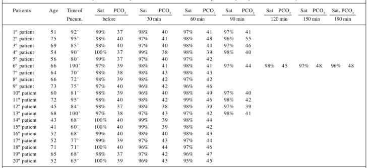
The 24 patients endured the surgery well. The mean time of LITA-LAD anastomosis was 9 minutes and the pneu-mothorax time ranged from 60-190 minutes. During the pro-cedure, no hemodynamic instability or arrhythmias occur-red. No important modifications occurred in blood satura-tion of PO2 and PCO2 (tab. I). One patient needed endotra-cheal intubation and a sternotomy with installation of CPB after a sudden cardiac arrest at the end of surgery when the thorax had already been closed. After the installation of the CPB, the heart began beating, and the ECG, obtained 15 mi-nutes after the procedure, was normal. This arrest was at-tributed to the heart coming out of the pericardial sack with a twisting of the vessels at the base. In this case, the pericar-dium had not been closed. The flux of the LITA was confir-med by a postoperative arteriograph.
Another patient had a thermic lesion of the LITA du-ring its dissection, at the incision angle. The wounded seg-ment was resectioned and a termino-terminal anastomosis was performed, which increased the pneumothorax time to 190 minutes.
No electrocardiographic, echocardiographic, or enzy-matic alterations revealed postoperative necroses. No neu-rologic or infectious complications or clinical pulmonary or radiologic alterations occurred.
All patients, with the exception to the ones that un-derwent CPB, were stable and released after 24 hours.
Discussion
During the last decade, percutaneous coronary bal-loon angioplasty and the implantation of a stent have emer-ged as the first-choice, offering quick myocardial revascu-larization and efficient and less aggressive surgery for patients with 1, 2, or 3 compromised vessels, in the absence of disease in the LAD and their indication seems to have expanded, but the use of CABG has been limited.
The literature shows that the benefits of surgery with LITA-coronary anastomosis is superior on a long-term basis 26. Some surgeons have hesitated to indicate CABG as
2 9 9 2 9 9
combined with CPB and a median sternotomy. On the other hand, with a relatively high incidence of restenosis after angioplasty and stent implantation 27, the need arose for
surgeons to simplify CABG. These simplified techniques have resulted in less aggression against the patient, lower cost, and more effective results.
The technique of myocardial revascularization without CPB has allowed an even greater advance in MID CAB. Initially, limiting factors for the expansion of MIDCAB were the lack of innovations in instrumentation and the discrepancy between the wish of cardiac surgeons to develop minimally invasive technology and the reception of such technology by the industry. Nowadays, new instruments are available, such as retractors for dissection of the mammary artery, stabilizers, internal shunts, CO2 blowers that keep the area stable and blood-free and the heart perfused, making it easy to perform the anastomosis of the coronary arteries, assuring a high rate of pervious flux. The development by Heigmen et al 28 of a new
stapler for coronary anastomosis reduced surgical time. The introduction of a video-assisted manual technique by Mack et al 29 and Benetti et al 30, and the recent emergence of robotics 31
have expanded the MID CAB technique.
Although CABG with the heart beating is already an established reality 20, its use for intramuscular coronaries and
for posterior arteries in large hearts with low ejection fract-ions is still limited. The lack of determination and preparation of surgical teams are handicaps that should be overcome.
The technique used in our series of patients is different from the mini-thoracotomy technique already established, because of the thoracic epidural block used and the specific psychological preparation of the patients for this procedure. The patient is kept awake the entire time and can talk.
The pneumothorax provoked by the thoracotomy has not caused any problems. The maximum time the thorax was
open was 190 minutes, and no important alterations occurred in saturation of O2, in arterial pressure or in PO2 and PCO2, through all the surgeries. The incision of the thoracotomy was larger, ranging from 14 to 16 cm, to permit complete dissection of the LITA to the subclavian artery. The time of 190 minutes that the thorax was open in 1 patient was because of the inadequate exposition of the LITA leading to a lesion and posterior reconstruction. This illustrates that the patient can withstand a long pneumothorax without major alterations in ventilation and without postoperative pulmonary complications.
The tourniquet was used only in cases of intense bleeding that could cut and fracture arterial wall plaques, which could be dissected and compromise the anastomosis. The partial collapse due to pneumothorax makes the dis-section of the LITA easier and also serves the surgical treat-ment for pulmonary and mediastinal diseases.
One patient, although having previous psychological preparation, got tired of lying in the same position and mo-ved, causing the surgeon certain discomfort. When the pa-tient breathes deeply, the heart tends to dislocate to below the sternum. This fact becomes less significant when the pericardium is well fixed at the edges, principally on the ster-num side, which does not permit dislocation. At the time of anastomosis, the anesthetist asks the patient to cooperate and avoid deep breathing and moving.
Respiratory exercises are done as soon as the patient arrives in the ICU, which is made easier by the type of anesthesia used permitting the exercises to be pain free, avoiding respiratory complications. Before the end of 24 hours after surgery, the patients were already in conditions for hospital discharge. The epidural catheter was removed 72 hours after surgery.
The case of reversion to sternotomy and installation of CPB, because of sudden cardiac arrest, was not believed to be in relation to the proposed operative techniques.
Table I - The first 20 patients: time of pneumothorax, saturation, and PCO2; before opening the thorax and every 30 minutes
Patients Age Time of Sat PCO2 Sat PCO2 Sat PCO2 Sat PCO2 Sat PCO2 Sat PCO2 Sat, PCO2 Pneum. before 30 min 60 min 90 min 120 min 150 min 190 min
1º patient 51 9 2 ’ 99% 37 98% 40 97% 41 97% 41
2º patient 75 9 5 ’ 98% 40 97% 41 98% 48 96% 55
3º patient 69 8 5 ’ 98% 40 97% 40 98% 44 97% 46
4º patient 54 9 0 ’ 100% 37 99% 38 98% 39 98% 40
5º patient 56 8 0 ’ 99% 37 97% 40 97% 42
6º patient 66 190’ 97% 39 98% 41 98% 41 97% 44 98% 45 97% 48 96% 48
7º patient 64 7 0 ’ 98% 38 98% 43 98% 43
8º patient 66 7 2 ’ 98% 39 98% 42 97% 42
9º patient 73 7 5 ’ 97% 40 96% 42 96% 46
10º patient 60 8 1 ’ 98% 39 96% 40 98% 49 97% 40
11º patient 72 9 5 ’ 98% 40 98% 42 99% 46 98% 42
12º patient 45 8 4 ’ 98% 37 98% 38 98% 39 97% 39
13º patient 68 100’ 97% 38 97% 43 97% 42 98% 41
14º patient 43 6 8 ’ 100% 40 99% 39 98% 44 15º patient 41 6 0 ’ 100% 40 99% 39 98% 42 16º patient 52 6 8 ’ 99% 40 98% 40 98% 43 17º patient 52 7 7 ’ 99% 39 97% 43 97% 44 18º patient 71 7 1 ’ 100% 40 96% 44 97% 46 19º patient 65 6 8 ’ 98% 37 97% 42 96% 47 20º patient 52 6 5 ’ 100% 39 96% 43 95% 45
3 0 0 3 0 0
1. Carrel A. On experimental surgery of the thoracic aorta and the heart. Ann Surg 1910; 52: 83-95.
2. Murray G, Porcheron R, Hilario J, Roschlau W. Anastomosis of a systemic artery to the coronary. Can Med Assoc J 1954; 71: 594.
3. Kahn DR, Mallina RF, Wilson WS, Sloan H. The use of the American and the Russian vascular staplers for coronary artery anastomosis in calves. J Thorac Cardiovasc Surg 1965; 50: 695-705.
4. Kolesov VI. Mamary artery-coronary artery anastomosis as method of treatment for angina pectoris. J Thorac Cardiovasc Surg 1967; 54: 535-44.
5. Olearchyk AS. Vasilie I Kolesov a pioneer or coronary revascularization by inter-nal mamary-coronary artery grafting. J Thorac Cardiovasc Surg 1988; 96: 13-8. 6. Kolesov VI, Kolesov EV. Twenty year results with internal thoracic
artery-coro-nary artery anastomosis. J Thorac Cardiovasc Surg 1991; 101: 306-61. 7. Garret HE, Dennis EW, De Bakey ME. Aorta coronary bypass with saphenous
vein graft seven year follow up. JAMA 1973; 223: 792-4.
8. Kirklin JK. Prospects for understanding and eliminating the deleterious effects of cardiopulmonary bypass. Ann Thorac Surg 1991; 51: 529-31.
9. Verrier ED. The vascular endothelium: Friends or Foe? Ann Thorac Surg 1993; 55: 818-9.
10. Gomes WJ, Carvalho ACC, Buffolo E, et al. Vasoplegic syndrome: a new dilema. J Thorac Cardiovasc Surg 1994; 07: 942-3.
11. Blauth CL, Arnold JV, Schulemberg WE, et al. Cerebral microembolism during cardiopulmonary bypass. J Thorac Cardiovasc Surg 1988; 95: 668-76. 12. Parker FB, Masvasti MA, Bove EL. Acute neuropsychological consequences of
coronary arter bypass surgery: the role of atherosclerose emboli. Thorac Cardio-vasc Surg 1985; 33: 207-9.
We believe that we can expand revascularization to more vessels (LAD and diagonal). Possibly in the future, like Benetti et al 32, we can perform a complete revascularization
of the left side of the heart with the patient fully awake, without an endotracheal tube and with normal breathing.
The use of epidural anesthesia and heparinization did not cause complications. We have been using this proce-dure on a large number of our patients, having operated on more than 560 patients with the epidural and CPB, without any problems. The formation of hematomas in the epidural catheter placement is very rare 33.
We avoided using drugs that could depress respi-ration. In this way, the patient maintained normal function keeping CO2 blood levels in acceptable limits. Epidural anesthesia can also reduce the incidence of graft thrombo-sis by preserving its fibrinolytic system 34, which can reduce
the tendency toward coagulation that exists in patients who undergo CABG without CPB 35 decreasing the
inciden-ce of postoperative arrhythmias 36. Hospital discharge 24
hours after surgery and release of the patient into a home care regime for several days is the ideal, because it avoids the inconveniences of atrial fibrillation that could even-tually occur.
Karagoz et al 25 in October 2000 were pioneers in CABG
with fully awake patients; however, they used an ‘H’ radial artery graft, with LITA in situ and the ADA without ope-ning the pleura. In our study, it was different. We opened the pleural cavity without consequences to the patient, elimina-ting the inconvenience of a possible spasm of the radial ar-tery and blood steal because of dissection from the
mam-mary artery to the subclavian and making the anastomosis of the LITA directly to the LAD.
In general, the progress of CABG began with the elimi-nation of the heart-lung machine and performance of CABG with the heart beating, followed by the reduction of the size of incision. The last important obstacle for comparison with percutaneous techniques, such as an angioplasty or stent implantation, was to maintain the patient fully awake during the procedure. MIDCAB allows elimination of this obstacle making this procedure efficient and safe.
In relation to the anxiety of the patient at the beginning of this procedure, we offer psychological preparation, whi-ch permits, in a pain-free environment, acceptance of the surgical procedure without hostility toward the form. We have had no refusals so far.
Concluding, even though the number of cases is small, the procedure is assuredly safe and viable. The pneu-mothorax did not cause postoperative morbidity and allo-wed the creation of a good anastomosis. The surgery could be useful for a large number of patients, and we believe that in the future, with more experience, it can be performed as a routine ambulatory procedure.
Acknowledgements
To the Drs. José Bruno Souza da Silveira, Hanry Bar-ros Souto, Alessandro Mota Franzini, João Batista de Paula, Gláucio Werneck Mozer and Cynthia Fernandes, for the collaboratrion received.
References
13. Trapp WG e Bisarya R. Replacement of coronary artery bypass graft without pump-oxygenator. Ann Thorac Surgery. 1975; 19: 1-9.
14. Ankeney JL. To use or not use the pump oxygenator in coronary bypass opera-tions. Ann Thorac Surg 1975; 19: 108-9.
15. Benetti FJ. Direct coronary with saphenous vein bypass without either cardio-pulmonary bypass or cardiac arrest. J Cardiovasc Surg 1985; 26: 217-22. 16. Buffolo E, Andrade JCS, Succi JE, et al. Direct myocardial revascularization
wi-thout cardiopulmonary bypass. Thorac Cardiovasc Surg 1985; 33: 06-9. 17. Buffolo E, Andrade JCS, Branco JNR, et al. Myocardial revascularization without
extracorporeal circulation. Eur J Cardiothoracic Surg 1990; 4: 504-8. 18. Buffolo E, Andrade JCS, Branco JNR, et al. Revascularização do miocárdio sem
circulação extra.corpórea: análise dos resultados em 15 anos de experiência. Rev Bras Cir Cardiovasc 1996; 11: 227-31.
19. Lima RC. Avaliação hemodinâmica intra-opertória na cirurgia de revasculariza-ção miocárdica sem circularevasculariza-ção extra-corpórea. Recife, PE. Tese Prof. Titular Uni-versidade de Pernambuco - Faculdade de Medicina, 1999.
20. Lobo JG, Albuquerque JMAC, Gomes CBG, et al. Revascularização cirúrgica das artérias posteriores do coração sem circulação extra-corpórea. Arq Bras Cardiol 1999; 72: 593-6.
21. Subramanian VA. Less invasive arterial CABG on a beating heart. Ann Thorac Surg 1997; 63(suppl): 568-71.
22. Robison MC, Gross DR, Zeman W, et al. Minimally invasive coronary artery bypass grafting: A new method using an anterior mediastinotomy. J Card Surg 1995; 10: 529-36.
3 0 1 3 0 1
24. Calafiore AM. Technique and results pre and post stabilization era. Symposium Facts and Myths of minimally invasive cardiac surgery. New Orleans, January 24, 1998: p 20.
25. Karagoz HY, Sommez B, Bakkaloglu B, et al. Coronary artery bypass grafting in the conscience patients without endotracheal general anesthesia. Ann Thorac Surg 2000; 70: 91-6.
26. Hueb WA, Belloti G, Oliveira AS, et al. The medicine angioplasty or surgery study (MASS): A prospective randomized trial of medical therapy balloon angio-plasty or bypass surgery for single proximal left anterior descendent artery ste-noss. J Am Coll Cardiol 1995; 26: 1600-5.
27. Schoming A, Kastrati A, Mudra H, et al. Four-year experience with Palmaz-schatz stenting in coronary angioplasty complicated by dissection with threatened or present vessel closure. Circulation 1994; 90: 2716-23.
28. Heigmen RH, Hinchliffe P, Borst C, et al. A novel one-shot anastomotic stapler prototype for coronary bypass grafting on the beating heart: feasibility in the pig. J Thorac Cardiovasc Surg 1999; 117: 117-25.
29. Mack M, Acuff TE Casimer Ahn H, et al. Video assisted coronary bypass grafting on the beating heart. Ann Thorac Surg 1997; 63(suppl 6): 5100-3.
30. Benetti FJ, Ballester C, Sani G, et al. Video assisted coronary bypass surgery. J Card Surg 1995; 10: 620-5.
31. Damiano R. Will robotic assistance enable us to perform totally endoscopic CABG? Symposium Facts and Myths of minimally invasive cardiac Surgery. New Orleans, January 24, 1998: p 26.
32. Benetti FJ. Experience with surgery on the beating heart. In: Emery RW, eds. Tech-nique for minimally invasive direct coronary artery bypass (MIDCAB) surgery. Philadelphia: Haley & Belfus Inc., 1997: 23-6.
33. Vandermeulen EP, Ake HV, Vermylen J. Anticoagulants and spinal-epifural anes-thesia. Anesth Analg 1994; 79: 1165-77.
34. Rosenfeld BA, Beattiec C, Christopherson R, et al. The effects of different anesthe-sia regimens on fibrinolysis and the development of post operative arterial throm-bosis. Anesthesiology 1993; 79: 435-43.
35. Mariani MA, Gu YJ, Boonstra PW, Grandjean JG, Van Oeverem W, Ebels T. Pro-coagulant activity after off.pump coronary operation. Ann Thorac surg 1999; 67: 1370-5.