Morphoquantitative study of the
submucous plexus (of Meissner)
of the jejunum-ileum of young
and old guinea pigs
Marcelo César Zanesco1, Romeu Rodrigues de Souza2
ABSTRACT
Objective: To study the aging of submucous plexus of the small intestine (jejunum-ileum) of the guinea pigs from the quantitative, structural and ultrastructural perspective. Method:
Chemical preparations of membrane of the jejunum-ileum of old and young animals with the use of light and electronic microscope. Results: The ganglia of young animals presented between 1 and 56 neurons and the old animals presented from 1 to 30 neurons. The mean density of the ganglia by cm2 in the young jejunum-ileum was of 551±36.89
and in the old one 413±11.86. The density of the neurons was 5011±291.11 neurons/cm2
average in young animals and 2918±120.70 neurons/cm2 in the old ones. The size of the
neurons varied in both age groups. The collagen fibers in the ganglia of old animals they were condensed. Degenerated mitochondrias in the interior of the cell were frequent in the old animals. Conclusion: In submucous plexus of the jejunum-ileum there is a loss of 38% of the neurons with aging.
Key words: aging, morphometrics, submucous plexus, jejunum-ileum, guinea pig.
Estudo morfoquantitativo do plexo submucoso (de Meissner) do jejuno-íleo de cobaias jovens e velhas
RESUMO
Objetivo: Estudar o envelhecimento do plexo submucoso do intestino delgado (jejuno-íleo) das cobaias do ponto de vista quantitativo, estrutural e ultra-estrutural. Método:
Preparados de membrana do jejuno-íleo de animais jovens e velhos com a utilização de microscopia de luz e eletrônica. Resultados: Os gânglios de animais jovens apresentaram entre 1 e 56 neurônios e os animais velhos apresentaram de 1 a 30 neurônios. A densidade média dos gânglios por cm2 no jejuno-íleo jovem foi de 551±36,89 e no velho foi de
413±11,86. A densidade dos neurônios foi de 5011±291,11 neurônios/cm2 em média nos
animais jovens e 2918±120,70 neurônios/cm2 nos velhos. O tamanho dos neurônios variou
em ambos os grupos etários. As fibras colágenas nos gânglios de animais velhos estavam mais condensadas. Mitocôndrias degeneradas no interior da célula foram freqüentes nos animais velhos. Conclusão: No plexo submucoso do jejuno-íleo há uma perda de 38% dos neurônios com o envelhecimento.
Palavras-chave: envelhecimento, morfometria, plexo submucoso, jejuno-íleo, cobaia.
Correspondence Marcelo César Zanesco University São Francisco Alameda Suíça 32
12919-170 Bragança Paulista SP - Brasil E-mail: [email protected]
Received 15 February 2010
Received in final form 21 September 2010 Accepted 28 September 2010
1Department of Anatomy, Institute of Biomedical Sciences of the University of São Paulo, São Paulo SP, Brazil; 2Department of Anatomy, São Judas Tadeu University, São Paulo SP, Brazil.
Aging of the nervous system provokes loss of neurons, both in the somatic and the autonomic nervous system1. he neu-rons of the autonomic nervous system that innervate the digestive tract, heart, trachea
(my-enteric plexus) and the other one in the submucous layer (submucous plexus). Together, these two plexuses inte-grate enteric nervous system. Studies on the aging of the myenteric plexus of the diferent segments of the diges-tive tract have been made since a long time ago2-5. How-ever, nothing has been mentioned in the literature on the aging of the submucous plexus of the small intestine. he submucous plexus acts in the control of the contrac-tile activity of the smooth musculature of the villi, in the transport of ions through the mucous and in the secreto-ry activity of glandular cells6-8. herefore, this work pres-ents a study on the aging of the submucous plexus (of Meissner) of the jejunum-ileum of the guinea pig, partic-ularly from the morphoquantitative point of view.
METHOD
Twenty two animals were used for this study. Six young male guinea pigs (around 5 months old) and six old ones (around 25 months old) were used. he animals were sacriiced with an overdose of ether. After opening the abdominal cavity and removing the small intestine, four equidistant segments of the jejunum-ileum were obtained from the duodenojejunal transition up to the cecum. Each segment was opened lengthwise along the mesenteri-al border and washed with a fixative solution9. Subse-quently, a 0.4 cm2 circular fragment was removed from each segment, from which the mucous was taken out.
he fragments remained in a ixative solution for 18 hours, after which they were transferred to the Giemsa staining solution5 where they remained for 24 hours. Af-ter being dehydrated in a series of absolute alcohol and diaphanized in a series of xylol, the pieces were dissected under a stereoscopic microscope (10× and 40×). hen the muscular layers were removed, keeping only the submu-cous layer, which was set up between sheet and small syn-thetic resin sheet, without microtomy, because the tissue was thick enough for the analysis. he neurons and gan-glions of the submucous plexus were counted in a light microscope, in the fragments of each segment of the jeju-num-ileum, with a 10× objective and a 40× objective. All the ganglions and neurons in each 0.4 cm2 of the mem-brane were counted, beginning with the upper left angle and scanning all the section in zig-zag. he neurons were also counted independently and in pairs. hese were con-sidered as ganglions for the sake of the calculation (re-gardless being isolated or not, they are part of gangli-ons). he densities of the ganglions and the neurons were calculated (number of ganglions and neurons per cm2) from the results obtained from the counting and the area of each section (0.4 cm2) already known, besides the av-erage number of neurons per ganglion respectively and the corresponding standard deviations. he area of the maximum cellular proile of the neurons was measured in
50 neurons obtained at random from each fragment, us-ing the computerized image (KS-300, ZEISS) for analysis. he averages obtained were statistically compared by the t Student test. he signiicant level employed was P<0.05. Six animals (3 young and 3 old) were used for the his-tological analysis of the plexus structure. After opening the abdominal cavity, the small intestine was removed, washed with saline solution and subsequently placed in a ixative Bouin solution for 24 hours. hen 2 cm long frag-ments were removed from the jejunum-ileum region of each animal. After including the blocks in parain, they were submitted to 5 µm thick histological sections, trans-versally and lengthwise to the axis of the digestive tract. he sections were alternately stained by Hematoxiline-Eosine and by Picrossirius9,10.
Four animals (2 young and 2 old) were used for the study of the plexus ultrastructure. After opening the ab-dominal cavity and removing the small intestine, frag-ments of the jejunum-ileum were placed in a glutaralde-id ixative solution at 3%, in a phosphate bufer solution (0.1 M (pH 7.3) for 2 hours and ixed in osmium tetrox-ide. Immediately after, the pieces were dehydrated in al-cohol, and included in Araldite resin. hick sections were made and stained with toluidine blue. Afterwards some small cuts were made, also in the microtome, which af-ter being stained with citrate of lead and acetate of ura-nila, they were placed under metal mesh of mesch 200 and examined with the Jeol transmission electronic mi-croscope (ICB-USP).
he total area of the jejunum-ileum of both age groups was obtained by tracing, with a black pencil, the small in-testine, from the transition duodenal-jejunal to the ileo-cecal transition area, placed on drafting paper, support-ed by negatoscope, and, later measursupport-ed with a planime-ter (OTT-30).
Ethical committee for animal research (CEEA): Proto-col nº 072/99 about “Morfoquantitative study on the sub-mucous plexus (Meissner) of the jejunum-ileum in ag-ing guinea-pig” with the Ethical Principles in Animal Re-search adopted by Brazilian College of Animal Experi-mentation (COBEA) and was approved by the Biomed-ical Sciences Institute/USP - EthBiomed-ical Committee for ani-mal research (CEEA) in 07/10/99 meeting.
RESULTS
Histological analysis
In both groups of animals (young and old) the sub-mucous plexus appeared in a similar manner: made up of small, elongated, circular and triangular ganglia, distrib-uted at random in the submucous layer (Fig 1).
Quantitative analysis
groups demonstrated a variation between 80227 gangli-ons and 99489 gangligangli-ons in young animals, and a varia-tion between 67933 ganglions and 72724 ganglions in old animals. he ganglia of the jejunum-ileum of the young animals showed between 1 and 30 neurons with an av-erage of 9±0.67 neurons/ganglion. On the other hand, the ganglia of the old animals presented between 1 and 30 neurons with an average of 7±0.32 neurons/ganglion. he average density of ganglia per cm2 in the young jeju-num-ileum was of 551±36.89 and in the old one it was of 413±11.86 /cm2. For the density of neurons, 5011±291.11 neurons/cm2 were observed in the jejunum-ileum of young animals and 2918±120.70 neurons/cm2 in the old. he statistic comparison of the density of neurons and the total number of neurons both in the jejunum segments and ileum both separately and together, showed signii-cant diferences in the two segments of the small intestine (jejunum and ileum separately and together), between the young and old groups (Tables 1 and 2).
With regard to the size (area of the cellular proile), the neuron population of the submucous plexus of the guinea pig’s small intestine is not even. In the young ani-mals, the size of the neurons varied between 76 µm2 and 704 µm2 with an average of 253±0.141µm2 and in the old
animals there was a variation between 87.5µm2 and 664 µm2 with an average of 262±7.990 µm2 (Table 3).
his same variation average was conirmed when the jejunum and ileum segments were analyzed separately. here was no signiicant diference in the size of the neu-rons between the age groups, but an apparently higher quantity of collagen ibers in the individuals of the old group (Fig 2) than in the young animals was revealed by the light microscope with the Picrossirius technique.
Ultrastrutural analysis
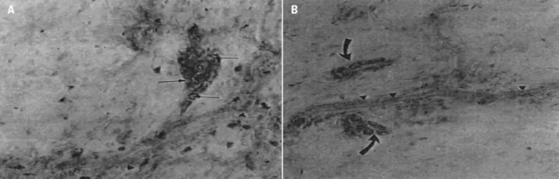
he analysis of the electronmicrographs showed that there was apparently a larger number of mitochondria in the group of old animals, denoting degenerative aspects (Fig 3). It can be summarized in the presence of
vacuol-Table 1. Mean values and standard deviations of the total number of ganglions of the jejunum-ileum of six guinea pigs in the two age groups.
Age group
Jejunum-ileum
Total number Density of ganglia (ganglia/cm2)
Young 90472±7748.8 551±36.89
Old 70103±1653.2 413±11.86
*p=0
Table 2. Mean values and standard deviations of the total number of neurons of the jejunum-ileum of six guinea pigs in the two age groups.
Age group
Jejunum-ileum
Total number Density of neurons (neurons/cm2)
Young 818362±60998.6 5011±291.11
Old 493924±27039 2918±120.70
*p=0
Table 3. Mean values and standard deviations of the neuron size (area of the cellular profile) of the jejunum-ileum (µm2) in six guinea pigs in the two age groups.
Age group Area of the cellular proile (µm2)
Young 253±0.141
Old 262±7.990
*p=0.172
ized mitochondrias and diference in the number of gran-ular and agrangran-ular synaptic vesicles.
DISCUSSION
he results of this work about the morphology of the neurons of the submucous plexus through the use of the staining techniques applied to the membrane prepara-tions, conirm observations made by other authors using histological sections11-13. he neurons of this plexus in the jejunum-ileum of the guinea pig are mostly found in gan-glion groups. hey can also appear isolated or in pairs. hese observations are in agreement with the indings of other authors in this species and also in others (Calomys callosus; newborn and adult rat)13-15.
he ganglia of the submucous plexus present small, medium and large sizes, containing a varied number of neurons, and arranged at random along the intestine, generally located close to blood vessels, with round, oval, triangular or elongated shapes conirming indings of other authors13,16. Apparently aging did not inluence the morphology of the neurons, which was similar
be-Fig 2. Transversal histological cuts to the jejunum-ileum of the guinea pig passing through the submucous ganglions (g). In [A,B] the ganglion of the young jejunum surrounded by collagen ibers type I (narrow ar-rows). In [C,D] the ganglion of old je-junum surrounded by collagen ibers type I (wide arrows) - polarized light - [A,B,C,D - 670×).
tween young and old animals. However, in ganglia of old animals, areas without neurons are sometimes observed, making up “clear” spaces inside of the ganglia similar to those found by Alves17, in the myenteric plexus of the co-lon of old guinea pigs. According to this author, this as-pect is due to the loss of neurons in the ganglia.
Structurally comparing the submucous plexus of the jejunum-ileum of old animals with that of the young an-imals, in the old group there is apparently a capsule of more compact collagen ibers, with an intense red color indicating the presence of a higher number of type I col-lagen ibers around the ganglion. his is a practical gener-al process that occurs in gener-all organs, by the substitution of cells, in this case, nervous cells, by collagen ibers.
According to Moratelli,18 and Gomes et al.19, in the case of myenteric ganglia, the collagen ibers of the gan-glia could act as protective elements for the gangan-glia and nervous ibers, preventing excessive distensions during the movements of the intestine. However, with aging, the increase of these ibers in the ganglia could damage its function, for example, making the metabolic exchanges between the capillary and the neurons of the ganglia dif-icult. his same reasoning could be applied to the gan-glia of the submucous plexus.
he ultrastructure of the ganglia and neurons of the submucous plexus of the guinea pig’s jejunum-ileum ap-peared similar to that of other species, both mammals, bullocks and ish, such as trout20,21. As Wilson et al.22, we also observed that in young adult guinea pigs, the gan-glia are surrounded by a continuous basal sheet, without changes with aging. Also as these authors, we did not ind other kinds of cells or blood vessels within the ganglia of the plexus of guinea pigs, except the neuronal cells, the Schwann cells and a neuropilus formed by the prolonga-tion entanglement of the neurons and the neuroglial cells. here are few indings on the ultrastructural aspects of aging, both in the myenteric and the submucous plex-us. A work carried out by Gabella1 describes that degen-erative signals are not common in the myenteric neu-rons in the aging process, showing that the ganglia keep the structures apparently well preserved in aged individ-uals. As Gabella1, we also found few degenerative chang-es with the transmission electron microscopic which can be summarized in the presence of vacuolized mitochon-drias and diference in the number of granular and agran-ular synaptic vesicles. However, other works must be car-ried out, including the use of immunohistochemical tech-niques to detect aging changes in neurotransmissors of the submucous plexus.
With regard to the quantitative aspects, there is a lower density of ganglia in the jejunum-ileum of the old group. hese results show a ganglion drop of about 20% for the submucous plexus of the jejunum-ileum of old
an-imals (P<0.05). he neurons of the jejunum-ileum of the animals of the old group presented a more sparse distri-bution within the ganglia, which conirms that the gan-glia of the plexus contained fewer neurons.
There was an important decrease in the number of neurons of the submucous plexus of the jejunum-il-eum of animals in the old group. The drop was about 38% compared to the plexus of the animals of the young group. his result is similar to the data obtained by Phil-ips7, who studied the aging of this plexus in the large intestine of rats.
Comparing our results with those regarding the ag-ing of the myenteric plexus both in the small intestine and in other parts of the digestive tract, we conirmed a reduction between 20 and 60% in the number of neu-rons for the myenteric plexus both in humans,2-5 and in animals1,17,23,24. herefore, the behavior of the submucous plexus follows a standard similar to that of the myenter-ic plexus with regard to aging.
According to Gabella11, the loss of neurons of the my-enteric plexus of the small intestine of the guinea pig was of 40 and 60%. On the other hand, for the human esopha-gus, Koberle26 and Wada27 observed a neuronal reduction of about 55% in the myenteric plexus due to age which would be responsible for malfunctions in the esophagus conirmed in the presbiesophagus. In a work on the aging of the myenteric plexus of the guinea pig colon, Alves17 observed a neuronal drop of around 46%. Santer and Bak-er25 observed a reduction of 64% in the rat colon for my-enteric neurons. According to these authors, the neuro-nal loss that occurs in aging promotes a reorganization of the remaining neuronal elements, suggesting that this loss cannot be considered a degenerative process.
Histophysiological studies on the submucous plexus show that it is involved in the control of the contractile activity of the smooth musculature within the villi, co-ordination of intestinal motility, control of electrolytes, transport of ions and contains sensitive integrating el-ements and motors of the secretory activity of several cells6-8. As we observed in this study, there was a signii-cant loss of nervous cells of the submucous plexus, with aging, which is possibly related to the functional altera-tions such as atrophy of the mucous, diverticulosis or terference in the absorption of nutrients in the small in-testine in the aged.
REFERENCES
1. Gabella G. Fall in the number of myenteric neurons in aging guinea pigs. Gastroenterology 1989;96:1487-1493.
2. Anaruma CA. Estudo morfoquantitativo do plexo mientérico do estôma-go humano em indivíduos jovens e idosos. [Tese de Doutorado - Institu-to de ciências Biomédicas da Universidade de São Paulo] São Paulo, 1994. 3. De Souza RR, Moratelli HB, Borges N, Liberti EA. Age-induced nerve cell
4. Gabella G. The Number of neurons in the small intestine of mice, guinea-pig and sheep. Neuroscience 1987;22:737-752.
5. Meciano Filho J, Carvalho V C, De Souza RR. Nerve cell loss in the myenter-ic plexus of the human esophagus in relation to age: a preliminary investi-gation. Gerontology 1995;41:18-21.
6. Furness JB, Costa M. The enteric nervous system. New York, Churchil Livingstone, 1987.
7. Philips RJ, Pairitz JC, Powley TL. Age-related neuronal loss in the submu-cosal plexus of the colon of Fischer 344 rats. Neurobiol Aging 2006;28: 1124-1137.
8. Stach W, Scheuermann DW. Concentration of external noradrenergic axo-nal nebwarks in the area of type III neuroaxo-nal aggregates and dense capillary nebworks of the external submucosal plexus (Schabadasch) in the guinea-pig small intestine. Z Mikrosk-Anat Forsch 1985;99:617-626.
9. Barbosa AJ. A técnica histológica para gânglios nervosos intramurais em preparados espessos. Rev Bras Pesq Med Biol 1978;11:95-97.
10. Junqueira LCU, Cossermelli WS, Brentani RR. Diierencial staining of col-lagens type, I,II and III by Sirius red and polarization microscopy. Arch Histol Jpn 1978;41:267-274.
11. Gabella G. Innervation of the gastrointestinal tract. Int Rev Cytol 1979;59: 129-193.
12. Meissner G. Uber die nerve der karmwand. Z Ration Med N F 1857;8:364-366; apud Furness, 1. B & Costa, M. 1987.
13. Sousa NB, LibertI EA, De Souza RR. Estudo Morfoquantitativo, histoquími-co e ultraestrutural do plexo submuhistoquími-coso do tubo digestivo do calomys callosus. [Tese de Doutorado - Universidade São Paulo] São Paulo, 1994. 14. Gabella G. Neuron size and number in the myenteric plexus of the
new-born and adult rat. J Anat 1971;109:81-95.
15. Gunn M. Histological and histochemical observation on the myenteric and submucous plexuses of mammals. J Anat 1968;102:223-239.
16. Ohkubo K. Studies on the intrinsic nervous system of the digestive tract. I the submucous plexus of guinea Pig. JPN Med Sci 1936;6:1-20.
17. Alves N. Estudo morfoquantitativo do envelhecimento no plexo mienté-rico do colo da cobaia. [Tese de Doutorado - Escola Paulista de Medicina] São Paulo, 1996.
18. Moratelli HB. Estudo morfológico e quantitativo do plexo mientérico do in-testino delgado humano em adultos e idosos. [Tese de Doutorado - Uni-versidade São Paulo] São Paulo, 1990.
19. Gomes OA, de Souza RR, Liberti EA. A preliminary investigation of the ef-fects of aging on the nerve cell number in the myenteric ganglia of the hu-man colon. Gerentology 1997;43:210-217.
20. Ezeasor DN. Ultrastructural observation on the submucous plexus of the large lntestine of the raimbow trout (salmo gairdneri Rich). Z Mikrosk Anat Forsch Leipzig 1979;93:803-812.
21. Mann Von A, Pospischil A, Dahme E. Der plexus submucosus (Meissner) beim kalb. I. Licht and elektronenmikrospische untersuchung der nermal-struktur. Zentralbl Veterinaermed Rihe A 1984;31:585-600.
22. Wilson AJ, Furness JB, Costa M. The ine structure of the submucous plex-us ofthe guinea-pig ileum II. Description and analysis of vesiculated nerve proiles. J. Neurocytol 1981;10:785-804.
23. De Souza RR, Ferri S, Ferraz De Carvalho CA, Paranhos G. Myenteric plex-us in a fresh water teleost intestine. Quantitative study of nerve cell. Anat Anz 1982;152:359-362.
24. Liberti EA, Queiroz LM, Pompeu E, et al. A quantitative and comparative study of the ganglionic neurons in the myenteric and submucous plexus-es of the small intplexus-estine, and in the intramural plexus of the gall bladder of the guinea-pig. Rev Bras Cienc Morfol 1994;12:106-114.
25. Santer RM, Backer DM. Enteric neuron numbers and sizes in Auerbach’s plexus in the small and large intestine of adult and aged rats. J Auton Nerv Syst 1988;25:59-67.
26. Koberle F. Patogenia do megaesôfago brasileiro e europeu. Rev Goiana Med 1963;9:79-116.