Hospital das Clínicas da Faculdade de Medicina de Ribeirão Preto - USP. Mailing address: José Antonio Marin-Neto - Div de Cardiologia, Depto de Clínica Médica - Hospital das Clínicas, FMRP-USP - Av. Bandeirantes, 3900 – 14048-900 - Ribeirão Preto, SP - Brazil.
The recovery of T. cruzi DNA from South American
human mummies shows that Chagas’ disease already afflicted mankind as early as 4000 years ago 1. Charles
Darwin probably contracted the disease during his expedition to South America, as suggested by his vivid description of the “benchuca” sting and the nature of his late life symptoms 2. However, it was in the early twentieth
century that Carlos Chagas, in a most unique accompli-shment in the History of Medicine, discovered the new morbid entity that carries his name, described its patho-logical and clinical features, the etiologic agent and its transmission mechanism through the inoculation of infected excreta of hematophagous insects of the family
Reduviidae (subfamily Triatominae) 3.
Although the mechanism by which T. cruzi invades
mammalian cells is not completely known, experimental work suggests that an essential step involves triggering of activation of the transforming growth factor (TGF)-beta signaling pathway. Therefore, adherent parasites cannot penetrate cells lacking TGF-beta receptors and replicate within them, whereas administration of TGF-beta potentiates
T. cruzi invasion ability in experimental models 4.
Virtually every organic system may be affected. Megaesophagus and megacolon occur in about 6% of the chagasic population and neurologic disorders in around 3%, but, by far, Chagas’ heart disease is the most serious complication, with clinical manifestations arising in nearly one third of Chagas infected people throughout their life span.
Epidemiological aspects
Conditions for vectorial transmission range between latitudes 42°N and 40°S of the American Continent, from Mexico to Argentina. On the basis of limited serological surveys, 4% to 7% of more than 200 million Latin Americans are estimated to be chagasic in extensive areas of 21 coun-tries, and 65-90 million are at risk of becoming infected 5.
Cross-sectional epidemiological studies in Brazil and Venezuela assessed the prevalence of clinical
manifes-José Antonio Marin-Neto, Marcus Vinícius Simões, Álvaro V. Lima Sarabanda
Ribeirão Preto, SP - Brazil
Chagas’ Heart Disease
tations and mortality due to Chagas’ heart disease. However, no clear-cut epidemiological picture of Chagas’ heart disease is yet available, due to the lack of appropriately designed large-scale studies to address this serious public health problem in extensive areas of the Latin American subcon-tinent. In addition, case reporting is not reliable even in areas of high endemicity. Probably because of marked variations in the genetic background, parasite strain, climate, socio-economic and related hygienic-alimentary conditions, and health care policies, the morbidity and mortality rates ascribed to Chagas’ disease are extremely variable even among endemic areas of each country 6.
Although the true prevalence of Chagas’ heart disease is unknown, these rough estimates clearly indicate that Chagas’ myocarditis is undoubtedly the most common form of cardiomyopathy in Latin-American countries 7. Also, due
to migratory currents between countries and far-distant regions, Chagas’ heart disease is likely to become ubi-quitous 7. A reflection of this tendency is exemplified by the
recent growing awareness regarding the occurrence of Cha-gas’ heart disease in the United States. Based on a pre-valence of 4.5% of T. cruzi serologically detected infection
in 205 Latin American immigrants to the USA, and on estimates of the number of such immigrants, approximately half a million infected people are believed to exist now in that country 8. Moreover, rural-urban migration from endemic
areas in Brazil is believed to have brought to large cities half a million infected people in the last three decades 6.
Other mechanisms of transmission - Infestation can
also, infrequently, occur by congenital and oral routes, breast feeding, laboratory contamination, and organ transplantation. Transfusional transmission of T. cruzi is
currently under close scrutiny in most places, since a survey carried out from 1988 to 1990 in 850 counties in of Brazil, revealed that serological screening for Chagas’ disease was performed in only two thirds of all blood donors 6. Also, a review of serological surveys for Chagas’
infection among blood donors, conducted over the last decade in several countries, disclosed a seropositivity rate ranging from 10 to 50% in endemic areas 9.
Prevention - Chagas’ heart disease carries a very high
These are figures that thoroughly substantiate the concept that elimination of Chagas’ disease vectorial (by improving the quality of housing and use of residual insecticides) and of transfusional transmission in both endemic and nonendemic areas 11-13, is a highly cost-effective public health policy 7,10.
Despite being hindered by financial limitations, these goals have been attained in scattered regions 14-16.
The Southern Cone Initiative program launched in 1991 has already produced impressive results at a cost of US$ 207 million that was allocated from national resources of the six countries involved 5,16,17. Thus, in Brazil, the 89%
reduction in the number of house-infested counties was accompanied by a drop in the rate of T. cruzi infected blood
donors from 6.5% to 1%, from 1982 to 1993 16,17. On the basis
of such figures, interruption of transmission is expected to occur between 1998 and 2000 in Brazil, Argentina, Chile and Uruguay 5,18. However, it may be too early for such high
expectations, as suggested by sporadic reports of trans-mission of the disease in areas previously consi-dered under epidemiological control 19.
Natural history and prognostic factors
Experimental, pathological and clinical evidence substantiate the conceptual division of Chagas’ heart disease into the acute myocarditis and the chronic phase, separated by the long period - 10 to 30 years - known as the indeterminate form of the disease, that constitutes its most intriguing conundrum 7,20,21. Reactivation of Chagas’
disease, with proteiform clinical expression, is now often seen in chronic chagasic patients with various causes of immunodeficiency, natural or iatrogenically induced 22-25.
Several observational studies mainly conducted in endemic areas in Brazil, Argentina and Venezuela, since the early 1940s disclosed the natural history of Chagas’ heart disease 7,26-43.
Also, many case-series studies describe the acute phase of Chagas’ disease acquired through nonvectorial transmission, but have limited value for the knowledge of its natural history 7,22-26.
Natural history studies of Chagas’ heart disease deri-ve predominantly from cross-sectional observations of infected people in rural areas of those countries 7. Very few
studies have described case-control populations of chagasic and non-chagasic people. Other observational investigations focused on the description and follow-up of hospital-based cohorts of chagasic patients 7,38-41.
Both the rural and hospital-based types of studies have clear limitations for the assessment of the influence of prognostic factors in Chagas’ heart disease’s natural history 7. Thus, no adequate identification of cardiac
involvement is usually provided in most of the rural-based studies 27-37. Conversely, in hospital-based studies the
heart disease is usually well characterized, but their results can not be extended to the whole spectrum of the chagasic population 8,38-41. Furthermore, because of the rather
protracted course of heart involvement, from the acute
myocarditis to the end-stage heart failure or malignant arrhythmia, no prospective studies encompassing the whole span of the disease in sizable populations are available 7,42.
Prognosis in the acute phase - Although cardiac
myocarditis is a constant finding in biopsy specimens or at necropsy examination 26, case series reported in endemic
areas using specific serological tests have shown that only around 10% of the acute cases have clinical manifestations consistent with a correct clinical diagnosis of Chagas’ disease 27. This is a major obstacle for gathering direct
insight into the transition from the acute to the chronic stages of human Chagas’ disease. Nevertheless, studies in experimental models of Chagas’ disease are in general agreement with such findings.
When the clinical diagnosis was possible (in the small subset of patients), cardiac involvement occurred in around 90% of 313 successive cases; in 70-80% cardiac enlargement was seen on X-rays, contrasting with only 50% of cases showing electrocardiogram (ECG) abnorma-lities. The severity of myocarditis was inversely propor-tional to age, with signs of heart failure being twice more intense in children aged up to two years than in those between the ages of three and five years 27. Mortality in the
acute phase of the disease, in this study, was 8.3%, a higher figure than the 3-5% reported in similar studies in other endemic areas in Brazil, Argentina and Uruguay. The ECG was normal in 63.3% of the nonfatal cases, and in only 14.3% of those who died in the acute phase of Chagas’ disease. Of all deaths, 75% occurred in children less than three years of age. Heart failure was the constant finding in all fatal cases, associated or not with encephalitis, and independent of age 27.
Of 172 patients whose acute phase of Chagas’ infec-tion had been diagnosed on the basis of general clinical signs and a positive serology, followed in Bambui (central Brazil) for up to 40 years, the development of chronic cardiac involvement - based on clinical signs, ECG and chest X-rays changes - occurred in 33.8%, 39.3% and 58.1%, respectively during follow-up periods of 10-20 years, 21-30 years and 31-40 years 27. In another review concerning the
same endemic area, for 268 patients whose acute phase of the disease had been diagnosed in an average of 27 years before, the overall mortality in the period was 13.8% 27.
Survival is characterized by disappearance of symp-toms and signs of heart failure within 1-3 months, and normalization of the ECG in over 90% of the cases after one year of the infection.
However, there is no evidence of spontaneous cure of the infection, as demonstrated by serial xenodiagnosis and serological tests in studies of several hundreds of chagasic patients 7.
Prognosis in the indeterminate phase - Although
requires that patients have positive serology and/or a positive xenodiagnosis test, no cardiovascular or diges-tive symptoms, a normal resting 12-lead ECG and no abnormalities detected by radiological examination of heart, esophagus and colon. Thus, the indeterminate phase ends and the chronic cardiac or digestive forms of the disease ensue only when symptoms appear or abnor-malities are shown on the ECG or by radiological cardiac or digestive scans.
The evolution potential at this stage of the disease, determined by as yet unknown factors, is shown by longitu-dinal cohort studies in endemic areas. A 1-3% per year rate of appearance of heart involvement has been observed in several studies 7.
Of 400 young adults followed for 10 years, 91 (23%) showed clinical and/or ECG or chest-X-ray markers of cardiac disease. Of note, eight deaths were recorded in that period, of which only one could be ascribed to recru-descence of chagasic cardiomyopathy 30.
Another longitudinal study in Bambui, central Brazil, contrasted the evolution of 885 young chagasic patients in the indeterminate phase, for 10 years, with that of 911 chagasic patients with initially abnormal ECG, in the same period. Survival after 10 years was 97.4% and 61.3%, respectively for the indeterminate group and the group with cardiac involvement 31.
A third longitudinal study in a rural Venezuelan community, with 47% prevalence of positive serology for Chagas’ disease, followed 364 patients for a mean period of four years. It revealed the appearance of heart disease at a rate of 1.1% per year in seropositive individuals. Mortality was 3% in the four years of follow-up and Chagas’ heart disease was the cause of death in 69% of all fatal cases 32.
In 1973 a longitudinal study was initiated in a rural community in northeast Brazil. In the initial cross-sectional study of 644 individuals aged >10 years, 53.7% were seropositive. The population initially described in 1973-1974 was re-examined in 1977, 1980 and 1983. The overall rate of development of abnormal ECG was 2.57% in seropo-sitive (248) as compared to 1.25% per year in seronegative (332) individuals, a relative risk of two for the same geographical area. The age-adjusted mortality rate was higher in seropositive (8.9/1000/year of 488 patients) than in seronegative individuals (7.8/1000/year of 509 individuals). However, mortality in this study was strongly associated with ventricular conduction defects and arrhythmias 33.
In summary, the results of these studies indicate that, as long as the patients remain in the indeterminate phase, their prognosis is fine 7. It must be emphasized that these
results were obtained in chagasic populations with >50% of the individuals younger than 20 years, and less indeter-minate cases are found in older age groups because of the evolutive nature of the disease. It is relevant to know that after 10 years almost 80% of the patients remain in the indeterminate phase of the disease and probably 50% of the entire population will have no signs of heart disease throughout their lives. What remains elusive are those
factors which determine the development of overt cardiac disease and cardiac failure in some patients who have been infected with T. cruzi as opposed to others with positive
serology but without cardiac involvement 7. It is likely that
the explanation will be multifactorial.
Prognosis of chronic Chagas’ heart disease - From
the studies mentioned above, analyzing prognostic factors of the indeterminate phase in rural populations in which only a superficial evaluation of the heart condition was carried out, it became apparent that the mere appearance of ECG changes entailed a bad prognosis 34,35. Also, a
retros-pective analysis of seropositive individuals followed over 18 years revealed that right bundle branch block was three times more common in fatal cases than in survivors 34.
In addition to ECG markers, the notion that the male gender is an important deleterious prognostic factor once the heart disease is manifest, is borne out from several studies carried out with long-term follow-up of hospital-based cohorts of chagasic patients and also by a case-control study 27-29. The later study also suggests possible
geographical clustering and/or familial aggregation of cases of Chagas’ heart disease in endemic areas 29.
Few case-control follow-up studies have been repor-ted in endemic areas 7,36,37. In central Brazil 36, two
cross-sectional clinical assessments spanning 10 years (1974 to 1984) were carried out including 12-lead ECG and radio-logical evaluation of heart size. Serum positive patients and controls were matched by age and gender. In the first cross-sectional study, 264 pairs of subjects were evaluated, of which 110 could be recomposed and reexamined after the 10-year follow-up period, with the same clinical, ECG and chest-X-ray assessment. The incidence of clinical heart disease, as diagnosed by the development of symptoms, ECG and/or radiological changes, in previously healthy but serologically positive individuals was 38.3% in the ten-year period. In those patients with previous heart involvement a rate of 34.5% of deterioration was observed in the same period. In the chagasic population the overall mortality was 23%, compared with 10.3% in the controls. Moreover, cardiac mortality, including sudden death and death in heart failure was 17% among chagasic patients, and only 2.3% in the control population. Again, the overall mortality was much higher in chagasic males and predominated in the group aged 30 to 59 years 36.
The same group of investigators, applying similar methods in northeastern Brazil showed that mortality rates were 1.6% and 0% for 125 matched pairs of respectively chagasic and nonchagasic patients followed for 4.5 years 37.
Progression of disease as assessed by ECG changes occurred in only 10.4% of patients, as compared to 4.8% of controls. The different morbidity and mortality rates between the two regions were hypothesized to mean possible differences in the pathogenicity of T. cruzi strains
in the two geographical regions, but no direct evidence for this was provided 7.
mortality associated with Chagas’ disease is strongly correlated with the severity of the myocardial dysfunction 7.
For example, survival two years after the first episode of heart failure was only 33.4% in 160 cases 38. Of note, 10% of
deaths were sudden. In addition 98 deceased people were autopsied, revealing <20% of prevalence of cardiac tissular forms of T. cruzi, with a clear predominance of this finding in
male patients 38.
In a study of 107 chagasic patients followed for 10 years, a significant reduction in life expectancy, as compared to that of 22 nonchagasic patients, was detected only in those with ECG and/or clinical changes. A mortality rate of 82% over the 10-year follow-up period was seen in the group of 34 patients with signs of heart failure at the beginning of the study. In contrast, a 65% 10-year survival was associated with ECG abnormalities but in absence of signs of heart failure 39.
Another study of 104 male patients admitted to the hospital with congestive heart failure revealed a mortality rate of 52% after five years. The strongest predictors of survival were left ventricular (LV) ejection fraction and maximal oxygen uptake during exercise 40.
In a series of 42 patients with Chagas’ heart disease in the USA, 11 deaths occurred during a mean follow-up of nearly five years, always in association with global or regio-nal LV dysfunction. Established or developing heart failure was a strong predictor of mortality, but, quite surprisingly, not aborted sudden death or the presence of sustained ventricular tachycardia 8. These results conflict with the
evidence that ventricular tachycardia detected during exercise testing is a marker of increased risk of sudden death in 44 chagasic patients followed for a mean period of two years 41. This discrepancy is likely related to the fact
that both studies are fraught with the same limitation of small numbers and a relatively short follow-up.
In summary, there is substantial evidence that the most important prognostic factor in established Chagas’ heart disease is the degree of myocardial dysfunction. Once overt cardiac failure is manifest, the prognosis is bleak, similarly to that reported in the heart failure Framingham cohorts, with mortality rates approaching 50% in four years. It is possible - but by no means proven by good evidence - that sudden death and related ventricular arrhythmias may play a more prominent role in mortality due to Chagas’ disease than in heart failure due to other etiologies 7.
Clinical features of Chagas’ heart disease
Following inoculation by the etiologic agent, there is an incubation period of approximately 7 to 10 days. Local skin or mucosa swelling produces the typical entry lesions known as chagomas (including the nonspecific Romaña’s
sign).
Cardiac abnormalities are always present in all stages of Chagas’ disease, but, characteristically, in the acute phase, there is a striking discrepancy between the severity of the myocarditis and the paucity of its clinical expression 7.
Gene-ral infective signs of disease (fever, myalgia, sweating, hepatosplenomegaly) at this stage are usually accompanied by nonspecific laboratory findings: leukocytosis with an absolute increase in lymphocyte count, chest X-rays cardiomegaly and ECG changes (sinus tachycardia, ven-tricular ectopic beats, low voltage, diffuse ST-T alterations, first degree atrioventricular block). Serologic tests for T. cruzi
infection are usually negative during the first weeks, but circulating parasites may be detected by xenodiagnosis. The diagnosis of acute Chagas’ disease due to blood transfusion requires a high level of awareness, particularly in nonendemic areas 42. This notion also applies regarding the problem of
recognizing recrudescence of Chagas’ disease in immuno-compromised patients who have the chronic form 42.
Necropsy studies and in vivo investigations, using
several methods to evaluate the ventricular performance, myocardial perfusion, cardiac autonomic function and rhythm, and findings on RV biopsy - demonstrate that virtually all patients, even in the indeterminate phase of the disease, have at least some subtle degree of cardiac invol-vement 7,43-48.
It is important to emphasize that all the anatomical and functional disturbances detected during life are consistent with the autopsy findings reported on several series of chagasic patients who died in the various stages of the disease 43,44,49,50.
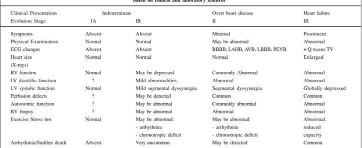
On the basis of the prognostic implications discussed above, it would be convenient to classify patients with Chagas’ heart disease according to the presence of symp-toms, physical and ECG abnormalities, cardiac enlargement, right and LV systolic and diastolic dysfunction, myocardial perfusion defects, cardiac autonomic impair-ment and RV biopsy findings (table I).
Symptoms and physical signs appearing in patients with the chronic stage of the disease arise from three basic syndromes that may coexist in the same patient: heart failure, cardiac arrhythmia, and systemic and/or pulmonary thromboembolism.
Heart failure of chagasic etiology is usually biven-tricular. However, the manifestations of right-sided failure (increased jugular venous pressure, peripheral edema, ascites, and liver enlargement) are usually more pro-nounced than those of left-sided failure (dyspnea and pulmonary rales) 51. Fatigue is also a common symptom.
Physical examination often reveals murmurs of mitral and tricuspid regurgitation, wide splitting of the second heart sound with an accentuated second component (P2), due to the combination of a right bundle branch block and pulmonary hypertension, and a prominent apical thrust.
Cardiac arrhythmias (such as bouts of ventricular tachycardia) cause palpitation, lightheadedness, dizziness, or syncope 52. The latter symptoms may also be caused by
tachy-cardia or complete AV block who have well preserved glo-bal ventricular performance (but usually regional wall motion abnormalities) 52.
Sudden unexpected death occurs with an undefined but not negligible frequency and can supervene (albeit rarely) even in patients previously asymptomatic. It is usually precipitated by physical exercise, and associated with ventricular tachycardia and fibrillation or, more rarely, with complete AV block. From autopsy studies, it is appa-rent that such patients have constant but variable degrees of inflammatory abnormalities and neuronal cardiac depopulation 44. In addition, necropsy and in vivo studies
show that most such patients have ventricular aneurysm at one or more sites (posterior, inferolateral, or apical) 42.
Systemic and pulmonary embolism, arising from mural thrombi in cardiac chambers and from deep venous throm-bosis due to low cardiac output, is a conspicuous com-plication of chronic Chagas’ heart disease. However, evidence from postmortem studies suggests that emboli are often overlooked. In a review of 1345 autopsy cases, the incidence of cardiac thrombus or thromboemboli was 44 percent; the right and left cardiac chambers were equally affected 50. Although thromboembolic phenomena were
more common in the systemic circulation, pulmonary embolism accounted for 14 percent of deaths. Chest pain, more often atypical for myocardial ischemia, is another common symptom 8,47,53,54. In a small but appreciable subset
of chagasic patients, it may mimic an acute coronary syndrome 55,56.
Diagnostic laboratory methods
It is remarkable that some chagasic patients with conspicuous ECG and ventricular regional abnormalities
may be asymptomatic hard workers 7,44,47. The appropriate
use of several diagnostic methods will detect the cardio-vascular dysfunction in virtually all patients, and help in establishing both the diagnosis and prognosis 42.
Serologic tests - The etiologic diagnosis is routinely
performed with methods that detect circulating antibodies that bind to parasite antigens 57. The most commonly used
tests are based upon complement fixation, immunofluo-rescence, or ELISA assays, that, carefully standardized, achieve sensitivity and specificity rates higher than 90 percent. Chagas’ disease is diagnosed with greater sensi-tivity by the detection of T. cruzi specific sequences of
DNA, using molecular biology approaches 58-60. These later
techniques also have the potential for improving the diagnostic and prognostic characterization of the disease, on the basis of parasite strain identification 60.
Electrocardiogram - The most common alterations on
the routine ECG are right bundle branch block, often associated with left anterior hemiblock, diffuse ST-T changes, ventricular premature beats that may be multiform, and runs of nonsustained ventricular tachycardia. Other frequent findings are abnormal Q waves and various degrees of atrioventricular block and, in more advanced stages of disease, atrial fibrillation and low QRS voltage 61.
Chest x-ray - The most common radiographic finding
is marked cardiomegaly with mild or absent pulmonary congestion 42.
Ambulatory electrocardiographic monitoring
-Virtually all types of atrial and ventricular arrhythmias can occur including sinus node dysfunction, intermittent
com-Table I - Pathophysiological classification of chronic Chagas’ heart disease, according to evolution stage, based on clinical and laboratory features*
Clinical Presentation Indeterminate Overt heart disease Heart failure
Evolution Stage IA IB II III
Symptoms Absent Absent Minimal Prominent
Physical Examination Normal Normal May be abnormal Abnormal
ECG changes Absent Absent RBBB, LAHB, AVB, LBBB, PEVB + Q waves TV
Heart size Normal Normal Normal Enlarged
(X-rays)
RV function Normal May be depressed Commonly Abnormal Abnormal
LV diastólic function ? Mild abnormalities Abnormal Abnormal
LV systolic function Normal Mild segmental dyssynergia Segmental dyssynergia Globally depressed
Perfusion defects ? May be detected Common Common
Autonomic function ? May be abnormal Commonly abnormal Abnormal
RV biopsy ? May be abnormal Abnormal Abnormal
Exercise Stress test Normal May be abnormal: May be abnormal: Abnormal:
- arrhythmia - arrhythmia reduced
- chronotropic deficit - chronotropic deficit capacity Arrhythmia/Sudden death Absent Very uncommon May be detected Common
* Modified from 7, with permission. LV - left ventricle; RV - right ventricle; ECG - electrocardiogram; AVB - atrioventricular block; LAHB - left anterior hemiblock;
plete atrioventricular block, and complex ventricular arrhythmias 52,62.
Echocardiography - In the early stages,
echocardio-graphy may reveal one or more areas of dyssynergia, including the typical, almost pathognomonic ventricular aneurysm. More advanced disease is revealed by global ventricular dilatation and diffuse hypokinesis, often associated with mitral and tricuspid regurgitation 63.
Exercise testing - Exercise testing is of limited
use-fulness for the evaluation of patients presenting with chest pain because, as noted above, most have baseline electro-cardiographic abnormalities 64. However, this test may
constitute an alternative to ambulatory monitoring for the detection of exercise-related ventricular dysrhythmia 41,65.
A deficient chronotropic response can also be detected in chagasic patients, due to parasympathetic denervation of the sinus node 66,67.
Radionuclide angiography - Although less frequently
used than echocardiography for clinical purposes, radio-nuclide angiography has been employed in inves-tigations aiming at the detection of early impairment of biventricular function. Regional wall motion abnormalities and global right ventricular dysfunction may be detected in patients with the indeterminate or isolated digestive form of the disease in whom LV performance is still preserved 68,69.
Myocardium perfusion scintigraphy - Both transient
(reversible or paradox) and irreversible perfusion defects are usually detected by myocardial perfusion scanning of patients who complain of angina-like pain. Perfusion disturbances occur in the presence of normal epicardial coronary arteries and probably represent abnormalities of the coronary microvasculature or areas of myocardial fibrosis 8,47,53.
Cardiac catheterization and angiography - Cardiac
catheterization and angiography may be mandatory in patients with symptoms but elusive electrocardiographic or scintigraphic signs of ischemia, to confirm or, more fre-quently, to exclude the presence of epicardial obstructive coronary artery disease 42,53-56. The method also depicts the
regional wall motion abnormalities that may be accom-panied by mural thrombosis 54.
Electrophysiologic testing - This test is indicated in
selected cases to assess sinus node function and atrio-ventricular conduction when the origin of symptoms remains uncertain after noninvasive evaluation 42.
Al-though definitive evidence of benefit is lacking, this procedure is also useful in two other subsets: survivors of sudden cardiac death; and those with sustained ventricular tachycardia to determine prognosis and to select the appropriate antiarrhythmic therapy (medical, surgical, or implantable device) 70,71.
Electrophysiologic testing does not have an impor-tant prognostic role in most patients with preserved LV function who have nonsustained ventricular tachycardia or in those without spontaneous arrhythmia. Programmed stimulation did not induce sustained ventricular tachy-cardia in any of 72 patients with 400 to 1200 ventricular extrasystoles/hour, of whom 35 percent had nonsustained ventricular tachycardia on Holter monitoring 71. The mean
LV ejection fraction in this group was 60 percent. During an average follow-up of 36 months only 1 of the 72 patients had spontaneous sustained ventricular tachycardia 71.
Signal averaged electrocardiogram - Preliminary
experience in patients not showing conduction distur-bances suggests that late potentials occur more frequently with sustained ventricular tachycardia than in its absence but the significance of this finding remains to be determined 72. The
presence of late potentials also seems to correlate with the degree of myocardial depression 73.
Magnetic resonance imaging - Although not yet
used for clinical purposes, this method has the potential to show the underlying myocarditis and also may provide accurate anatomic and functional characterization of cardiac involvement 74
Cardiac autonomic assessment - Cardiac autonomic
dysfunction, mainly parasympathetic, can be shown in chagasic patients (including those with isolated digestive disease) whose heart response to several autonomic tests (including the RR variability measurement) is impaired, as compared to control subjects 47,75,76,77. However, these
abnormalities are neither correlated with any symptoms, nor cause postural hypotension.
Pathological abnormalities
Necropsy findings in humans have been correlated with observations in several animal models of experimental Chagas’ disease reproducing the various stages of the disease 43,78-86. Endomyocardial biopsy has also been used
in subsets of the chagasic population, including patients in the indeterminate phase 46,87.
The main cardiac pathologic changes during the acute phase consist of four-chamber marked dilation and peri-cardial effusion.
Microscopic examination shows intense parasitism in virtually every organic system. The myocarditis is intense and diffuse, showing myocyte necrosis, interstitial edema, vasculitis, and mononuclear and polymorphonuclear infiltration. The inflammatory process may reach the endocardium, resulting in thrombus formation. The conduction system is also involved, as well as the intramural and extracardiac neuronal ganglia.
in the right chambers 84, and signs of systemic congestion
(ascites, hepatomegaly) predominate over lung congestion 51.
It is possible that this peculiar feature of Chagas’ heart disease could be explained by early severe damage of the right ventricle, a chamber frequently neglected in many investi-gations that included cardiac functional evaluation 68,69.
Intracardiac mural thrombosis in various stages of organization is found in nearly 50% of such cases; the right and left heart chambers are equally affected 50. The most
specific cardiac anatomic lesion is the ventricular apical aneurysm which, in one series, was noted in 52 percent of 1078 autopsied chagasic patients 88. The lesion does not
show the fibrosis usually seen in aneurysms due to myo-cardial infarction, and rarely undergoes rupture 78,88. There is
no relation between the frequency of apical aneurysm and age or heart weight, and aneurysms have been reported even in patients who died suddenly, with no apparent previous clinical manifestations of disease 44. Histologic
examination reveals mild chronic myocarditis, manifested by scattered mononuclear cell infiltrates with the surroun-ding myocytes undergoing various stages of degeneration and necrosis 82,89. These changes have been traditionally
interpreted as not being related to direct parasitism of myocardial cells, since intact parasites are rarely detected in humans and in experimental models of Chagas’ disease 82.
Focal and diffuse fibrosis is prominent, in the myocar-dium and the conduction system 90. Preferential
invol-vement of the right bundle branch and the left anterior fascicle of the left bundle by inflammatory and fibrotic changes correlates with the frequent occurrence of ECG block of these structures 89. Microvascular changes in
expe-rimental models consist of decapillarization, interstitial edema, intravascular platelet aggregation and thickening of the vascular basement membrane.
Similar findings are found on endomyocardial biopsy. Studies in patients with the indeterminate form have described changes in approximately 60 percent of patients, although the findings are less severe than those in patients with overt cardiac disease 46,87.
Striking autonomic neuronal depopulation and nerve degeneration, mostly in the cardiac, esophageal and colon tissues, is another typical feature of chronic Chagas’ disease
82,91,92. However, no correlation exists between the intensity of
neuronal destruction and dilation of the organ or other microscopic indices of myocarditis in the chronic phase 91.
Pathophysiology and pathogenetic mechanisms
The clinical manifestations and organ damage occur-ring duoccur-ring the acute phase are clearly linked to parasite presence in target organs like the gastrointestinal tract, cen-tral nervous system and heart. High grade parasitemia also correlates with lymphadenopathy, liver and spleen enlar-gement, as markers of widespread immunologic reaction. As the parasitemia abates, and the systemic inflammatory reaction subsides, it is believed that a silent relentless focal myocarditis ensues, during the indeterminate phase 7,78. This
causes cumulative destruction of cardiac fibers and marked reparative fibrosis. During this phase ventricular arrhy-thmias and sudden death may rarely occur as manifes-tations of the underlying focal inflammatory process 44,93.
This is also eventually responsible for myocardial mass loss attaining critical degrees, thereby leading to cardiac dilation and setting the anatomic substrate for malignant ventricular dysrhythmia 7,78. The support for this basic
conceptual framework comes from several investigations in various experimental models of Chagas’ heart disease using various animal species. Additional evidence has been provided by many studies correlating clinical and patho-logical findings in autopsied humans dying in all phases of the disease. All studies were observational and usually included case-series of dozens chagasic patients for the acute and indeterminate phases, and ranging from hun-dreds to thousands cases for the chronic phase of Chagas’ heart disease 7,78.
Complex ventricular arrhythmia constitutes one of the most important pathophysiological aspects, considering its implication on sudden death. It is believed that complex ventricular arrhythmia is more common in chagasic patients than in other dilated cardiomyopathies, but no adequate comparative study has been reported to support this gene-ral belief. As expected, there is reasonable evidence that more complex and frequent ventricular dysrhythmia parallels the worsening of ventricular function 94. However,
complex arrhythmias including nonsustained and sus-tained VT may also occur in chagasic patients with preser-ved global LV function. There is growing evidence that the electrophysiological substrate underlying sustained ventricular tachycardia in Chagas’ heart disease is a macroreentrant circuit within akinetic or dyskinetic areas in the posterobasal and/or posterolateral regions of the LV 95.
Despite recent advances in the understanding of Cha-gas’ heart disease pathophysiology, the main challenge still consists of the identification of the pathogenetic mecha-nisms acting during the indeterminate phase. The widely disparate clinical and pathological manifestations of the acute and chronic phases of a disease with a common infective basis also needs elucidation.
Basically, four main classes of mechanisms have been implied in the pathogenesis of chronic Chagas’ heart disease.
Neurogenic mechanisms - Necropsy studies in
hu-mans clearly demonstrated intense cardiac neuronal depo-pulation in the various Chagas’ disease stages 49,79-82. These
findings were reproduced by investigations in animal models of the disease 49,83-86,96-99. The histopathologic features are
foci of damaged nervous tissue arranged in a diffuse and irre-gular distribution. Neuronal parasitism, periganglionitis and degenerative abnormalities in Schwann cells and nervous fibers have also been observed. Not only cardiac parasym-pathetic nervous structures are involved but paravertebral sympathetic ganglia destruction has also been described 81.
parasym-pathetic and, to a lesser extent, of symparasym-pathetic cardiac autonomic control have been clearly documented by extensive laboratory investigation in humans 47,69,75-77,96.
This aspect seems to be a hallmark of this disease. In fact, less severe degrees of denervation have been found in rheumatic disease, endomiocardiofibrosis, and idiopathic dilated cardiomyopathy by direct comparative studies with Chagas’ disease 49.
Taking into account the early, intense, and largely predominant parasympathetic denervation in Chagas’ disease, that mostly explains the pathogenesis of chagasic megaesophagus and megacolon 100, a neurogenic theory of
Chagas’ heart disease has been proposed: a long lasting autonomic imbalance would lead to a catecholamine-induced cardiomyopathy 49,79.
However, various lines of evidence cast doubts on the participation of neurogenic derangements as main patho-genetic mechanisms of Chagas’ heart disease 7. Thus, the
frequency and the intensity of this abnormality are quite variable, and a mismatch between the presence of auto-nomic denervation and ventricular dysfunction is often seen 77,92,96,101-103. Differences in T. cruzi strain and/or
regio-nal environment are likely causes for variable neurotropism observed in several regions 99. More important, studies
aimed at investigating the presence of autonomic dys-function and early contractile abnormalities have failed to show any significant association 69.
However, a critical appraisal of this issue should consider that all the investigators mentioned above performed evaluation of autonomic cardiac function integrity by assessment of heart rate control. Hence, evidence of early nervous damage possibly occurring at the ventricular myocardial level, may have been overlooked 47.
A more appropriate insight into this aspect was obtained by recent investigations using 123 I-MIBG
scin-tigraphy for evaluation of myocardial sympathetic nerve terminals 104-106. Segmental areas of sympathetic
dener-vation were detected in a high proportion of patients even in the indeterminate phase of Chagas’ heart disease. This is the first functional evidence of cardiac sympathetic impairment preceding left wall motion abnormalities in the indeterminate phase of chronic Chagas’ disease.
Increased 123 I-MIBG washout rate was also observed
in patients with normal segmental ventricular function 106.
This could be due to early increased cardiac sympathetic activity, and lend support to the neurogenic theory as stated above. Alternatively, this abnormality could be caused by competition between the radiotracer and an endogenous substance for the neurotransmitter receptors of the sympathetic nerve terminals.
In plausible concordance with this last hypothesis, recent reports have documented in patients with Chagas’ disease the existence of circulating antibodies that bind to adrenergic and cholinergic receptors of lymphocytes and myocardium 107-111. Studies focusing on antibodies against
heart adrenergic and cholinergic receptors have shown their ability to trigger physiologic, morphologic, enzymatic
and molecular alterations, potentially leading to cardiac damage 109-111. Deposit of autoantibodies upon the
myocar-dial neurotransmitter receptor could induce its desensi-tization or down-regulation and cause progressive dener-vation. Such mechanisms could represent the elusive but significant link between denervation and autoimmune aggression as pathogenetic factors in Chagas’ heart disease. In conclusion, the neurogenic theory is still under de-bate. Its prognostic meaning has never been assessed, and the hypothesis implicating autonomic impairment in triggering sudden death remains entirely speculative 93,112.
Parasite-dependent inflammation - For decades no
significant pathogenetic role was attributed to T. cruzi
infection in the chronic phase of the disease, on the basis of histopathological evidence of low-grade fiber parasitism and an intriguing lack of topographic correlation between inflammatory foci and amastigote T. cruzi nests 82. This
classical view emphasized the presence of focal lym-phocytic myocarditis and myocytolytic necrosis in areas where no parasite could be seen 113, and seemed to be
supported by the finding of very low grade parasitemia that could be detected in only a minority of chronic Chagas’ disease patients 51,114.
However, more recent studies employing immune-histochemical techniques and monoclonal antibodies against
T. cruzi antigens have been performed on endo-myocardial
biopsy specimens retrieved from chronic Chagas’ heart patients and a relationship between parasite antigens and inflammatory foci was observed 113,115. Similar results were
obtained by detection of T. cruzi genomic fragments applying
the polymerase chain reaction method 116. In addition,
molecular biology techniques now permit the detection of circulating T. cruzi antigens in a much larger contingent of
chagasic patients in whom the conventional serologic methods fail for such purpose 117,118. It is plausible to
speculate that even low-grade persistent parasitism may lead to a continuous antigenic feedback loop to the auto-immune system, which may constitute the main damaging mechanism in the late phase 119-121.
A direct role of parasitism in the pathogenesis of chronic Chagas’ heart disease can have relevant thera-peutic implications. There is only very incipient evidence regarding the possible favorable impact of etiological treatment in the clinical outcome in the chronic phase of disease 122. In
parti-cular, some results have been reported on a nonrandomized, open label, placebo controlled trials of benznidazole in the clinical outcome of 131 treated and 70 nontreated chronic chagasic patients followed for eight years on average 123.
Significant reduction in the rate of new electrocardiographic abnormalities and in the incidence of clinical deterioration was observed in the treated group. Similar results were also recently obtained employing itraconazole and allopurinol 124.
Microvascular disturbances - Various classes of
evidence arising from clinical and experimental grounds suggest that transitory ischemic microvascular abnor-malities occur in Chagas’ heart disease.
The first order of evidence is related to morphological features of chronic myocarditis. The focal distribution of myocytolysis and interstitial reparative fibrosis (features also observed in nonchagasic experimental models of ischemia/reperfusion) is compatible with transient ischemic involvement at the microcirculatory level of discrete groups of fibers 125-127. Also, pathological involvement of coronary
vessels has been shown by necropsy studies 127-129 and is
reinforced by observations in murine experimental models of chronic Chagas’ disease 130-134. In the experimental setting
the detection of occlusive platelet thrombi in small epi-cardial and intramural coronary arteries indicates the occurrence of microcirculatory disturbances likely to produce ischemia detected by special histochemical techniques 133,134. Moreover, the administration of verapamil
(a calcium blocker with prominent vasodilator and anti-platelet effects) to T. cruzi infected-mice was accom-panied
by significant reduction in mortality and extension of tissue damage 135.
On clinical grounds, myocardial perfusion abnor-malities have been documented by several independent reports using various types of myocardial perfusion markers (thallium-201, 99mTc-Sestamibi, 99m
Tc-micros-pheres), during effort and at rest, in chagasic patients with angiographically normal coronary arteries 8,47,70,136-142.
Reversible, fixed and paradoxical perfusion defects, in areas with normal contraction, were found in a large proportion of patients even in the absence of other signs of myocardial involvement.
Abnormal response of coronary flow to acetylcholine administration has been reported in chronic Chagas’ heart disease patients, suggesting the occurrence of endothelial dysfunction 143. The demonstration of abnormal
sube-picardial coronary artery reactivity to hyperventilation and nitrate administration also supports the notion that func-tional abnormalities in myocardial flow regulation occur in chagasic patients 144.
Recent ultrastructural studies in canine experimental models suggest that small vessel involvement may be secondary to direct interaction of inflammatory cells and the endothelium 145. It is possible to speculate that substances like
thromboxane A2, cytokines, and prostaglandins, produced in the inflammatory infiltrate, may have important effects on vascular reactivity. According to this hypothesis, micro-vascular ischemia documented in Chagas’ cardio-myopathy may be secondary to the inflammatory response 126.
Conver-sely, the ischemic phenomena may represent a feedback loop that potentiates the primary damage mechanism.
It is also reasonable to assume that elucidation of the actual cause of ischemia in Chagas’ disease might improve the understanding of the precordial chest pain commonly experienced by the patients and help to establish thera-peutic strategies for treatment of this symptom.
Immune mediated cardiac damage - A wide array of
scientific investigations is considered to lend support to the theory of autoimmunity as the key process in the patho-genesis of chronic Chagas’ heart disease. The concept that the effector cells in the mononuclear chagasic myocarditis may damage nonparasited myocardial fibers has been suggested by histopathologic studies using light micros-copy; it was recently corroborated by ultrastructural studies in animal models 145, and is based on the postulate of
abnormal immune cross-reactivity between T. cruzi and
myocardial antigens.
Antigenic mimicry shared by the T. cruzi and the
myocardium has been shown for a series of cross-reactive antibodies directed against several host antigens 146-150.
However, the demonstration that this biological effect could have a clinical counterpart to support the auto-immune nature of this disease required the identification of an organ-specific autoantigen whose injection into the susceptible host could reproduce the lesion. Also, the myocardial damage in such circumstances, should be induced by passive transference of lymphocytes 151.
This kind of definite support of the lesion mechanism ascribed to the cellular immune infiltrate has been recently obtained by identification of myosin-specific TCD4+ lympho-cytes in the chronic murine Chagas’ disease model 152. Other
evidence was obtained by abrogation of the myocardial damage subsequently to depletion of TCD4+ in chronic infected mice 153. In addition, myocardial damage could be
reproduced in noninfected animals by passive transfer of TDC4+ lymphocytes, obtained from infected mice 153.
Moreover, specific epitopes associated with the host immune response and potentially able to produce myocar-dial damage have been recently identified 120,154-162. There is
also evidence that persistent T. cruzi antigen presentation
to macrophages could lead to cytokyne production, thus modulating the immune response and possibly causing the relative immunosuppressive state responsible for perpe-tuation of infection 121,163,164. All these findings can be
combined to support the theory of chronic chagasic focal fibrotic lymphocytic myocarditis being determined by autoimmune response to epitopes within myocardial proteins. This break of immune tolerance would be due to mimicry of myocardial antigens by T. cruzi, thus inducing
cross-reactive immune responses.
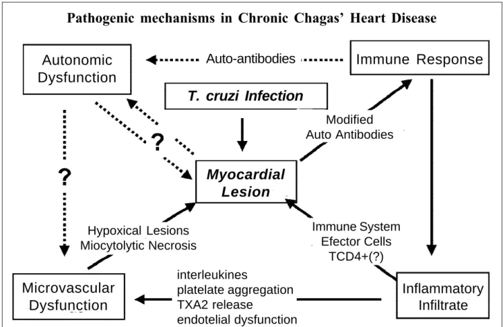
In essence, T. cruzi inflammatory and autoimmune
aggression seem to constitute the more prominent patho-genetic mechanisms. Autonomic disturbances and micro-vascular ischemia appear to play an ancillary role, acting as amplification loops and contributing to expand myocardial tissue damage. A unified overall pathogenetic framework may be constructed on the basis of such notions (fig. 1).
Management of Chagas’ heart disease
Etiologic treatment - Nifurtimox and benznidazol have
commonly responsible for discontinuation of treatment: dermatitis, polyneuritis, leukopenia, gas-trointestinal intolerance 165,166. A recent report on higher incidence of
cancer following antitrypanosoma therapy in heart trans-planted patients is also an indication that the search for more effective and better tolerated drugs seems clearly warranted 167. Recent reports of allopurinol and itraconazole
for treating Chagas’ disease reactivation after heart trans-plantation and chronic chagasic yielded intriguing results that require further serious scrutiny before any firm conclu-sions can be derived 124,168.
In the acute phase of Chagas’ infection, irrespective of the mechanism of transmission (vectorial, blood trans-fusion, laboratory accident, oral, or even reactivation of chronic disease during immunosuppressive conditions), it is virtually consensual that etiologic treatment is manda-tory to control symptoms and life threatening conditions, such as myocarditis and encephalitis, and, presumably, to prevent chronic organ damage. However, the efficacy of treatment regarding this later aspect has not been proved as no controlled long-term follow-up trials have been reported. Parasitologic evaluation shows negativity of xenodiagnosis in over 90% of cases and serologic tests are negative in 80%, after adequate treatment. The prognostic meaning of conversion to a negative serology has not been established, again due to lack of appropriately designed follow-up studies focusing on this relevant aspect.
Evidence for potential benefits of specific antitry-panosoma chemotherapy in chronic Chagas’ heart disease is lacking because misleading criteria have been used to assess therapeutic efficacy and also due to the fact that only small, nonrandomized, noncontrolled trials have been carried out 169-171. Thus, conversion from a serum-positive
to a serum-negative state following therapy is an unreliable marker of the impact of such treatment upon the course of Chagas’ disease, because in any event, negative serology is common in the chronic phase. Moreover, large fluc-tuations of parasitemia occur over time. Another negative aspect in those trials is the bias induced by selection of patients with persistent parasitemia in the pretreatment period 7. Furthermore, results of experimental studies have
shown that in the chronic phase the parasitemia is low or not detectable at all while there is a predominant tissular para-sitism by amastigote forms of T. cruzi 114.
Conversely, persistently positive serologic tests may merely reflect mechanisms of immunological memory, or be associated with cross-reactivity to altered host antigens. Hence, results of any of the serological criteria used to assess the therapeutic value of etiologic treatment in patients with the chronic form of Chagas’ heart disease are clearly unreliable. The reported rate of negativity of serological tests following treatment in the chronic phase is consistently very low (4-8%) in the trials suffering the epidemiological restrictions already pointed out 169-171.
Fig. 1 – Pathogenetic mechanisms in Chagas’ heart disease.
Pathogenic mechanisms in Chronic Chagas’ Heart Disease
Autonomic
Dysfunction
Auto-antibodies
Immune Response
T. cruzi Infection
Myocardial
Lesion
Modified
Auto Antibodies
Immune System
Efector Cells
TCD4+(?)
Hypoxical Lesions
Miocytolytic Necrosis
interleukines
platelate aggregation
TXA2 release
endotelial dysfunction
Microvascular
Dysfunction
Thus, until an adequate laboratory method is available for assessment of cure, the only acceptable criteria for any etiologic therapeutic intervention benefit must be based on the prevention of the appearance of the clinical form of disease or the arrest of progression of the damage already detected. For this, a very long follow-up period of large cohorts of chagasic patients is required to detect changes in the natural history of the disease.
No definitive recommendations are justifiable for etiologic treatment in the chronic phase of Chagas’ heart disease, until large randomized controlled studies encom-passing patients in different stages of disease have been performed 7.
Treatment of congestive heart failure - Since the
hemodynamic derangements in chronic chagasic patients with heart failure are comparable to those reported in dilated cardiomyopathies of other etiologies, classical therapeutic interventions (sodium restriction, diuretics, digitalis, and vasodilation with nitrates and hydralazine) are usually employed for relief of congestive symptoms in chagasic patients7. Several noncontrolled small studies documented
short-term hemodynamic beneficial effects of these agents, and, to a lesser extent, improvement in exercise tolerance in chronic chagasic patients. However, no studies reported improvement in survival, or even in long-term outcome based on hemodynamic and symptomatic benefit 7.
Preliminary studies involving therapy with ACE-inhibitors, enrolling small numbers of patients, have shown promising results in heart failure complicating Chagas’ disease 172,173, in regard to symptomatic control. Although
no long-term prospectively controlled study has been reported assessing the impact on survival of chagasic patients treated with ACE-inhibitors or any other pharma-cological interventions, there is no reason to expect that their beneficial effect would be any different from that observed in heart failure due to other etiologies. In fact, there seems to be a favorable acute neuromodulating effect of ACE-inhibition in chagasic patients 174.
As discussed earlier, early regional ventricular wall motion impairment and diminished contractile properties can be seen even in patients with the indeterminate form of the disease 63,175. Moreover, the mild dyssynergia thus
detected appears to reflect more extensive myocardial damage than the ECG changes classically interpreted as heralding more advanced cardiac involvement 176.
Further-more, there is recent preliminary evidence that these minor segmental wall motion abnormalities in chagasic patients may bear relevant prognostic implications 177. Therefore, it
is reasonable to conclude that it remains to be tested if chagasic patients, similar to what has been shown in other causes of heart failure, would benefit from early medical intervention, to detain the natural history and prevent the installation of overt cardiac dysfunction.
Surgical approaches to treatment - Heart transplan-tation - As in other cardiomyopathies, heart
transplan-tation has been performed in small groups of patients with refractory heart failure due to Chagas’ disease. However, wider application of this therapy is currently hindered by socioeconomical factors in endemic areas, and by the reactivation of infective manifestations associated with immunosuppression. Acute myocarditis, with marked transitory LV systolic depression occurred in five of the first nine patients included in the largest series 22 patients -operated in a single surgical center 178. Although the acute
reactivation was usually responsive to antiparasite therapy, the possibility of chronic damage to the allograft could not be ruled out. The results reported on the latest 13 patients of this series, using a reduced regimen of immunosuppression with cyclosporine, are promising as reactivation of disease supervened in only one patient. Also, a survival rate at 24 months posttransplantation of 80% in that later group, appears to compare favorably with those reported in clinical series. Nevertheless, the long-term impact of heart trans-plantation in chagasic patients remains to be determined by adequately controlled studies in large cohorts 7.
Dynamic cardiomyoplasty - Reported experience with
this palliative surgical procedure in chagasic patients is quite limited. Initial results showed encouraging symptom and LV function improvement in very few patients 179. A
recent survey of surgical centers in South America showed results pertaining to a total of 112 patients of whom 96 had heart failure due to dilated cardiomyopathy and 13 due to Chagas’ heart disease 180. Comparative analysis disclosed
survival rates of 86.1% and 49.8% for patients with dilated cardiomyopathy and 40% and 9.5% for chagasic patients, at one and five years follow-up, respectively. No clues from these data would point to any factors possibly involved in the worse prognosis thus suggested for chagasic patients. Clearly, large controlled randomized trials are necessary to define the issue of cardiomyoplasty as a temporary approach, before more radical interventions such as heart transplantation can be used in selected patients with refractory Chagas’ heart disease 7.
Prevention of thromboembolic events - There is very
limited clinical information concerning the risk of embolic phenomena in patients with detected mural thrombus or apical aneurysm 7. In 65 selected patients with apical
aneurysm a follow-up study ranging from 19 to 176 months documented 17 episodes of thromboembolism occurring in 14 patients (24.5%) 181. These patients also had congestive
heart failure, and 11 died in the period of observation. In eight of those patients the cause of death was related to heart failure, and in three it was a consequence of cerebral embolism. Another small study addressed the relative contribution of Chagas’ heart disease as the underlying cause of embolism in 69 patients with embolic strokes treated in an endemic region in South America 182. Of 13
fibrillation (29%) and rheumatic valvular heart disease (20.3%).
However, the real risk of thromboembolism in patients with Chagas’ heart disease is unknown, as no specific studies have addressed this problem. Furthermore, despite the preliminary evidence that thromboembolic events are relevant prognostic factors in the natural history of Chagas’ disease, no clinical studies focusing on adequate treatment and prevention of thromboembolism in chagasic patients have been conducted to date 7.
Current recommendations for anticoagulant therapy are based on information derived from other dilated cardio-myopathies. Chagasic patients presenting global LV dysfunction, atrial fibrillation, previous embolic episodes, dyskinetic areas with detected mural thrombus, are candidate to treatment with intravenous and/or oral anticoagulants 183. The issue is further complicated by the
fact that social and economic factors limit the implemen-tation of this strategy, even in chagasic patients with otherwise apparent clear indications for prevention of thromboembolic events 7.
Management of chagasic patients with precordial pain - This may be a difficult task, and is entirely empirically
based. The symptom is not related to vasotonic angina and recent studies show that chagasic patients do not have augmented responses to either coronary constrictor or endothelium-independent vasodilator stimuli, suggesting that no increased baseline coronary tonus occurs 144,184.
Also, therapeutic interventions aiming at the relief of symptoms as possibly derived from esophageal invol-vement are not usually justifiable. Although endoscopy evidence of esophagitis can be obtained in a substantial proportion of chagasic patients, their sensitivity to chemical or mechanical stimuli is typically depressed 185. Some
patients benefit from the use of nitrates and beta-adre-nergic or calcium channel blockers, but the individual response is unpredictable.
Management of rhythm disturbances - Management
of symptomatic bradyarrhythmias does not differ from that recommended for other cardiomyopathies, and in most situations relies on permanent pacemaker insertion, but no sound evidence based on large randomized controlled trials is available to support any specific treatment strategy 7.
Main indications for pacing are atrioventricular block and sinus node dysfunction 186. The evidence for a
bene-ficial effect of pacemaker implantation comes from the su-perior clinical outcome of patients with such rhythm disturbances in limited case-series reports, as compared with the natural history of patients in whom this treatment was not possible 187,188. Another relevant aspect is the
common association of atrioventricular disturbances and ventricular complex dysrhythmia in the same patient. The acceptable management of this clinical condition requires “prophylactic” artificial pacemaker implantation associa-ted with pharmacological antiarrhythmic therapy, even
though this combined strategy is not based on adequate evidence.
For patients with asymptomatic ventricular ectopic beats or nonsustained ventricular tachycardia no definite antiarrhythmic therapy was shown to improve survival 7.
Indeed, very scanty information has been published regarding this issue, but two moderately large randomized trials included chagasics among patients treated with amiodarone 189,190.
The GESICA (Grupo de Estudio de la Sobrevida en la Insuficiencia Cardiaca en Argentina) 189 concluded, after
two years of follow-up, that low-dose amiodarone was effective in reducing mortality and hospital admission in patients with severe heart failure, independent of the presence of complex ventricular dysrhythmia. Unfor-tunately, the contingent of chagasic patients was very small (48 of 516 patients), and subgroup analysis was neither provided nor would likely be useful.
An ongoing prospective multicenter randomized controlled study designed to evaluate the impact on survival of treatment of asymptomatic ventricular arrhy-thmia also included chagasic patients 190. In its pilot phase
this trial enrolled 127 patients (24 with Chagas’ heart disease) with LVEF <35%, presenting frequent ventricular premature complexes and/or repetitive forms of asymp-tomatic ventricular arrhythmia. The preliminary results after 12 months of follow-up showed a significant reduction in the incidence of sudden death in the amiodarone group (7.0% vs 20.4%). It must be pointed out that follow-up data were obtained in only 106 patients. This was a consequence of an excessively high drop-out rate (16%) which seriously limits the appraisal of the results. Nevertheless, we should await the final results of this trial, hopefully recruiting a larger contingent of chagasic patients, before the routine use of amiodarone could be recommended for chagasic patients with asymptomatic ventricular arrhythmias.
Although no prospective controlled studies with antiarrhythmic drugs have been performed for treatment of hemodynamically tolerated sustained VT in the setting of Chagas’ heart disease, those patients are generally treated with class III anti-arrhythmic drugs, either amiodarone (1000mg/day for 10 to 14 days followed by maintenance therapy at 200 to 600mg/day) 191 or sotalol (320mg/day) after
electrophysiologic study 192. The efficacy of empiric
treatment with amiodarone is strongly influenced by the grade of left ventricular dysfunction. This is reflected in one-year mortality rates of 0 and 40 percent and of recurren-ce of ventricular tachycardia of 30 and 100 perrecurren-cent after one year, respectively for groups of chagasics with class I-II and III-IV of the NYHA 42,191.
Patients at high risk of sudden death from lethal tachyarrhythmias would probably benefit from an implan-table cardioverter-defibrillator 8,193, but its widespread use is