424 Sao Paulo Med J. 2011; 129(6):424-7
CASE REPORT
Primary epidermoid carcinoma of the breast presenting as
a breast abscess and sepsis
Carcinoma epidermoide primário de mama se apresentando como abscesso
mamário e sepse
Andrea Pires Damin
I, Fernanda Costa Nascimento
II, João Batista Andreola
II, Talita Haubert Cerutti
II, Adriana Roehe
III,
Daniel Carvalho Damin
IVDivision of Breast Surgery, Hospital Fêmina, Department of Pathology, Universidade Federal de Ciências da Saúde, and Department of
Surgery, Universidade Federal do Rio Grande do Sul, Porto Alegre, Rio Grande do Sul, Brazil
ABSTRACT
CONTEXT: Squamous cell carcinoma (SCC) of the breast is an extremely rare form of cancer, accounting for approximately 0.04% of all malignant breast tumors. To date, only a limited number of cases of SCC of the breast have been reported, and most of them presented like the usual breast carcinomas.
CASE REPORT: A 39-year-old woman presented with a large breast abscess and signs of sepsis. After sur-gical debridement of the lesion, histopatholosur-gical examination of the abscess capsule revealed the pres-ence of SCC of the breast. The deinitive treatment for the tumor consisted of modiied radical mastectomy with resection of the residual lesion in the right breast.
CONCLUSIONS: This unusual case illustrates how an apparently benign disorder such as a breast abscess might be related to a clinically occult malignancy. A review of the literature on SCC of the breast is pre-sented.
RESUMO
CONTEXTO: O carcinoma de células escamosas da mama é um câncer extremamente raro, representando cerca de 0,04% dos tumores malignos da mama. Até o momento, apenas um número limitado de casos da doença foi relatado, a maioria se apresentando clinicamente como um carcinoma de mama usual.
RELATO DE CASO: Paciente feminina de 39 anos de idade se apresentando com grande abscesso de mama e sinais de sepse. Após debridamento cirúrgico da lesão, o exame histopatológico da cápsula do abscesso revelou a presença de carcinoma de células escamosas da mama. O tratamento deinitivo do tumor foi uma mastectomia radical modiicada com ressecção da lesão residual na mama direita.
CONCLUSÃO: O presente caso, de apresentação incomum, demonstra como uma lesão aparentemente benigna, como um abscesso mamário, pode estar relacionada a uma neoplasia maligna oculta. Uma revi-são da literatura a respeito do carcinoma de células escamosas de mama é apresentada.
IMD, PhD. Attending Physician, Division of Breast
Surgery, Hospital Fêmina, Porto Alegre, Rio Grande do Sul, Brazil.
IIMD. Attending Physician, Division of Breast
Surgery, Hospital Fêmina, Porto Alegre, Rio Grande do Sul, Brazil.
IIIMD, PhD. Adjunct Professor, Department of
Pathology, Universidade Federal de Ciências da Saúde, Porto Alegre, Rio Grande do Sul, Brazil
IVMD, PhD. Adjunct Professor, Department of
Surgery, Universidade Federal do Rio Grande do Sul, Porto Alegre, Rio Grande do Sul, Brazil.
KEY WORDS
Breast neoplasms. Carcinoma, squamous cell. Abscess.
Sepsis. Mastectomy.
PALAVRAS-CHAVE:
Neoplasias da mama.
Carcinoma de células escamosas. Abscesso.
Sepse. Mastectomia.
INTRODUCTION
Primary squamous cell carcinoma (SCC) of the breast is an exceedingly rare disease, accounting for approximately 0.04% of all breast malignancies.1 Its diagnosis is established when no other primary malignancy can be found in the resected breast specimen; presence of a breast metasta-sis from another primary tumor is ruled out; and the tumor does not arise from the breast skin. To date, only a limited number of cases of SCC of the breast have been reported, and most of them presented like the usual breast carcinoma.2,3 In this paper, we report on a case of SCC of the breast that atypically presented as a large and life-threatening breast abscess.
CASE REPORT
Primary epidermoid carcinoma of the breast presenting as a breast abscess and sepsis | CASE REPORT
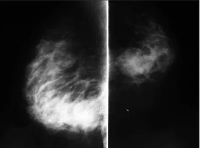
Sao Paulo Med J. 2011; 129(6):424-7 425 in the right breast, which was classiied as BIRADS 5 (Figure 1).
Breast ultrasound showed a 3.5 cm irregular mass containing a luid accumulation (Figure 2). An ultrasound-guided core biopsy was then performed, and this revealed atypical ductal hyperplasia with accentuated pleomorphism.
Ater admission to the hospital, the patient sufered rapid dete-rioration of her general clinical condition. Within 24 hours, she showed overt signs of sepsis, despite the use of systemic broad-spectrum antibiotics. Since no other source of infection was identi-ied, she was subjected to an urgent exploration of the right breast. During the procedure, a large mass extending from the lower mammary quadrants up to the retro-areolar region was identiied. he appearance of the lesion was atypical, with imprecise limits and a large central cavity containing necrotic material. A biopsy of the capsule of this lesion was obtained. Ater surgical debridement of this material, the patient started to present improvements in all the inlammatory signs and symptoms.
Histopathological analysis on the surgical biopsy revealed an undiferentiated carcinoma with extensive areas of necrosis. Additionally, immunohistochemical analysis demonstrated high expression of cytokeratin (34bE12) within the tissues, which was compatible with a diagnosis of epidermoid carcinoma of the breast. he breast tumor proile was negative for estrogen recep-tors, progesterone receptors and HER2/neu overexpression.
In view of the pathological indings, the patient underwent modiied radical mastectomy with resection of the residual lesion in the right breast three weeks ater the irst surgery. Histopatho-logical analysis on the surgical specimen demonstrated a poorly diferentiated epidermoid carcinoma, which did not involve the breast skin (Figure 3). No metastases were detected in the 13 axil-lary lymph nodes that were resected. Ater postoperative recovery, the patient was referred for complementary radiotherapy to the chest wall. She did not receive adjuvant chemotherapy. She remains free of disease two years ater the deinitive surgical treatment.
DISCUSSION
Primary squamous cell carcinoma of the breast is a very rare dis-ease. Its diagnosis is established when the malignant cells are all of squamous cell type, it does not arise from the skin of the breast and there is no other primary SCC anywhere else in the body.4
Although these tumors most frequently present like the usual breast carcinoma, SCC of the breast may, on very rare occasions, present as a breast abscess.5 A systematic survey of indexed arti-cles using the terms “epidermoid carcinoma of the breast” and “abscess” and using the terms “squamous cell carcinoma”, “breast” and “abscess” in the Lilacs (Literatura Latino-Americana e do Car-ibe em Ciências da Saúde), Embase (Excerpta Medica Database), Scirus, SciELO, Medline and Cochrane Library databases and MeSH (Medical Subject Headings) revealed that only nine articles have been published on this topic to this date. All of these papers
Figure 1. Radiological appearance of the lesion in the mediolateral and craniocaudal mammographic views.
Figure 2. Breast ultrasound showing an irregular lesion with a central cavity containing liquid material.
CASE REPORT | Damin AP, Nascimento FC, Andreola JB, Cerutti TH, Roehe A, Damin DC
426 Sao Paulo Med J. 2011; 129(6):424-7
were found in Medline (Table 1).5-13 A less speciic survey using the terms “epidermoid carcinoma” and “breast (MeSH terms)” and using the terms “squamous cell carcinoma” and “breast (MeSH terms)” was also conducted. However, this proved to be less pro-ductive, since only ive articles written in English since 1980 could be found.14-19 None of these articles (all found in Medline) describe any cases of epidermoid carcinoma of the breast presenting as abscess or sepsis.
It is important to consider SCC in the diferential diagnosis of breast abscess when there is no initial clinical response to drainage of a breast abscess or to administration of broad-spectrum antibi-otics. Wrightson et al. described three cases of SCC of the breast. One of them presented as a breast abscess that was initially man-aged by surgical drainage and debridement of necrotic material. Similarly to what happened in our case, the deinitive treatment for their patient required that mastectomy should be performed.6
It is important to highlight that SCCs of the breast should be distinguished from mixed tumors in which some areas con-taining squamous cells can be found within primary adenocar-cinoma of the breast. It is also essential to rule out the presence of SCC elsewhere in the body, which might represent the actual primary tumor from where the breast SCC is derived (metastatic breast involvement).1,4
SCC of the breast is a diicult tumor to diagnose through the subsidiary imaging examinations commonly used. here are no typical indings on the mammogram. Although ultrasound may show the presence of a complicated cyst or an inlamma-tory process, these tumors have not been reported as showing any speciic characteristics. A biopsy should be always obtained, as it deinitively conirms the diagnosis of SCC of the breast.20,21 In most of the reported cases, the diagnosis was based solely on the histopathological indings. Immunohistochemical anal-ysis showing tumor cells positively stained for cytokeratin was used by some authors to further support the pathological ind-ings.22 In the present case, immunohistochemistry was positive for cytokeratin, but negative for hormone receptors. According
to Siegelmann-Danieli et al., estrogen and progesterone recep-tors are negative in more than 90% of pure squamous cell carci-nomas of the breast.23
he treatment for SCC of the breast is similar to that of other malignant breast tumors. Although a conservative approach to the breast can be used, many of these patients already present locally advanced disease at diagnosis, thus precluding breast con-servation. In addition, it has been demonstrated that auxiliary dissection is a fundamental part of the treatment.21,24,25 Because of the rarity of this cancer, only limited data are available on the role of chemotherapy.4 he therapeutic combination most commonly used is 5-luorouracil and cisplatin, and some degree of success from this has been reported.4,26,27 he combination of docetaxel and doxorubicin has been reported to be an alternative in some cases.17,26-29 Complete clinical and pathological responses to plati-num agent-based regimens have been described in patients with either systemic metastases27 or locally advanced disease.17 he role of radiation has been described as unclear in many studies. Although SCC is generally radiosensitive, locoregional relapse occurs frequently and may even involve the irradiated ield.20,21,30 Since previous cases of SCC have predominantly been hormone receptor-negative, there only seems to be a limited role for hor-monal therapy in this type of cancer. 2,20,27
CONCLUSION
We reported an extremely rare case of SCC of the breast atypically presenting as a breast abscess that rapidly progressed to overt sep-sis. his case illustrates that an apparently benign disorder such as a breast abscess might occasionally be related to a clinically occult malignancy. Complicated cysts and breast abscesses should always be evaluated through histopathological examination.
REFERENCES
1. Weigel RJ, Ikeda DM, Nowels KW. Primary squamous cell carcinoma
of the breast. South Med J.1996;89(5):511-5.
2. Sheen-Chen S, Chen YS, Chou FF, Eng HL. Primary squamous cell
carcinoma of the breast. South Med J. 1992;85(2):207-9.
3. Grabowski J, Saltzstein SL, Sadler G, Blair S. Squamous cell carcinoma
of the breast: a review of 177 cases. Am Surg. 2009;75(10):914-7.
4. Hennessy BT, Krishnamurthy S, Giordano S, et al. Primary squamous
cell carcinoma of the breast. J Clin Oncol. 2005;23(31):7827-35.
5. Nair VJ, Kaushal V, Atri R. Pure squamous cell carcinoma of the breast
presenting as a pyogenic abscess: a case report. Clin Breast Cancer.
2007;7(9):713-5.
6. Wrightson WR, Edwards MJ, McMasters KM. Primary squamous cell
carcinoma of the breast presenting as a breast abscess. Am Surg.
1999;65(12):1153-5.
7. Gupta S, Usha. Primary squamous cell carcinoma of the breast arising
within an abscess. J Indian Med Assoc. 1982;79(1-2):12-3.
8. Melamed JB, Schein M, Decker GA. Squamous carcinoma of
Data base Search strategy Results*
PubMed
“epidermoid carcinoma of the breast” AND “abscess”
8 case reports5-12
No reviews of the literature “epidermoid carcinoma of
the breast” AND “sepsis”
No case reports or reviews of the literature
“squamous cell carcinoma of the breast “AND “abscess”
9 case reports5-13
No reviews of the literature “squamous cell carcinoma of
the breast” AND “sepsis”
No case reports or reviews of the literature
Table 1. Results from our reviews of medical databases using descriptors for the main clinical indings observed in our patient
Primary epidermoid carcinoma of the breast presenting as a breast abscess and sepsis | CASE REPORT
Sao Paulo Med J. 2011; 129(6):424-7 427 the breast presenting as an abscess. A case report. S Afr Med J.
1986;69(12):771-2.
9. Tan YM, Yeo A, Chia KH, Wong CY. Breast abscess as the initial
presentation of squamous cell carcinoma of the breast. Eur J Surg
Oncol. 2002;28(1):91-3.
10. Cappellani A, Di Vita M, Zanghì A, et al. A pure primary squamous
cell breast carcinoma presenting as a breast abscess: case report and
review of literature. Ann Ital Chir. 2004;75(2):259-62; discussion 262-3.
11. Gupta C, Malani AK. Abscess as initial presentation of pure primary
squamous cell carcinoma of the breast. Clin Breast Cancer. 2006;7(2):180.
12. Comellas N, Marin Gutzke M. Primary pure squamous cell carcinoma
of the breast presenting as a breast abscess. J Plast Reconstr Aesthet
Surg. 2009;62(6):e178-9.
13. Mokhtar GA. Squamous cell carcinoma of the breast. Saudi Med J.
2009;30(10):1346-9.
14. Annam V, Giriyan SS, Kulkarni MH. Diagnosis of pure squamous cell
carcinoma of the breast by ine needle aspiration cytology. Acta
Cytol. 2009;53(6):722-3.
15. Breuer A, Kandel M, Fissler-Eckhof A, et al. BRCA1 germline mutation
in a woman with metaplastic squamous cell breast cancer. Onkologie.
2007;30(6):316-8.
16. Zaconati F, Zanella M, Falconieri G, Di Bonito L. Gestational squamous
cell carcinoma of the breast: an unusual mammary tumor associated
with aggressive clinical course. Pathol Res Pract.
1997;193(11-12):783-7; discussion 789-90.
17. Dejager D, Redlich PN, Dayer AM, Davis HL, Komorowski RA. Primary
squamous cell carcinoma of the breast: sensitivity to
cisplatinum-based chemotherapy. J Surg Oncol. 1995;59(3):199-203.
18. Uzoaru I, Adeyanju M, Ray VH, Nadimpali V. Primary squamous cell
carcinoma of the breast presenting as a nipple discharge. Acta Cytol.
1994;38(1):112-3.
19. Tashjian J, Kuni CC, Bohn LE. Primary squamous cell carcinoma
of the breast: mammographic indings. Can Assoc Radiol J.
1989;40(4):228-9.
20. Behranwala KA, Nasiri N, Abdullah N, Trott PA, Gui GP. Squamous cell
carcinoma of the breast: clinic-pathologic implications and outcome.
Eur J Surg Oncol. 2003;29(4):386-9.
21. Aparicio I, Martínez A, Hernández G, Hardisson D, De Santiago J.
Squamous cell carcinoma of the breast. Eur J Obstet Gynecol Reprod
Biol. 2008;137(2):222-6.
22. Vera-Alvarez J, García-Prats MD, Marigil-Gómez M, et al. Primary pure
squamous cell carcinoma of the breast diagnosed by ine-needle
aspiration cytology: a case study using liquid-based cytology. Diagn
Cytopathol. 2007;35(7):429-32.
23. Siegelmann-Danieli N, Murphy TJ, Meschter SC, Stein ME, Prichard
J. Primary pure squamous cell carcinoma of the breast. Clin Breast
Cancer. 2005;6(3):270-2.
24. Cardoso F, Leal C, Meira A, et al. Squamous cell carcinoma of the
breast. Breast. 2000;9(6):315-9.
25. Shigekawa T, Tsuda H, Sato K, et al. Squamous cell carcinoma
of the breast in the form of an intracystic tumor. Breast Cancer.
2007;14(1):109-12.
26. Rostock RA, Bauer TW, Eggleston JC. Primary squamous carcinoma of
the breast: a review. Breast. 1984;10:27-31.
27. Stevenson JT, Graham DJ, Khiyami A, Mansour EG. Squamous
carcinoma of the breast: a clinical approach. Ann Surg Oncol.
1996;3(4):367-374.
28. Li Z, Li YT. Squamous cell carcinoma of the breast. Am J Surg.
1984;147(5):701-2.
29. Bhatt L, Fernando I. Primary squamous cell carcinoma of the breast:
achieving long-term control with cisplatin-based chemotherapy.
Clin Breast Cancer. 2009;9(3):187-8.
30. Pramesh CS, Chaturvedi P, Saklani AP, Badwe RA. Squamous cell
carcinoma of breast. J Postgrad Med. 2001;47(4):270-1.
Sources of funding: None
Conlict of interest: None
Date of the irst submission: October 1, 2010
Last received: February 24, 2011
Accepted: April 14, 2011
Address for correspondence:
Andrea Pires Damin
Avenida Lageado, 1099/202
Petrópolis — Porto Alegre (RS) — Brasil
CEP 90460-110
Tel. (+55 51) 3207-5688