Sao Paulo Med J. 2010; 128(4):211-4
211
Original article
Breast density in women with premature ovarian failure or
postmenopausal women using hormone therapy: analytical
cross-sectional study
Densidade mamária em mulheres com falência ovariana prematura ou na pós-menopausa e
em uso de terapia hormonal: estudo transversal analítico
Patrícia Magda Soares
I, César Cabello
II, Luis Alberto Magna
III, Eduardo Tinois
IV, Cristina Laguna Benetti-Pinto
IVDepartment of Obstetrics and Gynecology, Universidade Estadual de Campinas, Campinas, São Paulo, Brazil
IMD, Postgraduate student, Department of Obstetrics and Gynecology, School of Medical Sciences, Universidade Estadual de São Paulo (Unicamp), Campinas, São Paulo, Brazil. IIMD, PhD. Associate professor, Department of Obstetrics and Gynecology, School of Medical Sciences, Universidade Estadual de Campinas (Unicamp), Campinas, São Paulo, Brazil. IIIMD, PhD. Titular professor, Department of Medical Genetics, School of Medical Sciences, Universidade Estadual de Campinas (Unicamp), Campinas, São Paulo, Brazil. IVPhysicist and engineer, Biomedical Engineering Center, Universidade Estadual de Campinas (Unicamp), Campinas, São Paulo, Brazil.
VIMD, PhD. Professor, Department of Obstetrics and Gynecology, School of Medical Sciences, Universidade Estadual de Campinas (Unicamp), Campinas, São Paulo, Brazil.
ABSTRACT
CONTEXT AND OBJECTIVE: Studies on postmenopausal women have reported increased risk of breast cancer relating to the type and duration of hormone therapy (HT) used. Women with premature ovarian failure (POF) represent a challenge, since they require prolonged HT. Little is known about the impact of prolonged HT use on these women’s breasts. This study aimed to evaluate the effects of one type of HT on the breast density of women
with POF, compared with postmenopausal women.
DESIGN AND SETTING: Cross-sectional study at the Department of Obstetrics and Gynecology, Universidade Estadual de Campinas (Unicamp).
METHODS: 31 women with POF and 31 postmenopausal women, all using HT consisting of conjugated equine estrogen combined with medroxyprogesterone acetate, and matched according to HT duration, were studied. Mammography was performed on all subjects and was analyzed
by means of digitization or Wolfe’s classiication, stratiied into two categories: non-dense (N1 and P1 patterns) and dense (P2 and Dy).
RESULTS: No signiicant difference in breast density was found between the two groups through digitization or Wolfe’s classiication. From digitization, the mean breast density was 24.1% ± 14.6 and 18.1% ± 17.2 in the POF and postmenopausal groups, respectively (P = 0.15). Wolfe’s classiication identiied dense breasts in 51.6% and 29.0%, respectively (P = 0.171).
CONCLUSION: There was no difference in breast density between the women with POF and postmenopausal women, who had used HT for the same length of time. These results may help towards compliance with HT use among women with POF.
RESUMO
CONTEXTO E OBJETIVO: Estudos com mulheres na pós-menopausa relatam aumento no risco de câncer de mama relacionado ao tipo e duração da terapia hormonal (TH) utilizada. Mulheres com falência ovariana prematura (FOP) representam desaio por necessitarem de TH prolongada. Pouco se
conhece sobre ação da TH nas mamas dessas mulheres. Este estudo objetivou avaliar os efeitos de um tipo de TH sobre a densidade mamária de mulheres com FOP comparativamente à de mulheres pós-menopausa.
TIPO DE ESTUDO E LOCAL: Estudo de corte transversal no Departamento de Tocoginecologia, Universidade Estadual de Campinas (Unicamp).
MÉTODOS: Estudaram-se 31 mulheres com FOP e 31 mulheres na pós-menopausa, todas usando TH com estrogênio conjugado equino mais acetato
de medroxiprogesterona, pareadas pelo tempo de utilização da TH. Todas realizaram mamograia, analisada por digitalização e por classiicação de Wolfe, estratiicada em duas categorias: não densa (padrão N1 e P1) e densa (P2 e Dy).
RESULTADOS: Não houve diferença signiicativa entre a densidade mamária dos grupos analisadas por digitalização ou classiicação de Wolfe. Pela digitalização, calculou-se densidade mamária média em 24.1% ± 14.6 e 18.1% ± 17.2 nas com FOP e pós-menopausa, respectivamente (P = 0,15); pela classiicação de Wolfe identiicou-se mamas densas em 51,6% e 29,0%, respectivamente (P = 0,171).
CONCLUSÃO: Não se observou diferença na densidade mamária de mulheres com FOP comparativamente à de mulheres na pós-menopausa
utilizando TH pelo mesmo período de tempo. Estes resultados podem auxiliar na aderência à TH de pacientes com FOP.
KEY WORDS:
Hormone replacement therapy.
Mammography. Menopause.
Premature ovarian failure. Breast.
PALAVRAS CHAVE: Terapia de reposição hormonal Mamograia.
Menopausa.
Falência ovariana prematura.
Mama.
INTRODUCTION
Studies carried out on postmenopausal women have shown in-creased risk of breast cancer relating to the type of hormone therapy (HT) used and the duration of its use.1-7 It is still a matter for debate
whether HT causes a reduction in the sensitivity and speciicity of
mam-mographic screening as a result of the increase in breast density.8-10 he
Sao Paulo Med J. 2010; 128(4):211-4 Soares PM, Cabello C, Magna LA, Tinois E, Benetti-Pinto CL
212
Women with premature ovarian failure (POF) represent a challenge, since they require prolonged hormone therapy in view of their early loss of gonad function. However, little is known about the impact of pro-longed HT use on these women’s breasts.11 Concern about breast cancer
is one of the most frequent causes of discontinuation of HT.12
Recently, postmenopausal changes in breast density, as evaluated by mammography, have been considered to be a strong marker for the risk of breast cancer. Breast density has been shown to reveal information on the exposure to endogenous and exogenous hormones that afect the environment in which cancer originates and develops.10 Various
stud-ies have shown that women with denser breasts have a two to six-fold higher risk of developing cancer, compared with women with less dense breasts.13-17
Much information linking HT and breast density in postmeno-pausal women has been published in the literature.7,18-23 However, to the
best of our knowledge, no papers evaluating the breast density of wom-en with POF have bewom-en published, evwom-en though these womwom-en are fre-quently treated with HT in the same way as postmenopausal women.
OBJECTIVE
Considering the scientiic evidence, the questions regarding the ef-fect of estrogen-progestin hormone therapy on postmenopausal breast density and the lack of information on women with premature ovarian failure regarding this subject, a study was carried out to compare breast densities between women with POF who were using estrogen-progestin HT and postmenopausal women using the same type of HT for simi-lar lengths of time.
MATERIAL AND METHODS
his cross-sectional pilot study evaluated 31 women between 30 and 40 years of age with a diagnosis of POF shown by secondary amen-orrhea with hypergonadotropic hypoestrogenism, with follicle-stimu-lating hormone (FSH) > 40 mIU/ml at two diferent times.24 hese
women were receiving care at the gynecological endocrinology outpa-tient clinic of the Department of Obstetrics and Gynecology, Univer-sidade Estadual de Campinas (Unicamp), and they had been using an estrogen-progestin HT regimen composed of 0.625 mg of conjugat-ed equine estrogen (CEE) combinconjugat-ed with mconjugat-edroxyprogesterone ace-tate (MPA), cyclically or continuously, for at least 12 months. Women who had some form of pathological condition and/or had undergone previous breast surgery, women who smoked more than 20 cigarettes/ day and those with body mass index (BMI) > 30 kg/m2 were excluded
from the study.
his group was compared with a control group of 31 postmeno-pausal women who were using the same hormone therapy as a cyclic or continuous regimen. he control group women were matched with the women in the study group according to duration of hormone use (± 11 months). hey were selected from the menopause clinic of the Depart-ment of Obstetrics and Gynecology, Unicamp. Women over 50 years of age for whom the menopause had been diagnosed at least 12 months
previously were eligible for inclusion in the study. he exclusion criteria were identical to those of the study group.
All the women in both groups underwent mammography, and the data were analyzed and compared using both the technique of mammo-graphic digitization25 and Wolfe’s classiication.26 However, the patients
were stratiied into only two groups: non-dense breasts (N1 and P1 pat-terns) and dense breasts (P2 and Dy patpat-terns).
he study protocol was approved by the Institutional Review Board of the Department of Obstetrics and Gynecology, Unicamp, and by the Ethics Committee of the School of Medicine, Unicamp.
Mammography was carried out using a high-resolution scanner (CGR Senographe 500T, GE Medical Systems) with a Kodak RPX-OMAT processor. Kodak diagnostic ilms were used, and the left mid-lateral oblique incidence was used for digitization.
he mammogram ilms were placed on a negatoscope-type appara-tus and covered by a sheet of transparent tracing paper. he outlines of the images corresponding to the ibroglandular and fatty portions were sketched by a specialist in mammography. Areas with the same density as the pectoralis major muscle were considered to be ibroglandular, while the remainder was considered to be fatty tissue. he drawings were digi-tized using a Hewlett Packard scanner and an IBM 486 desktop computer (DX4, 8 RAM, 540 HD). Digitization fragmented the igure into small areas referred to as pixels (picture elements). In the computer, the images were opened using an image editing software program (Paintbrush, Mi-crosoft), in which the fatty areas were colored light grey and the ibroglan-dular areas were colored black. A numerical value of 250 was attributed to the light grey areas and zero to the black areas. he images were evaluated using the Mathlab4 software program by a specialist in physics, to quan-tify the percentage of glandular tissue in relation to the total volume of the breast, thus resulting in the dependent variable of breast density.
Evaluation of breast density was also carried out in accordance with Wolfe’s classiication. However, it was subdivided into only two catego-ries: non-dense (N1 and P1 patterns, i.e. ibroglandular tissue accounting for < 25% of the breast volume in the left mid-lateral oblique incidence) and dense (P2 and Dy patterns, i.e. ibroglandular tissue accounting for ≥
25% of the breast in the left mid-lateral oblique incidence). he special-ist in mammography and the physicspecial-ist who performed the analyses were blinded with regard to the identities of the groups.
Means and standard deviations were calculated to analyze the vari-ables of age, parity and BMI. For the comparison of breast density be-tween groups (digitization technique), Student’s t-test for independent groups was used after performing the Kolmogorov-Smirnov test for nor-mality. For analysis on Wolfe’s classiication, stratiied into two catego-ries, proportions were compared using the chi-square test in contingen-cy tables.27 he signiicance level was established at 5%. he software
used for the statistical analyses was the Statistical Package for the Social Sciences (SPSS), version 15.0 for Windows 2006.
RESULTS
Breast density in women with premature ovarian failure or postmenopausal women using hormone therapy: analytical cross-sectional study
Sao Paulo Med J. 2010; 128(4):211-4
213
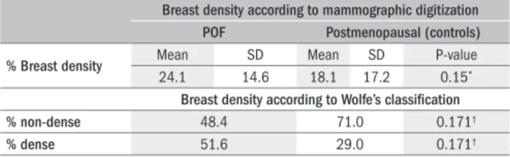
Table 1. Breast density of women with premature ovarian failure (POF) and
postmenopausal women (n = 31 in each group) analyzed according to digitization and Wolfe’s classiication
Breast density according to mammographic digitization
POF Postmenopausal (controls)
% Breast density Mean SD Mean SD P-value
24.1 14.6 18.1 17.2 0.15*
Breast density according to Wolfe’s classiication
% non-dense 48.4 71.0 0.171†
% dense 51.6 29.0 0.171†
P-value < 0.05; *Student’s t test; †Chi-square test (χ2). SD = standard deviation.
approximately normally distributed in both groups: P = 0.388 and P = 0.652 respectively). he two groups were paired according to the dura-tion of hormone therapy use (± 11 months). Hormone therapy (CEE +
MPA) had been used for a mean of 50.3 ± 39.0 months by women in
the POF group and for a mean of 50.1 ± 38.4 months by the women
in the postmenopausal control group (variable also approximately nor-mally distributed in both groups: P = 0.386 and P = 0.403 respectively). here was no statistically signiicant diference in duration of HT use between the two groups (P = 0.98). he mean time since diagnosis of POF was 85.9 ± 44.4 months, whereas the mean time since diagnosis of menopause was 117.7 ± 57.6 months (this variable also being approxi-mately normally distributed in both groups: P = 0.870 and P = 0.680 respectively).
With regard to the other variables evaluated, the women with POF had had fewer pregnancies than had the postmenopausal women (1.5 ±
1.7 and 3.7 ± 2.6 pregnancies, respectively, P < 0.001; variable approxi-mately normally distributed in both groups: P = 0.290 and P = 0.192 respectively). Fewer of the women with POF had breastfed than had the women in the control group (54.8% of the women with POF and 81.7% of the control group, P = 0.005).
Although women with BMI > 30 kg/m2 had been excluded from
enrollment in the study, the diference in BMI between the group with POF (24.1 ± 3.2) and the control group (25.8 ± 3.3) was statistically signiicant (P = 0.04). BMI was approximately normally distributed in both groups (P = 0.809 and P = 0.480 respectively).
he percentage of breast density analyzed using mammographic digitization, a variable that was approximately normally distributed in both groups (P = 0.976 and P = 0.268 respectively), was 24.1 ± 14.6% in the POF group and 18.1 ± 17.2% in the control group. his difer-ence was not statistically signiicant (P = 0.15). Wolfe’s classiication, stratiied into two subgroups (non-dense: ibroglandular tissue ing an area < 25% of the breast; and dense: ibroglandular tissue occupy-ing an area ≥ 25% of the breast), also failed to detect any statistically sig-niicant diference in breast density between the two groups, although 51.6% of the women with POF had dense breasts, compared with only 29% of the postmenopausal group (P = 0.171) (Table 1).
DISCUSSION
No diference in breast density was found between the women with POF and the postmenopausal women using hormone therapy with conjugated equine estrogens and medroxyprogesterone acetate for similar periods of time, either when analyzed objectively using digitization or subjectively in accordance with Wolfe’s classiication. However, in both groups, the percentage of ibroglandular tissue in re-lation to the total area of the breast was low (24.1 ± 14.6% and 18.1
± 17.2%, respectively).
In the POF group, which was composed of younger women (mean age 36.9 ± 2.9 years), the mean time elapsed since gonad failure was shorter than in the control group. Other factors that could have protec-tively contributed towards the reduction in breast density were in fact less frequent in the POF group, in which the women had had fewer
pregnancies and fewer women had breastfed. Despite these characteris-tics, breast density in the study group was no diferent from that of the control group.
his study does not enable conclusions to be reached regarding the causes of this inding, but one hypothesis may be that the hypoestro-genism following gonad failure, which would cause regression of i-broglandular tissue and its progressive replacement by fatty tissue,18,20
may have greater repercussions in reducing breast density when present at a younger age.
One concern regarding increased breast density resulting from estro-gen-progestin replacement therapy relates to impaired mammographic sensitivity and speciicity. his would result in a higher number of false-positive results, since the dense glandular tissue tends to make identii-cation of tumorous masses more diicult,9,10 thereby compromising the
early diagnosis of breast cancer.8
Hormone therapy is known to increase the density of the breast parenchyma. However, this does not occur in the majority of women,18,20,22,28 and this stimulus is also known to vary according to
the type of hormone therapy used. In view of these factors, the groups were matched for duration of HT use and for the type of hormone used (CEE + MPA), Nonetheless, matching for exclusively cyclic or continu-ous use was not possible, since younger women with POF often want to menstruate, whereas postmenopausal women prefer continuous regimes in order to avoid bleeding.
Although age has been shown to have an inverse correlation with breast density,29 this association was not found in the study group or in
the control group of postmenopausal women. Although the women in the study group were younger, their breast density was similar to that of the older postmenopausal women.
No signiicant diference in breast density was found between the two groups in this study, despite the fact that the groups had very dif-ferent characteristics. his latter point has encouraged us to proceed with designing a new, prospective study involving data correlation that would enable greater precision of control over the variables that afect each woman participating in the study.
Finally, based on the results from this pilot study, and considering the likelihood of type I error as 0.05 (alpha = 0.05) and a test power (type II error) of 80% (1 - beta = 0.80), we recommend that for fur-ther studies, a sample size of at least 92 patients in each group should be used.
Sao Paulo Med J. 2010; 128(4):211-4 Soares PM, Cabello C, Magna LA, Tinois E, Benetti-Pinto CL
214
his ratio may make it hard to achieve bigger samples than the pres-ent one.
Although the authors are aware of the limitations of the present sample size, this was a pilot study. Hence, the indings described here are important because this is the irst paper reporting on the efects of estrogen-progestin HT on the breast density of women with POF com-pared to that of postmenopausal women using the same type of HT for similar periods of time.
he patients enrolled in this study are being followed up for pro-spective evaluation proposals.
Finally, it should be stressed that the question of hormone treat-ment remains open. New studies would be necessary, with bigger sam-ple sizes if possible, in order to answer women’s concerns and thus pro-mote better compliance with treatment.
CONCLUSIONS
here was no diference in breast density between the women with POF and the postmenopausal women who had used the same HT dur-ing similar periods.
REFERENCES
1. Rossouw JE, Anderson GL, Prentice RL, et al. Risks and beneits of estrogen plus progestin in healthy postmenopausal women: principal results From the Women’s Health Initiative randomized controlled trial. JAMA. 2002;288(3):321-33.
2. Beral V; Million Women Study Collaborators. Breast cancer and hormone-replacement the-rapy in the Million Women Study. Lancet. 2003;362(9382):419-27.
3. Conner P, Svane G, Azavedo E, et al. Mammographic breast density, hormones, and growth factors during continuous combined hormone therapy. Fertil Steril. 2004;81(6):1617-23. 4. Chen FP, Cheung YC, Teng LF, Soong YK. The relationship between mammographic density
and duration of hormone therapy: effects of estrogen and estrogen-progestin. Hum Reprod. 2005;20(6):1741-5.
5. Harvey J, Scheurer C, Kawakami FT, et al. Hormone replacement therapy and breast density changes. Climacteric. 2005;8(2):185-92.
6. Junkermann H, von Holst T, Lang E, Rakov V. Inluence of different HRT regimens on mammo-graphic density. Maturitas. 2005;50(2):105-10.
7. Christodoulakos GE, Lambrinoudaki IV, Vourtsi AD, et al. The effect of low dose hormone therapy on mammographic breast density. Maturitas. 2006;54(1):78-85.
8. Kavanagh AM, Mitchell H, Giles GG. Hormone replacement therapy and accuracy of mam-mographic screening. Lancet. 2000;355(9200):270-4.
9. Banks E. Hormone replacement therapy and the sensitivity and speciicity of breast cancer screening: a review. J Med Screen. 2001;8(1):29-34.
10. Warren R. Hormones and mammographic breast density. Maturitas. 2004;49(1):67-78. 11. Armitage M, Nooney J, Evans S. Recent concerns surrounding HRT. Clin Endocrinol (Oxf).
2003;59(2):145-55.
12. Mann RD. Hormone replacement therapy and breast cancer risk: studies of the last ifteen years. In: Mann RD, editor. Hormone replacement therapy and breast cancer risk. New Jer-sey: Parthenon Publishing Group; 1992. p. 1-8.
13. Boyd NF, Byng JW, Jong RA, et al. Quantitative classiication of mammographic densities and breast cancer risk: results from the Canadian National Breast Screening Study. J Natl Cancer Inst. 1995;87(9):670-5.
14. Byrne C, Schairer C, Wolfe J, et al. Mammographic features and breast cancer risk: effects with time, age, and menopause status. J Natl Cancer Inst. 1995;87(21):1622-9. 15. Maskarinec G, Meng L. A case-control study of mammographic densities in Hawaii. Breast
Cancer Res Treat. 2000;63(2):153-61.
16. Vachon CM, Kuni CC, Anderson K, Anderson VE, Sellers TA. Association of mammographically deined percent breast density with epidemiologic risk factors for breast cancer (United States). Cancer Causes Control. 2000;11(7):653-62.
17. Noh JJ, Maskarinec G, Pagano I, Cheung LW, Stanczyk FZ. Mammographic densities and circula-ting hormones: a cross-sectional study in premenopausal women. Breast. 2006;15(1):20-8.
18. Laya MB, Gallagher JC, Schreiman JS, et al. Effect of postmenopausal hormonal re-placement therapy on mammographic density and parenchymal pattern. Radiology. 1995;196(2):433-7.
19. Breast cancer and hormone replacement therapy: collaborative reanalysis of data from 51 epidemiological studies of 52,705 women with breast cancer and 108,411 women without breast cancer. Collaborative Group on Hormonal Factors in Breast Cancer. Lancet. 1997;350(9084):1047-59.
20. Marugg RC, van der Mooren MJ, Hendriks JH, Rolland R, Ruijs SH. Mammographic changes in postmenopausal women on hormonal replacement therapy. Eur Radiol. 1997;7(5):749-55. 21. Koukoulis GN. Hormone replacement therapy and breast cancer risk. Ann N Y Acad Sci.
2000;900:422-8.
22. Lundström E, Wilczek B, von Palffy Z, Söderqvist G, von Schoultz B. Mammographic breast density during hormone replacement therapy: effects of continuous combination, unoppo-sed transdermal and low-potency estrogen regimens. Climacteric. 2001;4(1):42-8. 23. Greendale GA, Palla SL, Ursin G, et al. The association of endogenous sex steroids and
sex steroid binding proteins with mammographic density: results from the Postmeno-pausal Estrogen/Progestin Interventions Mammographic Density Study. Am J Epidemiol. 2005;162(9):826-34.
24. de Moraes-Ruehsen M, Jones GS. Premature ovarian failure. Fertil Steril. 1967;18(4):440-61.
25. Cymberknoh M. Mamograia digital. In: Dias EN, Calefi M, Silva HMS, Figueira-Filho ASS, eds. Mastologia atual. Rio de Janeiro: Revinter; 1994. p. 75-8.
26. Wolfe JN. Breast patterns as an index of risk for developing breast cancer. AJR Am J Roent-genol. 1976;126(6):1130-7.
27. Snedecor WG, Cochram WG. The comparison of two samples. In: Snedecor WG, Cochram WG, editors. Statistical methods. 8thed Ames: Iowa State University Press; 1989. p.
83-102.
28. Persson I, Thurfjell E, Holmberg L. Effect of estrogen and estrogen-progestin replacement regi-mens on mammographic breast parenchymal density. J Clin Oncol. 1997;15(10):3201-7. 29. Harvey JA, Bovbjerg VE. Quantitative assessment of mammographic breast density:
rela-tionship with breast cancer risk. Radiology. 2004;230(1):29-41.
Sources of funding: None
Conlict of interest: None
Date of irst submission: October 28, 2009
Last received: June 17, 2010
Accepted: June 23, 2010
Address for correspondence:
Cristina Laguna Benetti-Pinto Rua João Simões da Fonseca, 598 Loteamento Residencial Barão do Café Campinas (SP) — Brasil