b
1 Integrin in a Novel RGD-Independent Manner
Luisa F. Jime´nez-Soto1, Stefan Kutter1, Xaver Sewald1, Claudia Ertl1, Evelyn Weiss1, Ulrike Kapp2, Manfred Rohde3, Torsten Pirch4, Kirsten Jung4, S. Francesco Retta5, Laurent Terradot2, Wolfgang Fischer1, Rainer Haas1*
1Max von Pettenkofer-Institute for Hygiene and Medical Microbiology, Ludwig-Maximilians-Universita¨t, Mu¨nchen, Germany,2Macromolecular Crystallography Group, European Synchrotron Radiation Facility, Grenoble, France,3Helmholtz Center for Infection Research, Department of Microbial Pathogenesis, Braunschweig, Germany, 4Munich Center of integrated Protein Science, CiPSM, at the Department of Biology, Microbiology, of the Ludwig-Maximilians-Universita¨t Mu¨nchen, Planegg-Martinsried, Germany,5Molecular Biotechnology Centre, Department of Genetics, Biology and Biochemistry, Torino, Italy
Abstract
Translocation of the Helicobacter pylori (Hp) cytotoxin-associated gene A (CagA) effector protein via the cag-Type IV Secretion System (T4SS) into host cells is a major risk factor for severe gastric diseases, including gastric cancer. However, the mechanism of translocation and the requirements from the host cell for that event are not well understood. The T4SS consists of inner- and outer membrane-spanning Cag protein complexes and a surface-located pilus. Previously an arginine-glycine-aspartate (RGD)-dependent typical integrin/ligand type interaction of CagL witha5b1 integrin was reported to be essential for CagA translocation. Here we report a specific binding of the T4SS-pilus-associated components CagY and the effector protein CagA to the host cellb1 Integrin receptor. Surface plasmon resonance measurements revealed that CagA binding to a5b1 integrin is rather strong (dissociation constant, KD of 0.15 nM), in comparison to the reported
RGD-dependent integrin/fibronectin interaction (KDof 15 nM). For CagA translocation the extracellular part of theb1 integrin
subunit is necessary, but not its cytoplasmic domain, nor downstream signalling via integrin-linked kinase. A set ofb1 integrin-specific monoclonal antibodies directed against various definedb1 integrin epitopes, such as the PSI, the I-like, the EGF or theb-tail domain, were unable to interfere with CagA translocation. However, a specific antibody (9EG7), which stabilises the open active conformation of b1 integrin heterodimers, efficiently blocked CagA translocation. Our data support a novel model in which thecag-T4SS exploits theb1 integrin receptor by an RGD-independent interaction that involves a conformational switch from the open (extended) to the closed (bent) conformation, to initiate effector protein translocation.
Citation:Jime´nez-Soto LF, Kutter S, Sewald X, Ertl C, Weiss E, et al. (2009)Helicobacter pyloriType IV Secretion Apparatus Exploitsb1 Integrin in a Novel RGD-Independent Manner. PLoS Pathog 5(12): e1000684. doi:10.1371/journal.ppat.1000684
Editor:Guy Tran Van Nhieu, Institut Pasteur, France
ReceivedJune 10, 2009;AcceptedNovember 5, 2009;PublishedDecember 4, 2009
Copyright:ß2009 Jime´nez-Soto et al. This is an open-access article distributed under the terms of the Creative Commons Attribution License, which permits
unrestricted use, distribution, and reproduction in any medium, provided the original author and source are credited.
Funding:Funded by grants from the Deutsche Forschungsgemeinschaft (HA 2697/7-1 and 7-2) and (SFB576, project B1) to RH. The funders had no role in study design, data collection and analysis, decision to publish, or preparation of the manuscript.
Competing Interests:The authors have declared that no competing interests exist.
* E-mail: [email protected]
Introduction
Infection with the gastric pathogen Helicobacter pylori (Hp) is associated with a spectrum of pathologies, ranging from mild gastritis to peptic ulcers and gastric cancer [1]. However, the molecular mechanisms underlying the development ofHp -associ-ated gastroduodenal diseases are still poorly defined. Two major virulence factors of Hp that have been associated with disease induction are the vacuolating cytotoxin (VacA) and the cytotoxin-associated antigen A (CagA), both of which are delivered into eukaryotic target cells. VacA, a secreted multifunctional protein toxin, induces intracellular vacuoles in epithelial cells, inhibits T lymphocyte proliferation and modulates T cell function [reviewed in 2]. Using theb2 integrin subunit CD18 as a cellular receptor for uptake [3], VacA efficiently down-regulates transcription of several cytokines or chemokines in T cells [4]. CagA, an immunodominant protein of 120–170 kDa, is encoded on thecagpathogenicity island (cag-PAI). Thecag-PAI comprises a total of 27 genes, encoding the cag-Type IV Secretion System (T4SS) inHp[5].
would refer to these structures as type IV secretion system pili. Furthermore, the a5b1 integrin heterodimer has recently been identified as a receptor for theHppilus-associated adhesin CagL [14].
Integrins represent a family of about 24 different ab heterodimeric receptors that mediate cell-cell, cell-extracellular matrix and cell-pathogen interactions and govern migration and anchorage of almost all kinds of cells. Each of the non-covalently associated subunits contains a large N-terminal extracellular domain, a transmembrane segment and a short C-terminal cytoplasmic tail. Affinity for biological ligands is regulated by inside-out and outside-in signalling. The bent conformation of the integrin heterodimer represents the physiological low-affinity state, whereas inside–out signalling and ligand binding induces a large-scale conformational rearrangement, in which the integrin extends from the bent into an extended, open conformation [15]. Hp T4SS-pilus-associated CagL was suggested to bind via its arginine-glycine-aspartate (RGD) motif to a5b1 integrin, a process described as essential for CagA translocation and activation of focal adhesion kinase (FAK) and Src kinase [14]. In the present study we show that further components of thecag-T4SS, such as CagY (HP0527) and the effector protein CagA interact with distinct extracellular domains ofb1 integrin. These components are located along or at the tip of the T4SS-pilus. We propose a model that suggests conformational changes of the integrin heterodimer as a basis for CagA translocation.
Results
CagA Translocation into Epithelial Cells is Dependent on
b1 Integrin Heterodimers
Hptranslocates its effector protein CagA via thecag-T4SS into a number of different cell typesin vitro[16]. Recently it was shown that CagA translocation is dependent on the interaction of theHp T4SS-pilus-associated protein CagL, binding in an RGD-depen-dent way toa5b1 integrin on the host cell [14]. However, nothing is known about the mechanism of CagA translocation and the involvement of other T4SS components in this process. Using a
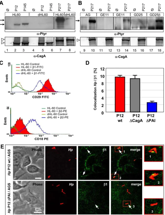
different approach, we also identified b1 integrin as a cellular receptor for the cag-T4SS. Three independent Hp strains (P12, P145, P217) were applied to study host cell requirements for CagA translocation using several human and animal cell lines (data not shown). Of special interest were human promyelocytic leukaemia (HL60) cells, which were fully competent for CagA translocation (Figure 1A, lanes 1–3 and 7), whereas, HL60 cells differentiated to a granulocyte-like phenotype (dHL60 cells) revealed only a very weak CagA-P signal (Figure 1A, lanes 4–6 and 8). Thus, the capacity ofHpto translocate CagA varies considerably, even for the same type of cell, dependent on its cellular differentiation stage. Flow cytometry revealed elevated b2, but significantly reducedb1 integrin levels on the surface of dHL60, as compared to HL60 cells (Figure 1C). We therefore concentrated on b1 integrin as a potential receptor for the T4SS. CagA translocation was completely absent for epithelial (GE11) or fibroblast-like (GD25) b1 integrin knockout cells, but was functional in genetically complemented GE11b or GD25b cells (Figure 1B) [17]. In agreement with Kwok et al. [14], these data indepen-dently confirmed the important finding thatb1 integrin is essential for CagA translocation.
CagA, CagY and CagI areb1 Integrin Interaction Partners
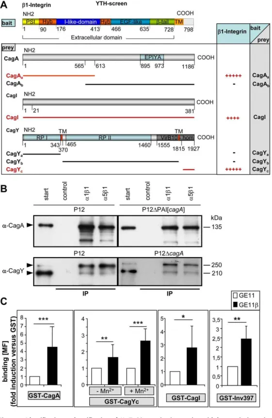
CagL is the only protein encoded on thecag-PAI which carries an RGD motif and therefore might be recognized by thea5b1 integrin receptor in a typical integrin/ligand–like fashion. Whereas Kwok et al. [14] specifically concentrated on the CagL/a5b1 integrin interaction, we chose a systematic approach to identify possible T4SS-integrin interactions and applied a yeast two-hybrid (YTH) screen using the GAL4 Matchmaker system (Clontech) (Figure 2A). Since various cell lines expressing different a/b1 integrin combinations proved successful for CagA translo-cation (data not shown), the b1 subunit was considered as important and the extracellular portion of the humanb1 integrin gene was used as bait. As prey for the YTH screen each of the 27 cag-PAI-encoded proteins were assayed [18]. Positive interactions were obtained for the extracellular part ofb1 integrin with the N-terminal region of CagA (HP0547a), the C-terminal
(VirB10-homologous) portion of CagY (HP0527c) and with CagI (HP0540)
(Figure 2A, Figure S1). Similar results were obtained when bait and prey were exchanged (data not shown).
To confirm the YTH data, pulldown experiments using Hp T4SS-associated proteins were performed (see Figure S2A for preparation of extracts) using functionala1b1 anda5b1 integrin heterodimers (Chemicon) coupled to magnetic beads. CagA was specifically pulled down from wt cell lysates by a1b1 or a5b1 integrin beads, but not by controls (Tris-blocked beads) (Figure 2B). The ectopic expression of cagA from the shuttle plasmid pJP66 [19] in a P12DPAI strain demonstrated that CagA alone is able to interact withb1 integrin without any other component of thecag -PAI. Preferentially the upper band of CagY and only small amount of the lower band of CagY was pulled down by the same procedure (Figure 2B, lower panel). Again, precipitation of CagY from acagA-negativeHpbackground confirmed an interaction of CagY with integrin, independent of CagA.
The putative interaction ofb1 integrin with CagI could not yet be verified by pulldown assays, due to the lack of a specific functional antibody against CagI. We therefore used as a further method a cell-based assay to determine binding of the corre-sponding GST-Cag fusion proteins to b1 integrin-proficient (GE11b), versus b1 integrin-deficient cells (GE11) by flow cytometry (Figure 2C and Figure S2B,C). GST did not bindb1– integrin-dependent, but purified GST-CagA, GST-CagI and GST-CagYc bound significantly more efficiently to GE11b as Author Summary
Integrins are single transmembrane proteins present on almost all types of cells. They are composed of anaand a
bsubunit, which together form the ligand binding pocket, able to interact with extracellular matrix proteins. The best known binding domain on integrin ligands is the RGD domain. Many bacterial, but also viral pathogens exploit this ligand-binding domain to interact with integrins on the host cell. Helicobacter pylori, a common bacterial pathogen associated with gastric diseases, was recently added to this list. One ofH. pylori’smost important factors associated with gastric pathologies is the CagA protein. This protein is directly injected into host cells through the Cag Type IV Secretion System (cag-T4SS). Previous studies demonstrated that thecag-T4SS requires integrins for the injection (translocation) of CagA into cells. We provide evidence that three proteins, CagA, CagI and CagY, interact with integrins in an RGD-independent way. Additionally, our data point out that the Cag apparatus needs the physical capacity of ab1 integrin heterodimer to change from an active/extended conformation to a closed/ bent conformation. This novel kind of integrin interaction opens a new way in which pathogens can use receptors on cells.
Figure 1. Host cell b1 integrin is essential for Hp to translocate and tyrosine-phosphorylate CagA in different cell lines.(A) Immunoblots to determine translocation of CagA in HL60 versus dHL60 cells. (B) Immunoblots ofb1 integrin knockout fibroblasts (GD25), epithelial cells (GE11) orb1 gene-complemented cells (GD25b, GE11b) infected withHpstrains or media (control). The upper panel shows CagA translocation using a phosphotyrosine-specific antibody (mAb PY99), the lower panel shows CagA production byHp. Bands corresponding to Phospho-CagA (CagA-P) or CagA are indicated by arrows or open arrowheads, respectively. (C) FACS analysis quantifyingb1 andb2 integrin surface localization on HL60 and dHL60 cells using antibody CD29 FITC (b1 integrin) or CD18 PE (b2 integrin). (D) Quantification of co-localization events ofHpwt,DcagA or
DPAI bacteria andb1-integrin by laser scanning confocal microscopy. (E) Confocal micrographs, showing binding ofHpP12-gfpwt (upper panel) or P12DPAI (lower panel) andb1-integrin-labelled AGS cells (4B7-AlexaFluor568) and areas of co-localization ofHpandb1 integrin (arrows).
Figure 2. Identification and verification ofHpT4SS proteins interacting with humanb1 integrin.(A) Domain organization of theb1 integrin clone used as bait in a YTH screen and identification of interacting proteins encoded by thecag-PAI used as preys. (+, interaction;2, no interaction), tenfold dilution steps still allowing growth of yeast on selective media are indicated by number of ‘‘+’’ signs (see Figure S1). (B) For pulldown assays, magnetic beads (SiMAG) were loaded with purified functionala1b1 ora5b1 integrin or Tris buffer (control) and incubated with processedHpP12 wt or defined mutant strains, as indicated (fraction Soluble II, Figure S2A). Beads were recovered by magnetic forces, washed, boiled and run for SDS-PAGE. Immunoblotting witha-CagA ora-CagY antibody detects precipitated proteins (arrowheads). (C) Quantification of binding of purified GST-CagA, GST-CagYc, GST-CagI or GST-Inv397 (30mg/16106cells) to integrin-deficient GE11 versus integrin-proficient GE11bcells by flow cytometry. Binding is analysed bya-GST antibody (fold increase in binding versus GST, the values for binding of GST alone has been set to 1). RPI, repeat region I, RPII, repeat region II, (*, p,0.05; **, p,0.01; ***, p,0.001; students T-test). MF, mean fluorescence, (n = 20). Oligonucleotides for construction of GST fusion proteins see Table S2.
doi:10.1371/journal.ppat.1000684.g002
compared to GE11 cells (Figure 2C). Interestingly, GST-CagY, but not the other GST fusion proteins, bound more efficiently when the integrins were activated by Mn2+. TheYersinia invasin (GST-Inv397) [20], known to specifically bindb1 integrin, showed a similar behaviour in these binding assays as the GST-Cag proteins (Figure 2C). To exclude that these GST fusion proteins would bind unspecifically to the GE11bcells, we also generated unrelated cag-PAI GST fusion proteins, such as GST-Cagb (HP0524), GST-CagG (HP0542) and GST-CagZ (HP0526), which did not show b1 integrin-specific binding to the cells (Figure S2B). These data confirmed the b1 integrin-specific binding of CagI from the YTH assay and verified CagI as an additional cag-T4SS component interacting with b1 integrin. Unexpectedly, neithera1b1, nora5b1 integrin beads specifically pulled down CagL from membrane or soluble fractions ofHpwt cells using our precipitation conditions (data not shown). We therefore also generated GST-CagL and GST-CagL-RAD mutant protein. Although both purified proteins revealed a rather weak interaction tob1 integrin, the binding was completely independent from the RGD motif of CagL (Figure S2C).
Taken together, these data verified CagA (the translocated effector protein), CagY and CagI as direct interaction partners of different b1 integrin heterodimers. The fact thatb1 integrin in combination with different integrinachains (a1,a5) precipitated the Cag proteins confirmed that these proteins bind to the b1 subunit, rather than theasubunit of the heterodimer and strongly support the YTH results.
Localization of CagY and CagA Along the T4SS Pilus or Tip
To allow binding of CagA, CagY and CagI to the integrin receptor, these T4SS components should be accessible at the surface of the T4SS pilus. CagY is known as an essential component of the membrane-spanning T4SS complex, but in addition its surface- or T4SS pilus-association has also been demonstrated [12,13]. In addition to CagY, we also verified CagA on the pilus by field emission scanning electron microscopy (FESEM) (Protocol S1) (Figure S3) [12]. Anti-CagA-coupled gold particles preferentially labelled the pilus tip, with one or rarely two gold grains only, but no staining of the pilus base and only rare background staining of the bacterial or eukaryotic cell surfaces was visible (Figure S3A-I). To investigate a binding of the cag-T4SS pilus to b1 Integrin during the infection process, confocal laser scanning microscopy (CLSM) and life cell imaging were applied. Thegfp-expressingHpP12 wild type (wt) strain, but not the equally well binding P12DPAI mutant strain, showed a rapid co-localization with b1 Integrin upon infection of AGS cells (Figure 1E). Quantification data revealed that roughly 10% of HpP12 wt or P12DcagA, but only 2.5% of P12DPAI bacteria co-localized with b1 Integrin (Figure 1D). A P12DcagA deletion mutant still showed co-localization tob1 integrin, which can be explained by its binding via CagY or CagI.
The RGD Motif in CagL is not Essential for T4SS Function
We wondered why CagL was detected neither in our YTH screen, nor the pulldown assays. To reassess the described RGD-dependent interaction of CagL and a5b1 integrin [14], we first generated a definedcagLdeletion mutant in HpP12. The strain was genetically complemented in the Hp recA locus by mutated cagL genes encoding RAD, RGA or DRGD versions of CagL (Figure 3A). AGS cells infected with the P12DcagLstrain failed to translocate CagA and to induce IL-8, as described earlier [11,14], but surprisingly all three distinct Hp cagL mutant strains (cagL -RAD,cagL-RGA andcagLDRGD) behaved identical to the P12 wt
strain concerning CagA translocation, as well as IL-8 induction (Figure 3B). This clearly indicated that under infection conditions an RGD-mediated interaction of CagL witha5b1 integrin either is not existent, or not necessary for CagA translocation, or for IL-8 induction.
Binding of CagA tob1 Integrin with High Affinity
To judge the specificity and the strength of CagA binding tob1 integrin, surface plasmon resonance measurements were per-formed. The expression of recombinant CagA is problematic because the protein is rapidly degraded [21,22]. We considered this property as we purified a stable N-terminal fragment (100 kDa) of CagA, lacking the C-terminal 33kDa domain [22]. The protein was used for binding studies with purified a5b1 integrin andaVb3 integrin (Clontech) as a negative control. CagA binds with high affinity to a5b1 integrin (KD= 0.153+/
20.096 nM) (Figure 3C) and with a 2-log higher KD value to
aVb3 integrin (KD= 33.4+/218 nM) (Figure 3D), demonstrating
the avidity and the specificity of CagA forb1 integrin binding. Thus, the KD value of a5b1 and CagA is approximately
hundredfold lower as compared to the same integrin with its cognate ligand, fibronectin, which is dependent on an RGD motif (KD= 15 nM) [23]. This strong and specific binding suggests an
important function for this interaction.
Integrin Clustering is Essential for CagA Translocation, but not Signalling via theb1 Integrin Cytoplasmic Tail
Specific binding of CagA or CagY to theb1 integrin subunit of the heterodimer on the host cell surface might stimulate integrin clustering and internalization. Cholesterol depletion of AGS cells by methyl-b-cyclodextrin strongly reduced CagA translocation in a dose and time-dependent manner (Table 1). Calpeptin inhibits the Ca2+-dependent protease calpain, which is required for the release of integrins from the cytoskeleton and for clustering in lipid rafts. Calpeptin treatment completely abrogated CagA transloca-tion (Figure S4, Table 1) and together with the methyl-b -cyclodextrin data strongly suggested that the organization ofb1 integrins into lipid rafts and integrin clustering is essential for CagA translocation, as recently confirmed [24].
To clarify whether b1 integrin-mediated signalling might be necessary for CagA translocation, we used CHO cells stably transfected with either a full-length humanb1A gene, a deletion comprising the complete cytoplasmic tail (b1TR) or constructs containing only the transmembrane and the common region of the b1A tail (b1COM) [25]. Surface expression ofb1A integrin was verified by flow cytometry (data not shown). With exception of the CHO vector control, all cell lines were competent for CagA translocation by HpP217 and with lower efficiency in the P12 strain (Figure 4A). The generally low efficiency of CagA translocation intob1 reconstituted CHO cells might be due to a combination of humanb1 integrin with the endogenous hamstera integrin chains. Nevertheless, these data suggest that the cytoplasmic tail ofb1 integrin and therefore outside-in signalling via the integrinb1 chain is not essential for CagA translocation, although these data do not exclude that such a signalling occurs under in vivo conditions.
no effect on the capacity of CagA translocation by any of the three Hpstrains used (Figure 4B), providing strong evidence that CagA translocation solely depends on the extracellular part of b1 integrin and its clustering in lipid rafts, but does not necessarily need theb1 integrin/ILK downstream signalling pathway.
Interference with CagA Translocation
b1 integrin heterodimers binding to ligands, such as fibronectin, collagen or laminin, involves thea chain and the b chain [27]. Integrin head domains are able to adopt two alternative conformations, termed open (high affinity) and closed (low affinity), which are modulated via binding of metal ions, such as Ca2+ (stabilising closed conformation, deactivating), or Mg2+or
Mn2+(stabilising open conformation, activating). [27]. Deactiva-tion of integrins by treatment with Ca2+ or EDTA did not interfere, but the intracellular Ca2+-chelator BAPTA significantly reduced either translocation or tyrosine-phosphorylation of CagA in AGS cells. In contrast, extracellular activation of integrins (Mn2+) significantly enhanced CagA translocation and its tyrosine-phosphorylation (Table 1). Treatment of AGS cells with proteases, such as trypsin or thrombin, which leads to detachment of the cells, but not cleavage of integrin heterodimers, resulted in slightly enhanced, rather than abrogated CagA translocation efficiency (Table 1). This observation might possibly be explained by an indirect activation of b1 integrin through Proteinase-Activated Receptor-2 (PAR-2) [28]. Natural ligands ofa5b1 integrin, such as Figure 3. Construction and functional characterization of Hp P12 strains producing CagL proteins with defined amino acid exchanges in the RGD motif and CagA binding tob1 integrin.(A) Immunoblot of P12 wt and P12DcagLstrains genetically complemented by acagLwt or a mutated gene carrying a RAD or a RGA exchange, or an RGD empty site (DRGD). (B) Sandwich ELISA to determine the IL-8 secretion of AGS cells infected by P12 wt or specificcagLmutant strains. (C) Surface plasmon resonance sensograms of the interaction of immobilized CagA (stable 100 kDa N-terminal fragment) with integrina5b1 (left) andaVb3 (right) (0.07–1.12 ng/ml) shown in resonance units (RU). The following
concentrations of integrin were used: magenta, 0.07 ng/ml; green, 0.14 ng/ml; red, 0.28 ng/ml; grey, 0.56 ng/ml; blue, 1.12 ng/ml. (n = 10 individual
measurements and KDcalculations). doi:10.1371/journal.ppat.1000684.g003
fibronectin,Yersinia enterocoliticainvasin [29] or RGD peptides, even in high quantities, did not alter CagA translocation efficiency (Table 1).
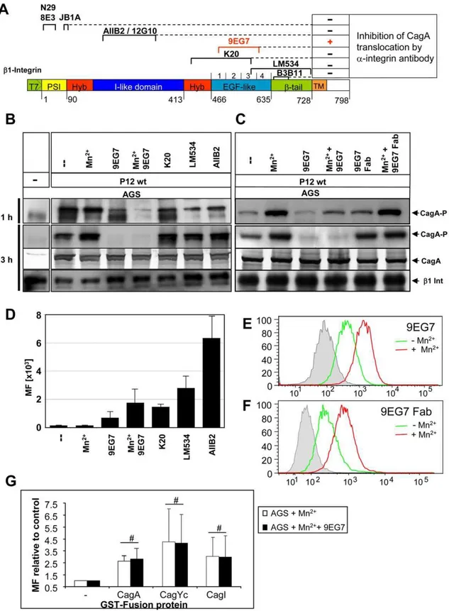
We next used a set of specific anti-b1 monoclonal antibodies (mAbs), including stimulatory (N29, 8E3, 12G10, 9EG7, B3B11), inhibitory (JB1A, AIIB2), and neutral ones (K20, LM534), to check for interference with the binding of Cag proteins to b1 integrin and thus eventually block CagA translocation (Table 1). Antibodies targeting different domains ofb1 integrin were shown to bind to AGS cells (Figure 5A,D). These well-defined mAbs cover essentially all domains and conformations of theb1 integrin chain (Figure 5A), but with the exception of 9EG7, none of them was able to interfere with CagA translocation (Figure 5B,C and Table 1). According to its function, mAb AIIB2 detached AGS cells from the tissue culture plate. This is due to its interaction with the ligand binding domain and its b1 integrin deactivation, but this binding did not block CagA translocation (Figure 5B). Thus, our data show a novel type of interaction of thecag-T4SS withb1 integrins, which is independent of the integrin a chain and a
typical integrin/ligand interactions, as well as RGD-motifs in any of the Cag proteins.
Locking Integrin in its High Affinity Conformation Blocks CagA Translocation
Several studies have indicated that a close apposition of theaand bsubunits in the membrane-proximal region and the so-called bent structure of the heterodimer are characteristic for the low affinity state of integrins [30,31]. In contrast, the extended conformation, characterized by separated legs (comprising theb1 I-EGF1-4/b-tail and theachain Calf-1/Calf-2 domains), represents the high affinity state [15]. Certain allosteric b1 integrin antibodies are able to modulate integrin activity rather specifically by stabilizing a distinct affinity state of the integrin [32]. 9EG7 is ab1-specific mAb with a binding epitope in the I-EGF2-4 region, which is strongly exposed upon manganese treatment or ligand binding (Figure 5E) [33]. The mAb 9EG7 stabilizes and probably fixes conformational changes in the integrin heterodimer, which means that 9EG7 binding of activated integrin will no longer allow its inactivation/bending. Table 1.Ligands or inhibitors of integrin signalling and their effect on CagA translocation.
Treatment Concentration Target CagA Translocation Ref
None - - +++
-Integrin activating and
-inactivation Ions this study
CaCl2 10 mM Extracellular inhibitor of Integrin, binding of fibronectin +++ this study
MnCl2 2 mM Extracellular activator of Integrin, binding of fibronectin ++++ this study
EDTA 0.8–1.2 mM Extracellular divalent cation chelator (Ca2+& Mg2+)
+++ this study
BAPTA 60mM Intracellular calcium chelator + this study
Proteases, Integrin ligands, antibodies
-8E3 30mg/ml Stimulatory mab, binds inactive form of theb1 integrin PSI domain
+++1 [40]
N29 30mg/ml Stimulatory mab, bindsb1 integrin PSI domain +++1 [41]
JB1A 30mg/ml Inhibitory mab, bindsb1 integrin PSI domain +++1 [42]
AIIB2 30mg/ml Inhibitory mab, inactivatesb1 integrin binding to
substrate
+++1 [43]
12G10 30mg/ml Stimulatory/inhibitory mab, binds to active form ofb1 +++1 [44]
K20 30mg/ml Neutral mab, binds to hybrid/EGF-like region ofb1 +++1 [45]
LM534 30mg/ml Neutral mab, binds to hybrid/EGF-like/b-tail region ofb1 +++1 [46]
B3B11 30mg/ml Stimulatory mab, binds tob-tail region ofb1 +++1 [41]
9EG7 30mg/ml Stimulatory/inhibitory mab, binds EGF-like region of
activatedb1
21 [33]
Trypsin 10mg/ml Digestion of extracellular proteins ++++ this study
Thrombin 4 U/ml Digestion of extracellular proteins ++++ this study
RGD peptide 5–100mg/ml Binding to vWF domain of integrin Competition of
binding to fibronectin
+++1 this study
RAD peptide 5–100mg/ml Peptide control of RGD +++1 this study
Invasin 197 & 397 5mg/ml Binding tob1 integrin (CD29), induces uptake +++1 this study
Fibronectin 100–10mg/ml Ligand ofa5b1 integrin +++1 this study
Inhibitors membrane signaling & lipid raft formation
-Methyl-b-cyclodextrin 0.1 g/ml Cholesterol depletion from membranes. Lipid rafts disturbance
(+)2 this study
Calpeptin 280mM Serine/threonine protease inhibitor, inhibits calpain
proteolytic activity
2 this study
1Cells synchronized before treatment. 2Strain-dependent.
Interestingly, mAb 9EG7 completely blocked CagA transloca-tion in AGS cells, whereas Mn2+ alone enhanced, rather than reduced CagA translocation (Figure 5B). A papain-generated Fab fragment of mAb 9EG7 binds in a Mn2+-dependent way to the integrin receptor (Figure 5F), but is unable to block CagA translocation (Figure 5C). A direct competition for binding of 9EG7 and GST-Cag proteins to the same integrin epitope was excluded, as measured by FACS analysis (Figure 5G).
The important function of the integrin activation state was supported by the human cervix cell line HeLa. This cell line produced normal levels of b1 integrin on the cell surface, as determined by flow cytometry (data not shown), but was only very inefficiently, or not at all able to act as host cell for CagA translocation by certainHpstrains (Figure 6A). Anin vitrophosphorylation assay ruled out a possible defect in CagA tyrosine phosphorylation (data not shown). The binding capacity of mAb 9EG7 revealed a 20% difference between non-activated and activated (Mn2+) state of the cells, whereas for AGS cells the difference was approx. 65%. These data suggest that, due to an unknown mechanism, HeLa cells apparently produce constantly activatedb1 integrin, which might be locked in the active state unable to switch back to the closed, inactive conformation, similar to the situation obtained by 9EG7 binding. This could explain the limited ability ofHpto translocate CagA.
Discussion
Thecag-Type IV Secretion System (cag-T4SS) ofHpconstitutes one of the most important virulence factors of this gastric bacterial pathogen. The mechanism by which Hptranslocates CagA into host epithelial cells is still not well understood. An important finding was that thecag-T4SS apparently does not inject its effector protein CagA randomly into target cells, but uses the a5b1 integrin as a cellular receptor for the pilus-associated adhesin CagL [14]. CagL is the onlycag-PAI encoded protein carrying an RGD sequence, which is present in certain extracellular matrix proteins and known as a typical integrin/ligand interaction motif [34].
In the present study, we describe the mammalianb1 integrin in different combinations with integrinachains as a receptor for the HpT4SS. Convincing data for a functional role of b1 integrin were obtained by the promyelocytic HL60 cell line. Non-differentiated promyelocytic HL60 cells, producing high levels of b1 integrin on their cell surface, but not differentiated dHL60 cells, with low levels of surface-associatedb1, translocated CagA very efficiently (Figure 1A,C). These data were substantiated by using integrin-deficient murine fibroblast (GD25) or epithelial (GE11) cells, which were completely resistant to CagA transloca-Figure 4.b1 Integrin signalling is dispensable for CagA translocation.(A) CagA translocation assay usingHpstrains P12 and P217 and CHO cells stably expressing either no (CHO K1), theb1A (complete gene), the b1COM (only common region of cytoplasmic tail) or the b1TR (no cytoplasmic tail) version of the humanb1 integrin gene. For a full description of the truncated b1 gene constructs please refer to [25]. Filled arrowheads depict CagA-P, open arrowheads CagA. ext, integrin external region, TM, transmembrane region. (B) Lysates of AGS epithelial integrin linked kinase (ILK) knockdown cells (siRNA-ILK) and cells transfected with GC-matched oligonucleotides (siRNA-Control) or lipofectamine-transfection (Mock-Transfection) were immunoblotted with a-ILK (Sigma) and anti-tubulin antibodies (loading control). The level of ILK knockdown was determined to be 86% by densitometry. AGS wt, ILK knockdown or control cells were infected withHpstrains, as indicated. Cell lysates were immunoblotted witha-phospho-tyrosine (PY99) ora-CagA antibodies (AK257). Bands representing CagA-P or CagA are marked by filled arrowheads, open arrowheads depict tyrosine-phosphorylated host cell proteins.
doi:10.1371/journal.ppat.1000684.g004
Figure 5. Interference with CagA translocation usingb1 integrin-specific monoclonal antibodies.(A) Mapped binding sites of anti-b1 integrin mAbs and their capacity to block CagA translocation are indicated (see also Table 1). (B) Pre-treatment of synchronized AGS epithelial cells by
b1-specific mAbs (30mg, 1h or 3h), or (C) Pre-treatment of synchronized AGS epithelial cells by mAb 9EG7 or its Fab fragments generated by papain
digestion (15mg, 1h or 3h). AfterHpinfection CagA translocation was determined (CagA-P, tyrosine-phosphorylated CagA, PY99), CagA andb1
Integrin were used as loading controls. (D) Verification ofb1 integrin antibody binding to AGS cells, as determined by FACS (MF, mean fluorescence (a-mouse and a-rat AlexaFluor488). (E) Quantification of 9EG7 binding to AGS cells with or without Mn2+ treatment by flow cytometry. (F) Quantification of 9EG7 Fab fragment binding to AGS cells with or without Mn2+treatment by flow cytometry. (G) Quantification of binding of
GST-CagA, GST-CagYc or GST-CagI to AGS cells treated with Mn2+
or Mn2+
and mAb 9EG7 by FACS analysis, to determine a possible competition in binding of GST fusion protein and 9EG7.#indicates no significant difference.
tion byHp, but could be functionally restored upon re-expression of theb1 integrin (GD25b, GE11b) (Figure 1B).
Here, we performed a systematic YTH screen to identify all proteins of thecag-PAI interacting with theb1 integrin receptor. We identified the translocated effector protein CagA, the C-terminal domain of the secretion apparatus component CagY and CagI as binding partners of the integrin receptor. Biochemical evidence for a receptor function ofa1b1 (a collagen and laminin receptor) ora5b1 integrin (a fibronectin receptor) was obtained by (i) pulldown experiments using Hplysates (Figure 2B), (ii) direct binding of the corresponding GST-Cag fusion proteins to b1 integrin as determined by FACS analysis (Figure 2C), or (iii) by surface plasmon resonance measurements (Figure 3C,D). CagA binds b1 integrin with a significantly lower KD value as a5b1
integrin binds in a RGD dependent way to fibronectin, its natural ligand. CagA affinity foraVb3 is significantly lower (approx. 100-fold) as compared toa5b1, demonstrating the high specificity of CagA for theb1 heterodimer. CagA also binds with significantly higher affinity as postulated for CagL to a5b1 integrin. CagA
carries a C-terminal translocation signal, but also the N-terminus is essential [19]. So far, the role of the N-terminal portion of CagA had remained elusive, but this specific binding tob1 could explain its important role in the translocation process. The exceptionally high affinity of CagA for the integrin receptor might compensate for the relative low abundance of CagA at the tip of thecag-T4SS pilus and suggests an important function for the surface-associated CagA. The integrin binding might have a structural role in triggering integrin rearrangements, whereas only the cytoplasmic (non-pilus-associated) CagA might act as the translocated effector molecule when a translocation-competent configuration has been established (see model Figure 7).
Whereas our data support the essential role ofb1 integrin for the process of CagA translocation, the RGD-dependent binding of CagL to the integrin receptor could not be verified. Using Hp extracts, we were able to show here a direct interaction of native (non-recombinant) CagA or CagY with integrin heterodimers, however we could not confirm an interaction of native CagL with thea5b1 integrin. It is possible that in the native pilus-associated CagL the RGD motif is buried within the protein and not accessible to interaction. In recombinant overexpression systems, this motif could be exposed, due to partially incorrect folding. Our genetic complementation data support this theory. We are able to successfully rescue CagA translocation with the complementation of CagL mutants, independently of the RGD status of the protein. Possible failures in complementation are known for Hp due to frequent secondary mutations, often in the cag-PAI [35]. In support of these genetic data we also showed that binding of GST-CagL protein tob1 integrin is very low, as compared to CagA, CagI or CagYc. More important, the binding of purified GST-CagL tob1 integrin was completely independent from its RGD motif. Thus, we show on the functional as well as on the binding level that the RGD motif of CagL is not essential for the protein function.
To further study the type of interaction between theb1 integrin heterodimer and the Cag proteins, we used typical b1 integrin ligands, such as RGD peptide, fibronectin or Yersinia invasin protein, to possibly interfere with CagA translocation (Table 1). None of these known ligands was able to block CagA translocation, indicating that the Cag proteins use different sites on the integrin for interaction. Kwok et al showed thatEscherichia colistrain HB101 that expresses Yersinia invasin inhibited CagA phosphorylation in AGS cells. It might be possible that E. coli expressing invasin on the surface could sterically inhibitH. pylorito bind and translocate CagA, just by blocking the access to the integrins.
Interaction of integrins with its ligands results in integrin clustering. Inhibition of integrin clustering into lipid rafts using methyl-b-cyclodextrin or calpeptin strongly reduced CagA trans-location, indicating that Hp-mediated clustering of b1 integrin heterodimers on the cell surface might be essential for this process. To determine whether integrin signalling might play a role for CagA translocation, signalling-deficient, truncated versions ofb1 integrin receptor were used. Unexpectedly, neither theb1 integrin cytoplasmic tail, nor signalling via the integrin linked kinase was necessary for CagA translocation, indicating that only the extracellular domains of theb1 integrin is important. CHO cells derived from hamster generally showed a lower CagA transloca-tion efficiency as compared to human AGS cells (Figure 4A). We assume that human/hamster integrin heterodimers, generated upon transfection are the reason, an effect also seen for murine GE11 cells (human/mouse integrin heterodimers) (Figure 1B).H. pyloriP217 shows a very strong CagA phosphorylation in AGS cells due to its high number of EPIYA motifs (8 motifs as Figure 6. HeLa cells are inefficient for CagA translocation and
produce constitutively activeb1 integrin.(A) Human gastric AGS and HeLa cells were infected withHpstrains P12 P217 and P145 and CagA translocation was determined by immunoblotting with antibody PY99 (CagA-P). Equal loading of bacteria and cells was determined by CagA (AK257) andb1 integrin detection (LM534). (B) Quantification of binding of mAb 9EG7 to AGS or HeLa cells with or without Mn2+
treatment by flow cytometry. doi:10.1371/journal.ppat.1000684.g006
compared to 4 in P12), resulting in more efficient tyrosine phosphorylation as P12 in CHO cells.
To obtain insight which domains of the extracellular part of the integrin receptor are important, a set of defined monoclonal antibodies against various b1 integrin domains were applied (Figure 5A, Table 1). None of these antibodies, even those blocking the integrin ligand interaction (AIIB2, 12G10), were able to block CagA translocation, except mAb 9EG7. This is in contrast to many viruses, which use integrins as receptors or co-receptors for entry into different host cells [36–38]. Most viruses known to use integrins as entry receptors have been shown to do so by extracellular matrix (ECM) protein mimicry, which means that viral proteins contain an RGD or any other conserved integrin recognition motif. Thus, specific antibodies, which block integrin ligand interaction, usually abrogate virus infectionin vitro[36–38]. Taken together our data suggest that thecag-T4SS uses the extracellular portion of integrin to mediate entry of the effector protein into the cell by a different mechanism, probably independent from ECM protein mimicry and the usual integrin ligand interaction.
What is the difference in the effect of mAb 9EG7 in comparison to all the otherb1 specific mAbs used in this study? 9EG7 binds an epitope in theb1 integrin which is close to the fulcrum at the genu and is buried in the inactive (bent) state of the integrin receptor. Mn2+ treatment or ligand binding opens the integrin into the extended conformation and the epitope is free for antibody binding. When 9EG7 is bound, the integrin cannot move back into the bent conformation, probably due to sterical problems with the bulky Fc part of the antibody or its ability to crosslink integrin chains. In addition, we cannot exclude the possibility that 9EG7 might prevent
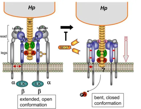
the interaction of theb1 integrin subunit with a co-receptor necessary for the translocation process, although there is no evidence for a co-receptor being involved. The 9EG7 Fab fragment still needs activation of the integrin for binding (Figure 5E,F). This indicates that its binding is unchanged, but the lack of the Fc chain will not cause the effects presumed for the complete mAb, due to the smaller size of the Fab fragment and its inability to crosslink. Our data lead us to propose a model whereby the rearrangement of the integrin from the extended, open conformation, binding the T4SS components, to a bent conformation (bent closed) is an essential step in the process of CagA translocation (see Figure 7 for a model). We propose that this mechanics of the integrin, which is associated with a closer approximation of the integrin head to the cellular membrane [27], brings the pilus closer to the cellular membrane. When this conformational change is inhibited, CagA translocation cannot occur.
Materials and Methods
Bacterial strains, cell lines and culture conditions
HpStrains. Hpstrains P12, P145 and P217 and the isogenic knockout mutants P12DcagA, P12DcagE, P12DcagY as well as P12DPAI and P217DPAI, lacking the entire cag pathogenicity island, have been constructed as previously described for the corresponding mutant strains inHp26695 [11].Hpstrains were grown on GC agar plates (Difco) as described previously [11].
Eukaryotic cell lines. Eleven different eukaryotic cell lines were analyzed for their capacity to tyrosine-phosphorylate Hp translocated CagA and IL-8 induction (Table S1). Cells were grown in media as indicated and subcultured every 2 to 3 days. Figure 7. Proposed working model forb1 integrin acting as a receptor of thecag-T4SS pilus for translocation of CagA protein.Hp T4SS pili (consisting of the pilus subunit CagC) are decorated by proteins CagA, CagY, CagL and probably CagI. The pilus associated proteins contact
b1 integrin in the open extended conformation of the head, leading to the clustering of the integrin heterodimers. These binding events may induce a change at the legs of the integrina/bchains, moving both legs together, which results in a closing and downward movement of the integrin head domains (bent, closed conformation) [39], which seems to be essential for CagA delivery. The mAb 9EG7 binds to the EGF-like domains of theb1 legs and stabilizes the open conformation by preventing a close interaction between theaandbintegrin legs (disulfide bridges, indicated by red bars), which is suggested to prevent the downward movement and therefore a possible membrane insertion of the T4SS pilus and thus CagA delivery.a, alpha integrin subunit;b, beta integrin subunit; ILK, integrin-linked kinase; A, CagA; Y, CagY; L, CagL protein; E, EGF-like1-4;b,b-tail domain; H, Hybrid; I, I-like; P, PSI domain, 9EG7, mAb 9EG7.
Antibodies and reagents
Antibodies against phosphotyrosine were obtained from Santa Cruz (PY99) or Upstate (4G10), polyclonal horseradish peroxidase (HRP) and alkaline phosphatase-conjugated mouse IgG, anti-rat IgG and anti-rabbit IgG antisera, HRP-conjugated streptavi-din, Heptakis(2,6-di-O-methyl)-b-cyclodextrin (Heptakis), fibro-nectin from human plasma, RGD (Gly-Arg-Gly-Asp-Ser-Pro-Lys) and RAD (Gly-Arg-Ala-Asp-Ser-Pro-Lys) peptides, protease inhibitors PMSF, Leupeptin and Pepstatin were obtained from Sigma. Purified humana1b1 (from smooth muscle),a5b1 (from placenta) and aVb3 (from placenta) were purchased from Clontech (Millipore). Integrin a5b1 and monoclonal anti-b1 integrin Clone LM534 were purchased from Chemicon Interna-tional. CD29 FITC and CD18 PE were purchased from BD Biosciences, and anti-b1 integrin antibody (Clone 4B7) was from Calbiochem. Rat anti-human b1 integrin inactivating antibody AIIB2 was extracted from hybridoma cells supernatant. For antibodies and their sources see Table 1. To detect CagL, a rabbit antibody against a purified CagL fusion protein was used [18].
Yeast two hybrid assay
The Invitrogen system consisting of the entry vector pDONR207 and the destination vectors pDEST-GADT7 (prey vector) and pDEST-GBKT7 (bait vector) were used. Yeast two-hybrid bait and prey libraries were generated comprising the externalb1 integrin gene sequence and thecag-PAI genes. For the cag-PAI genes, 22 full-length open reading frames (excluding N-terminal signal sequences), and 10 partial open reading frames were amplified from chromosomal DNA of strain 26695 by nested PCR, and cloned in the bait and prey vectors exactly as described [18]. Bait and prey plasmids were transformed into the haploid Saccharomyces cerevisiaestrains Y187 and AH109. Diploid yeast cells were selected after mating and selection on SD medium lacking tryptophan (Trp2) and leucine (Leu2), thus generating all possible combinations of bait and prey plasmids. After growth on SD-Trp2Leu2 medium, yeast colonies were transferred to SD-Trp2Leu2His2 medium in order to select for interactions. Growth after 3 to 6 days indicated bait-prey interactions. Additionally, the stringency of this screen was enhanced by selection on SD-Trp2Leu2His2medium containing the compet-itive inhibitor 3-aminotriazole (5 mM).
Phosphorylation assays
Cells were infected with Hp at 70–90% confluency with a multiplicity of infection (MOI) of 60. For synchronization experiments, cells were detached with PBS/2 mM EDTA, seeded and after 24 hours synchronized overnight in serum free media. Before infection, RPMI (GIBCO) complete media (CM) containing 10% Fetal Calf Serum (GIBCO) was added to cells, counting this point as time 0. To test different inhibitors, 1, 2 and 4h infections were performed after 60 min from addition of CM. After infection, supernatants from 2 & 4h experiments were collected, cells were harvested in PBS with protease inhibitors (pepstatin 1mM, leupeptin
1mM, PMSF 1mM) and phosphatase inhibitor sodium vanadate (1mM). Harvested cells were centrifuged at 5006g for 10 min at 4uC and pellets lysed in RIPA buffer with protease and phosphatase inhibitors and DNase I for later SDS-PAGE and immunoblot analysis under non-reducing conditions.
Pull-down assays and immunoblotting
SiMAG magnetic beads (Chemicell) were coated with 50mg a5b1 integrin/10 mg beads following the manufacturer’s instruc-tions. Beads were saturated with 1 M Tris-HCl (pH 7.5). 1 mlHp
(OD550 of 2) in PBS with protease inhibitors was treated with
lysozyme (10 mg/ml, 4 mM EDTA) for 30 min at RT, DNase I was added (1mg/ml) and bacteria were lysed by ultrasonication on ice. Soluble proteins (Soluble I) and membranes were separated by ultracentrifugation. Membranes were resuspended in 1 ml HSL (High Salt Lysis, 25mM Tris-HCl, pH 7.4, 0.05% Triton-X100, 4 mM MgCl2, 3 mM MnCl2, 150 mM NaCl) buffer with protease
inhibitors, sonicated and centrifuged at 4uC, 13.000 rpm for 1min to collect the soluble fraction (Soluble II). Soluble II was used for protein pulldown. After pulldown, 3ml beads were incubated at 4uC for 1 h, washed 3 times with HSL 400 (HSL with 400 mM NaCl) buffer, boiled and used for SDS-PAGE (non-reducing conditions). Proteins were transferred to PVDF membranes, and blotted with the antisera indicated. Blots were routinely stripped and reprobed with the indicated antisera (a-actin ora-b1 integrin antibodies) as loading controls. Blots shown are representative of three independent experiments.
Live cell imaging
For co-localization experiments the integrinb1-specific mono-clonal antibody 4B7 was labelled with AlexaFluor568according to
the manufacturer’s instructions (10 mol Alexa Fluor568/mol antibody). AGS cells were grown in 35 mm glass bottom dishes (MatTek) to 60–70% confluency. Cells were washed once with PBS (without Ca2+and Mg2+) and infected with GFP-expressing P12 wt or P12DPAI grown on serum-free media at an MOI of 60. 2mg/ml Alexa Fluor 568-labelled antibody againstb1 integrin was added. Infection was performed for 7 min. at 37uC and PBS was exchanged before microscopy studies. For quantification of co-localization, assays were recorded over a time range of 50s and picture sequences were analyzed for co-localization events of single bacteria and integrin b1 clusters. Percent co-localization was calculated from ratio of bacteria co-localizing with integrin to total adherent bacteria.
Imaging was done using an UltraView LCI spinning disc confocal system (PerkinElmer) fitted on a Nikon Eclipse TE300 microscope equipped with a temperature- and CO2-controllable
environment chamber. Images were taken with the black/white ORCA ER Camera (Hamamatsu). Pictures were taken and edited using LCI UltraView software. For immunofluorescence assays, an Olympus BX 64 microscope and Cell‘P software were used.
Purification of CagA 100 kDa fragment
CagA gene was cloned into a vector expressing an TEV cleavable His-tag fusion CagA (pHAR3011–CagA) as described in [22]. Briefly, the protein was expressed in BL21 cells induced o/n at 20uC with 1mM isopropyl-b-D-galactopyranoside (IPTG). Harvested cells were lysed by sonication in buffer A (10 mM Na Phosphate pH7.5, 5mM immidazole, 500 mM NaCl, 10% glycerol) containing DNaseI, lysozyme, one mini complete EDTA-free protease inhibitor cocktail tablet (Roche). After centrifugation, the supernatant was loaded onto a His-trap column (GE Healthcare) and eluted with a linear gradient of immidazole (5 to 500 mM) in buffer A. Two major N-terminal fragments (29 kDa and 100kDa, assessed by the His-tag presence) were eluted together with minor degradation products. Fractions were concentrated and the buffer was exchanged to 10mM Tris pH 7.5, 150mM NaCl. The two fragments were separated by gel filtration using Superdex S75 10/300 GL column (GE Healthcare). Fractions containing ,95% pure 100 kDa fragment (residue 1 to approximately 885) were pooled, concentrated and reloaded on the same column to ascertain stability. The protein was finally concentrated to 1.8 mg/ml.
Surface plasmon resonance
Using a BiacoreX unit, the purified 100 kDa N-terminal fragment of CagA was attached to a CM5 chip (BiaCore) using the standard amine coupling procedure. The flow-cell 1 was treated similarly without coupling of a protein and was used as a reference. Integrin binding to the reference was negligible. For the evaluation of the interaction of proteins, a Tris buffer was used containing 24 mM Tris-HCl pH 7.5, 137 mM NaCl and 2.4 mM KCl. Injection of the integrin proteins was for 1 min using a flow of 60ml/min. Subsequently, dissociation was evaluated for 200 sec. Regeneration of the chip took place between each measurement using a solution of Tris 20 mM pH 7.4, 150 mM NaCl, 1 mM MgCl2, 1 mM CaCl2and 0.0125% Triton, resulting
in a stable baseline and retaining activity. BiaEvaluation software (version 4.1) was used for the evaluation of the dissociation constant using a 1:1 langmuir model of binding.
Generation of 9EG7 Fab fragments
1 mg of 9EG7 mAb (rat) (BD Biosciences) was digested using beads coupled to Papain (Pierce) following the manufacturers’ instructions. Fab fragments were detected by western blotting using a rabbit anti rat Fab (Rockland Immunochemicals). Binding capacity of the Fab fragments to AGS cells was evaluated by flow cytometry. Secondary antibodies anti-rat Alexa488and anti-rabbit
Alexa488were from Molecular Probes.
Statistical analysis
Data are presented as mean+/2SEM. Differences between groups were assessed by the paired, two-tailed Student’st-test, or by the Mann-WhitneyUtest for unpaired groups depending on the data set of concern (see figure legends).
Supporting Information
Protocol S1 Supporting methods file
Found at: doi:10.1371/journal.ppat.1000684.s001 (0.04 MB DOC)
Table S1 Cell lines used in this study and their growth conditions
Found at: doi:10.1371/journal.ppat.1000684.s002 (0.04 MB DOC)
Table S2 Oligonucleotides used for the generation of GST fusion proteins
Found at: doi:10.1371/journal.ppat.1000684.s003 (0.05 MB DOC)
Figure S1 Protein-protein interactions amongH. pylori cag-PAI proteins andb1 integrin. Yeast cells co-transformed with plasmids expressing b1 integrin and either HP0527a (CagYa), HP0527b (CagYb), HP0527c (CagYc), HP0540 (CagI), HP0547a (CagAa) or HP0547b (CagAb), growing on minimal SD medium lacking tryptophan and leucine (SD-Trp-Leu) or on selective minimal SD
medium lacking tryptophan, leucine, and histidine (SD-Trp-Leu-His). Negative and positive controls were used as described. Found at: doi:10.1371/journal.ppat.1000684.s004 (0.77 MB TIF)
Figure S2 Integrin binding assays. (A) General procedure to generate Hpextract for pulldown assay with b1 integrin beads. Total lysate depicts the presence of CagA and CagY in P12 wt strain. (B) Quantification of binding of purified GST-Cagb, GST-CagG, GST-CagZ (negative controls for integrin binding) or GST-Inv397 (positive control) (30mg/16106 cells) to
integrin-deficient GE11 versus integrin-proficient GE11b cells by flow cytometry (n = 4). (C) Quantification of binding of purified GST-CagL or GST-GST-CagL(RAD) (30mg/16106 cells) to
integrin-deficient GE11 versus integrin-proficient GE11b cells by flow cytometry (n = 6). Binding was analysed by anti-GST antibody (fold increase in binding versus GST, the values for binding of GST alone has been set to 1). (*, p,0.05; **, p,0.01; ***, p,0.001; students T-test). MF, mean fluorescence.
Found at: doi:10.1371/journal.ppat.1000684.s005 (0.63 MB TIF)
Figure S3 High resolution FESEM micro-graphs of AGS cells infected withH. pylori. (A–F) CagA labelling is repeatedly found on the tip region of the secretion pilus using immunogold labelled antibody AK273 (arrows) (G–I) P12DPAI strain showing only few gold-particles on the AGS cell surface (arrows in G and H); in control experiments withH. pylori-infected AGS cells incubated with the gold marker alone no labelling in the tip region of the secretion apparatus is detectable. The background labelling was comparable to theDPAI strain (arrows in I). (K–L) Localization of CagY protein on the OM and thecag-T4SS pilus of Hp 26695 wt, but not 26695DPAI using immunogold-labelled antiserum AK273 by FESEM. Bars represent 200 nm (A–F,K), 1mm (G), 500 nm (H,I,L).
Found at: doi:10.1371/journal.ppat.1000684.s006 (6.78 MB TIF)
Figure S4 Pharmacological Iinhibitors interfering with CagA translocation in AGS epithelial cells. CagA translocation into AGS cells is inhibited by calpeptin (CP), an inhibitor of the Ca2+
-dependent protease calpain. CP, calpain. Filled arrowheads mark CagAP-tyr, open arrowheads mark CagA bands.
Found at: doi:10.1371/journal.ppat.1000684.s007 (0.79 MB TIF)
Acknowledgments
We thank Sven Hammerschmidt and Simone Bergmann for help with ILK knockdown experiments, M.J. Humphries for providing anti-integrin antibodies 12G10 and 8E3, John Wilkins for mAbs N29, JB1A, B3B11 and Reinhard Fa¨ssler for providing GD25/GD25b, GE11/GE11bcell lines. We are grateful to S. Hammerschmidt, J. Heesemann and W. Goebel for critical reading of the manuscript.
Author Contributions
Conceived and designed the experiments: LFJS XS WF RH. Performed the experiments: LFJS SK XS CE EW MR RH. Analyzed the data: LFJS SK XS TP KJ RH. Contributed reagents/materials/analysis tools: UK SFR LT RH. Wrote the paper: LFJS WF RH.
References
1. Suerbaum S, Michetti P (2002)Helicobacter pyloriinfection. N Engl J Med 347: 1175–1186.
2. Gebert B, Fischer W, Haas R (2004) TheHelicobacter pylorivacuolating cytotoxin: from cellular vacuolation to immunosuppressive activities. Rev Physiol Biochem Pharmacol 152: 205–220.
3. Sewald X, Gebert-Vogl B, Prassl S, Barwig I, Weiss E, et al. (2008) Integrin subunit CD18 Is the T-lymphocyte receptor for theHelicobacter pylorivacuolating cytotoxin. Cell Host & Microbe 3: 20–29.
4. Gebert B, Fischer W, Weiss E, Hoffmann R, Haas R (2003)Helicobacter pylori
vacuolating cytotoxin inhibits T lymphocyte activation. Science 301: 1099–1102.
5. Censini S, Lange C, Xiang ZY, Crabtree JE, Ghiara P, et al. (1996) Cag, a pathogenicity island of Helicobacter pylori, encodes type I-specific and disease-associated virulence factors. Proc Natl Acad Sci USA 93: 14648– 14653.
6. Odenbreit S, Pu¨ls J, Sedlmaier B, Gerland E, Fischer W, et al. (2000) Translocation ofHelicobacter pyloriCagA into gastric epithelial cells by type IV secretion. Science 287: 1497–1500.
8. Poppe M, Feller SM, Romer G, Wessler S (2007) Phosphorylation ofHelicobacter pyloriCagA by c-Abl leads to cell motility. Oncogene 26: 3462–3472. 9. Hatakeyama M (2004) Oncogenic mechanisms of theHelicobacter pyloriCagA
protein. Nat Rev Cancer 4: 688–694.
10. Rieder G, Hatz RA, Moran AP, Walz A, Stolte M, et al. (1997) Role of adherence in interleukin-8 induction inHelicobacter pylori-associated gastritis. Infect Immun 65: 3622–3630.
11. Fischer W, Pu¨ls J, Buhrdorf R, Gebert B, Odenbreit S, et al. (2001) Systematic mutagenesis of theHelicobacter pylori cagpathogenicity island: essential genes for CagA translocation in host cells and induction of interleukin-8. Mol Microbiol 42: 1337–1348.
12. Rohde M, Pu¨ls J, Buhrdorf R, Fischer W, Haas R (2003) A novel sheathed surface organelle of theHelicobacter pylori cag type IV secretion system. Mol Microbiol 49: 219–234.
13. Tanaka J, Suzuki T, Mimuro H, Sasakawa C (2003) Structural definition on the surface ofHelicobacter pylori type IV secretion apparatus. Cell Microbiol 5: 395–404.
14. Kwok T, Zabler D, Urman S, Rohde M, Hartig R, et al. (2007)Helicobacter
exploits integrin for type IV secretion and kinase activation. Nature 449: 862–866.
15. Takagi J, Petre BM, Walz T, Springer TA (2002) Global conformational rearrangements in integrin extracellular domains in outside-in and inside-out signaling. Cell 110: 599–11.
16. Bauer B, Moese S, Bartfeld S, Meyer TF, Selbach M (2005) Analysis of cell type-specific responses mediated by the type IV secretion system ofHelicobacter pylori. Infect Immun 73: 4643–4652.
17. Gimond C, van Der FA, van DS, Brakebusch C, Kuikman I, et al. (1999) Induction of cell scattering by expression of beta1 integrins in beta1-deficient epithelial cells requires activation of members of the rho family of GTPases and downregulation of cadherin and catenin function. J Cell Biol 147: 1325–1340. 18. Kutter S, Buhrdorf R, Haas J, Schneider-Brachert W, Haas R, et al. (2008) Protein subassemblies of theHelicobacter pyloriCag type IV secretion system revealed by localization and interaction studies. J Bacteriol 190: 2161–2171. 19. Hohlfeld S, Pattis I, Puls J, Plano GV, Haas R, et al. (2006) A C-terminal
translocation signal is necessary, but not sufficient for type IV secretion of the
Helicobacter pyloriCagA protein. Mol Microbiol 59: 1624–1637.
20. Wiedemann A, Linder S, Grassl G, Albert M, Autenrieth I, et al. (2001)Yersinia enterocoliticainvasin triggers phagocytosis via beta1 integrins, CDC42Hs and WASp in macrophages. Cell Microbiol 3: 693–702.
21. Pattis I, Weiss E, Laugks R, Haas R, Fischer W (2007) TheHelicobacter pylori
CagF protein is a type IV secretion chaperone-like molecule that binds close to the C-terminal secretion signal of the CagA effector protein. Microbiology 153: 2896–2909.
22. Angelini A, Tosi T, Mas P, Acajjaoui S, Zanotti G, et al. (2009) Expression of
Helicobacter pyloriCagA domains by library-based construct screening. FEBS J 276: 816–824.
23. Zimmermann D, Guthohrlein EW, Malesevic M, Sewald K, Wobbe L, et al. (2005) Integrin alpha5beta1 ligands: biological evaluation and conformational analysis. Chembiochem 6: 272–276.
24. Lai CH, Chang YC, Du SY, Wang HJ, Kuo CH, et al. (2008) Cholesterol depletion reduces Helicobacter pylori CagA translocation and CagA-induced responses in AGS cells. Infect Immun 76: 3293–3303.
25. Retta SF, Balzac F, Ferraris P, Belkin AM, Fassler R, et al. (1998) beta1-integrin cytoplasmic subdomains involved in dominant negative function. Mol Biol Cell 9: 715–731.
26. Brakebusch C, Fassler R (2003) The integrin-actin connection, an eternal love affair. EMBO J 22: 2324–2333.
27. Takagi J, Springer TA (2002) Integrin activation and structural rearrangement. Immunol Rev 186: 141–163.
28. Miyata S, Koshikawa N, Yasumitsu H, Miyazaki K (2000) Trypsin stimulates integrin alpha(5)beta(1)-dependent adhesion to fibronectin and proliferation of human gastric carcinoma cells through activation of proteinase-activated receptor-2. J Biol Chem 275: 4592–4598.
29. Isberg RR, Leong JM (1990) Multiple beta-1-chain integrins are receptors for invasin, a protein that promotes bacterial penetration into mammalian cells. Cell 60: 861–871.
30. Beglova N, Blacklow SC, Takagi J, Springer TA (2002) Cysteine-rich module structure reveals a fulcrum for integrin rearrangement upon activation. Nat Struct Biol 9: 282–287.
31. Luo BH, Carman CV, Springer TA (2007) Structural basis of integrin regulation and signaling. Annu Rev Immunol 25: 619–647.
32. Luo BH, Strokovich K, Walz T, Springer TA, Takagi J (2004) Allosteric beta1 integrin antibodies that stabilize the low affinity state by preventing the swing-out of the hybrid domain. J Biol Chem 279: 27466–27471.
33. Bazzoni G, Shih DT, Buck CA, Hemler ME (1995) Monoclonal antibody 9EG7 defines a novel beta 1 integrin epitope induced by soluble ligand and manganese, but inhibited by calcium. J Biol Chem 270: 25570–25577.
34. Takagi J (2004) Structural basis for ligand recognition by RGD (Arg-Gly-Asp)-dependent integrins. Biochem Soc Trans 32: 403–406.
35. Odenbreit S, Kavermann H, Puls J, Haas R (2002) CagA tyrosine phosphorylation and interleukin-8 induction byHelicobacter pyloriare indepen-dent fromalpAB, HopZ andbabgroup outer membrane proteins. Int J Med Microbiol 292: 257–266.
36. Cseke G, Maginnis MS, Cox RG, Tollefson SJ, Podsiad AB, et al. (2009) Integrin alphavbeta1 promotes infection by human metapneumovirus. Proc Natl Acad Sci U S A 106: 1566–1571.
37. Seo NS, Zeng CQ, Hyser JM, Utama B, Crawford SE, et al. (2008) Inaugural article: integrins alpha1beta1 and alpha2beta1 are receptors for the rotavirus enterotoxin. Proc Natl Acad Sci U S A 105: 8811–8818.
38. Maginnis MS, Forrest JC, Kopecky-Bromberg SA, Dickeson SK, Santoro SA, et al. (2006) Beta1 integrin mediates internalization of mammalian reovirus. J Virol 80: 2760–2770.
39. Xiao T, Takagi J, Coller BS, Wang JH, Springer TA (2004) Structural basis for allostery in integrins and binding to fibrinogen-mimetic therapeutics. Nature 432: 59–67.
40. Mould AP, Travis MA, Barton SJ, Hamilton JA, Askari JA, et al. (2005) Evidence that monoclonal antibodies directed against the integrin beta subunit plexin/semaphorin/integrin domain stimulate function by inducing receptor extension. J Biol Chem 280: 4238–4246.
41. Wilkins JA, Li A, Ni H, Stupack DG, Shen C (1996) Control of beta1 integrin function. Localization of stimulatory epitopes. J Biol Chem 271: 3046–3051. 42. Stupack DG, Stewart S, Carter WG, Wayner EA, Wilkins JA (1991) B
lymphocyte fibronectin receptors: expression and utilization. Scand J Immunol 34: 761–769.
43. Hall DE, Reichardt LF, Crowley E, Holley B, Moezzi H, et al. (1990) The alpha 1/beta 1 and alpha 6/beta 1 integrin heterodimers mediate cell attachment to distinct sites on laminin. J Cell Biol 110: 2175–2184.
44. Mould AP, Garratt AN, Askari JA, Akiyama SK, Humphries MJ (1995) Regulation of integrin alpha 5 beta 1 function by anti-integrin antibodies and divalent cations. Biochem Soc Trans 23: 395S.
45. Ni H, Li A, Simonsen N, Wilkins JA (1998) Integrin activation by dithiothreitol or Mn2+induces a ligand-occupied conformation and exposure of a novel NH2-terminal regulatory site on the beta1 integrin chain. J Biol Chem 273: 7981–7987.
46. Takada Y, Puzon W (1993) Identification of a regulatory region of integrin beta 1 subunit using activating and inhibiting antibodies. J Biol Chem 268: 17597–17601.