Submitted25 August 2016
Accepted 7 December 2016
Published19 January 2017
Corresponding author
Zhen-peng Kai, [email protected]
Academic editor
Dezene Huber
Additional Information and Declarations can be found on page 15
DOI10.7717/peerj.2881
Copyright
2017 Li et al.
Distributed under
Creative Commons CC-BY 4.0
OPEN ACCESS
Lepidopteran HMG-CoA reductase is a
potential selective target for pest control
Yuan-mei Li1, Zhen-peng Kai1, Juan Huang2and Stephen S. Tobe2
1School of Chemical and Environmental Engineering, Shanghai Institute of Technology, Shanghai, China 2Department of Cell and Systems Biology, University of Toronto, Toronto, Ontario, Canada
ABSTRACT
As a consequence of the negative impacts on the environment of some insecticides, discovery of eco-friendly insecticides and target has received global attention in recent years. Sequence alignment and structural comparison of the rate-limiting enzyme HMG-CoA reductase (HMGR) revealed differences between lepidopteran pests and other organisms, which suggested insect HMGR could be a selective insecticide target candidate. Inhibition of JH biosynthesis in vitro confirmed that HMGR inhibitors showed a potent lethal effect on the lepidopteran pestManduca sexta, whereas there was little effect on JH biosynthesis in Apis mellifera and Diploptera punctata. The pest control application of these inhibitors demonstrated that they can be insecticide candidates with potent ovicidal activity, larvicidal activity and insect growth regulatory effects. The present study has validated that Lepidopteran HMGR can be a potent selective insecticide target, and the HMGR inhibitors (especially type II statins) could be selective insecticide candidates and lead compounds. Furthermore, we demonstrated that sequence alignment, homology modeling and structural comparison may be useful for determining potential enzymes or receptors which can be eco-friendly pesticide targets.
SubjectsAgricultural Science
Keywords HMG-CoA reductase, Sequence alignment, Structural comparison, Selective insecticide target, Statins
INTRODUCTION
The traditional insecticides have made a major contribution to agriculture and health. However, as a result of improper use and the inherent shortcomings of some insecticides, many showed negative impacts on the ecological environment. Therefore, eco-friendly insecticides have received global attention in recent years. How to predict and avoid potential ecological risk in the initial phase of insecticide discovery is a problem that has not been fully resolved to date.
in the regulation of the MVA pathway in insects (Feyereisen, Pratt & Hamnett, 1981). JH biosynthesis in insect CA is inhibitedin vitroby compactin (Monger et al., 1982); mevinolin (Feyereisen & Farnsworth, 1987;Couillaud, 1991); or fluvastatin (Debernard, Rossignol & Couillaud, 1994). However, compactin shows poor inhibition of JH biosynthesisin vivo. Only repeated injections into Manduca sexta larvae induced the black pigmentation characteristic of JH deficiency; the black pigmentation is always followed by death within approximately 24 h. In addition, compactin treatment by topical application has no effect onM. sextalarvae (Monger et al., 1982). Fluvastatin injected into locusts inhibited JH biosynthesisin vivo, but by 12 h, JH biosynthesis had almost fully recovered, with no discernible effects on either JH-regulated metamorphosis or oocyte maturation (Debernard, Rossignol & Couillaud, 1994). However, the use of HMGR inhibitors for pest control has not been fully explored. In addition, because HMGR is an enzyme which exists in most organisms, its status as an eco-friendly insecticide target remains unclear.
In the present study, we predict the possibility of HMGR as an eco-friendly insecticide target by using sequence alignment, homology modeling, and structural comparison. The effects of three commercial HMGR inhibitors on JH biosynthesis was assayed by using M. sexta,Apis mellifera, andDiploptera punctataas experimental animalsin vitroto validate our predictions. Finally, the possible applicability of these compounds for pest control was demonstrated in this paper.
MATERIALS AND METHODS
Insects
Larvae of the tobacco hornworm,M. sexta, were raised from eggs provided by Carolina Biological Supply Company (Burlington, NC, USA) and reared on an artificial diet (Bio-Serv, NJ, USA) at 25◦C under a long-day (16 h light/8 h dark) photoperiod (Bell &
Joachim, 1976). Pharate 5th instar larvae were set aside 4–7 h before lights off. The larvae molted within a few hours and were designated day 0. At the start of wandering, indicated by the appearance of a prominent dorsal vessel, the larvae were transferred to plastic vials containing vermiculite until pupation. Freshly ecdysed pupae were transferred to a chamber containing a tobacco plant and 10% sucrose under a long-day photoperiod into which the adult moths emerged (Lee, Chamberlin & Horodyski, 2002).
Newly emerged mated femaleD. punctata(day 0) were isolated from stock cultures. Mating was confirmed by the presence of a spermatophore. Stocks and isolated females were fed Lab Chow and water ad libitum, and were kept at 27±1◦C and 50±5% relative
humidity with a 12 h light/12 h dark cycle (Kai et al., 2009).
Worker larvae of A. melliferawere collected from apiaries in Shanghai, China, and placed in an incubator at 34◦C and 80% relative humidity, fed a diet that was prepared
Chemicals
L[14C-S-methyl] methionine was purchased from Amersham Biosciences (Piscataway, NJ, USA). HMGR inhibitors fluvastatin, pitavastatin and lovastatin and high-performance liquid chromatography (HPLC)-grade isooctane was purchased from Sigma-Aldrich (St. Louis, MO, USA).
Bioassays
Assays for JH biosynthesis assays in vitro
Rates of JH biosynthesis were determinedin vitroby using the modified radiochemical assay (Tobe & Clarke, 1985;Tobe & Pratt, 1974). The radiochemical assays for JH biosynthesis were performed with CA from unfed day 1 fifth instars ofM. sexta, day 7 adult female D. punctataand fourth instar workers ofA. mellifera, respectively. HMGR inhibitors were dissolved in medium 199 (GIBCO) for assay as described previously (Lee, Chamberlin & Horodyski, 2002;Kai et al., 2009) and used on the same day that the inhibitors were prepared. Each pair of CA was incubated for 3 h at 30 ◦C in 100µL of medium 199
with Hanks’ salts, L-glutamine, 25 mM HEPES buffer (pH 7.2), 1.3 mM Ca2+ and 2%
Ficoll, containing L[14C-S-methyl] methionine (40 µM, specific radioactivity 1.48–2.03
GBq/mmol) in the dark with gentle shaking. After incubation, both medium and CA were extracted with isooctane. The isooctane phase was removed and its radioactivity determined by liquid scintillation spectrometry. Inhibition of JH biosynthesis was calculated as percent activity compared with the control group (i.e., no HMGR inhibitor added). The IC50values
for the test compounds were calculated by using GraphPad Prism version 5.0.
Assays for JH biosynthesis in vivo
Injection. Injections of HMGR inhibitors (2µL volume, and 1µM concentration) in newly molted fifth instarM. sexta(day 0) were carried out using a 10µL Hamilton-syringe. The final concentrations of the injected inhibitor in the hemolymph were approximately 4 nM. Control larvae were similarly injected, but with 2µL of double distilled water. Larvae were first anesthetized by cooling on ice and then injected between the seventh and eighth spiracles near the horn, close to the posterior heart chamber. These animals were assayed for JH biosynthesis at day 1 using the method described in ‘Assays for JH biosynthesis assaysin vitro’. Each group of inhibitor-injected animals was compared with a group of water-injected animals treated concurrently.
Topical application. Solutions of HMGR inhibitors (5 µL) were applied to the dorsal abdomen ofM. sextafifth instars at day 0, and animals were assayed for JH biosynthesis at day 3 as described (see ‘Assays for JH biosynthesis assaysin vitro’). The concentration of the inhibitors (in 20% DMSO and 80% acetone) used in the bioassays was 100µM. Each larva received 0.5 nmol inhibitor in the topical cuticular assays. Controls were treated with the solvent.
normal diet. Control larvae were similarly fed, but with 5 µL of double distilled water. JH biosynthesis in these treated animals was assayed one day 1ater by using the radiochemical assay.
Assays for ovicidal activity on M. sexta
M. sextaeggs that had been deposited on a paper filter were briefly immersed in solutions of the HMGR inhibitor (H2O containing 0.2% DMSO as co-solvent, concentrations ranged
from 1µM to 1,000µM). After the test solution had dried, eggs were maintained in Petri dishes. Five days later, the mortality (numbers of eggs that failed to hatch) was determined, relative to untreated controls (No eggs hatched after five days in either the treatment or control groups.).
Assays for impact of feeding on M. sexta larval growth and mortality Three groups of larvae were used for feeding assays. Newly hatched or newly molted M. sextalarvae were fed with HMGR inhibitor solution (2µL for first and second instars, 3µL for third instars, and 5µL for fourth and fifth instars) at the beginning of the stadium, and then reared on the normal artificial diet until the next ecdysis. Larval mortality and insect growth were recorded after treatment.
Statistics
Data presented as percentages were log-transformed before statistical analyses. Data were analysed by using a one-way analysis of variance (ANOVA) with Dunnett’s multiple comparison test as the post hoc determination of significance by using GraphPad Prism version 5.0. Dose–response curves were prepared with GraphPad Prism. Values are expressed as mean±standard errors (S.E.M.) withN indicating the number of samples
measured (N is 8–20).
Sequence alignment of HMGR
A sequence database of all known HMGR was collected from the literature and GenBank by using a combination of BLAST and keyword searches. Amino acid multiple sequence alignments for HMGR were constructed with ClustalW (Thompson, Higgins & Gibson, 1994) and adjusted by eye to ensure structural motifs were maintained. Poorly aligned regions and major gaps were deleted.
Homology modeling
0.05 kcal/(mol Å) was reached. The qualities of these models were analyzed by PROCHECK (Laskowski et al., 1993).
Docking calculations
A ligand lovastatin used for the docking studies with HMGRs from different organisms was constructed by using the 2D sketcher module in Sybyl. Minimum energy conformations of all structures were calculated with the Minimize module of Sybyl. The force field was MMFF94 with an 8 Å cutoff for nonbonded interactions, and the atomic point charges were also calculated with MMFF94 (Halgren, 1999). Minimizations were achieved with the steepest descent method for the first 100 steps, followed by the Broyden–Fletcher– Goldfarb–Shanno (BFGS) method until the Root-Mean-Square (RMS) of the gradient became less than 0.005 kcal/(mol Å) (Head & Zerner, 1985;Kai et al., 2006).
The Surflex-Dock (Spitzer & Jain, 2012) module implemented in the Sybyl program was used for the docking studies. The 3-D structures ofM. sexta,A. melliferaandD. punctata were performed with homology modeling. Each inhibitor was docked into the binding site of the corresponding protein by an empirical scoring function and a patented search engine in Surflex-Dock applied with the automatic docking. Other parameters were established by default in the software.
Molecular dynamics simulations
Docking calculations as described above were performed for each inhibitor in complex with HMGR in which energy was minimized and used the minimized program in Sybyl-X 2.0 with the optimization algorithm BFGS (Head & Zerner, 1985). The force field was AMBER7 FF99 and the atomic point charge was Gasteiger-Huckel for 500 steps to remove bad contacts (Kai et al., 2006). The system was equilibrated at 400 K for 0.1 ns followed by data collection, at regular intervals, for 10 ns. Each structure collected was subjected to 0.1 ns of simulated annealing to 300 K. The final 100 structures were energy-minimized and clustered using cut-off distance of <0.2 nm. AMBER7 FF99 was used for force field and Gasteiger-Huckel for charges in molecular dynamics simulation using the dynamics program of Sybyl.
RESULTS
Sequence analysis
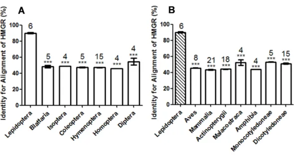
Figure 1 Comparison of the identity value of per species HMGR sequence relative toManduca sexta. (A) Asterisks indicate significant differences between Lepidoptera and other orders of Insecta as deter-mined by Dunnett’s multiple comparison test following one-way ANOVA: ***,P<0.001. (B) Asterisks indicate significant differences between Lepidoptera and other organisms other than Insecta as determined by Dunnett’s multiple comparison test following one-way ANOVA: ***,P<0.001.
Homology modeling, docking and molecular dynamics optimization With the goal of discovery of an eco-friendly insecticide target, the three-dimensional structures (especially the active site structures) of different species were analyzed. The HMGR ofM. sexta,A. melliferaandD. punctatawere selected for the structural comparison. The homology models ofM. sexta,A. melliferaandD. punctatawere generated using the crystal structure ofH. sapiens(PDB ID:1HWI) as the template. To select the best model, we checked the structural validity by PROCHECK (http://services.mbi.ucla.edu/SAVES). The geometry of the final refined models were evaluated with Ramachandran plot calculations computed using the PROCHECK program. The torsion angles ofϕandψ(the two torsion angles of the polypeptide chain, also called Ramachandran angles, describe the rotations of the polypeptide backbone around the bonds between N-Cα calledϕand Cα-C called ψ.) in the generated model was represented in the Ramachandran plot as shown inFig. S1. The Ramachandran plot showed 89.7% of the residues ofM. sexta, 88.2% residues of A. melliferaand 90.5% residues ofD. punctataexisted in the most favored regions. The percentages of residues in disallowed regions ofM. sexta,A. melliferaandD. punctataare 0.0%, 0.1% and 0.3%, respectively. This indicated that the backbone dihedral angles, phi and psi, of the three homology models were reasonably accurate.
conformations. By reporting the root mean square deviation (RMSD) of the protein structure from the starting model, the receptor changes in structure and reaches a relatively stable conformational minimum after approximately 3 ns. The conformations with the lowest energy of the final 100 structures from the MD simulation were selected as the final structures.
Structure comparison
Both subunits that constituted the binding pocket ofA. melliferaandD. punctataformed hydrogen bonds with lovastatin, whereas only one subunit ofM. sextacan form hydrogen bonds with the ligand (Arg 579 and Lys 680 of chain A). This suggested that the binding pocket ofM. sextais more flexible.
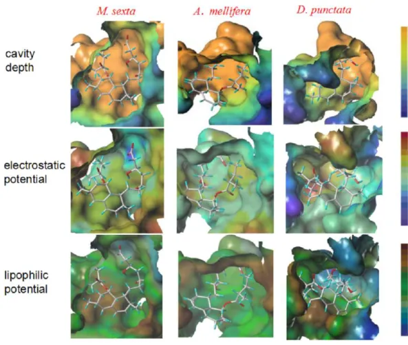
The surface properties of the binding pocket of the above three structures were defined using MOLCAD calculations (an interactive visualization of molecular scenarios) in Sybyl to analyse these binding pockets.Figure 2sketches the molecular surfaces of the pockets of these four HMGRs. The cavity ofM. sextawas smooth and did not penetrate deep into the structure compared with the cavities ofA. melliferaandD. punctata(Fig. 2). This result was in accordance with the results of hydrogen bond interaction. The electrostatic potential of the binding pockets ofM. sextawas positively-charged, because the whole surface of the pocket was colored in yellowish green, whereas that ofA. melliferawas electroneutral (Fig. 2). The front of the binding pocket ofD. punctatawas colored with blue, which suggests that some part of this pocket is electronegative. The lipophilic potential of the binding pocket of M. sextawas lipophilic, colored with brown, whereas that pocket of D. punctatawas more hydrophilic, colored with blue. The green color suggested that the pocket ofA. melliferais neutral (Fig. 2). These results show that the active pockets of the three species are different, suggesting that it is possible to design eco-friendly insecticides using differences in surface properties. Thus, the insect HMGR may represent a potential eco-friendly insecticide target.
The surface properties of these three pockets suggests that increasing the molecular volume, electronegativity and lipophilicity of the ligand can strengthen the binding affinity between ligand and HMGR ofM. sexta, whereas it also can weaken the binding affinities with other species.
Effects of HMGR inhibitors on JH biosynthesisin vitroandin vivo
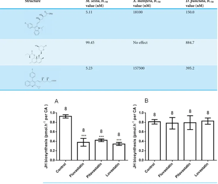
To validate the results of sequence alignment and structure comparison, three commercial human HMGR inhibitors (statins) were used for the assay of JH biosynthesisin vitroand in vivoinM. sexta,A. melliferaandD. punctata. Lovastatin is a type I statin. Fluvastatin and pitavastatin are type II statins. The IC50value of each compound is shown inTable 1.
Figure 2 Structure comparison of binding pockets ofM. sexta,A. melliferaandD. punctataHMGR. The molecule in the pocket is lovastatin. In the presentation of cavity depth, the deep blue colour repre-sents the outermost surface of the structure, whereas the orange colour reprerepre-sents the deepest part of the cavity. In the presentation of molecular electrostatic potential, the deep blue colour represents the most negative potential, whereas the deep red colour represents the most positive potential. In the presentation of the molecular lipophilic potential, the deep blue colour represents the most hydrophilic parts of the surface, whereas the deep brown colour represents the most lipophilic parts of the surface.
Type II statins have a greater effect than type I for the inhibition of JH biosynthesis inM. sexta. The IC50 value of lovastatin was 99.4 nM, which is much higher than that of
fluvastatin or pitavastatin (their IC50 values are 5.1 nM and 5.2 nM, respectively). These
results suggest that type II statins should be good lead compounds for new insecticide design. In addition to their effectsin vitro, the statins also showed significant effects on JH production byM. sextafollowing treatmentin vivo.
Injection
Following injection of the statin into newly molted fifth larval instar M. sexta, JH biosynthesis was assayed after 3 h with significant inhibitory effects apparent. After 3 h, the inhibition of fluvastatin, pitavastatin and lovastatin was 64.1±5.2%, 61.4±5.8%
Table 1 The IC50values of HMGR inhibition of JH biosynthesisin vitro.
Compound Structure M. sexta, IC50
value (nM)
A. mellifera, IC50
value (nM)
D. punctata, IC50
value (nM)
Fluvastatin 5.11 18100 150.0
Lovastatin 99.45 No effect 884.7
Pitavastatin 5.23 157500 395.2
Figure 3 JH biosynthesis following oral treatment of various inhibitors (statins) (A) and topical
cutic-ular application of the same inhibitors (B).Each bar represents the mean±SEM. Asterisks indicate
sig-nificant differences between inhibitor- and water-fed groups of animals as determined by Dunnett’s mul-tiple comparison test following one-way ANOVA: ***,P<0.001.
Effects on JH biosynthesis following oral administration
In addition to the effects in the injection bioassays, the statins also showed a significant effect on JH production following oral administration (Fig. 3A). In bioassays at 1 µM, inhibition of JH biosynthesis by fluvastatin, pitavastatin and lovastatin was 58.9±8.9%,
54.4±3.1% and 62.6±3.4%, respectively. This suggests that HMGR inhibitors can also
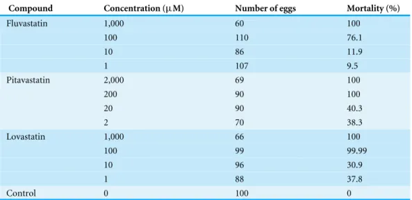
Table 2 Ovicidal effect of HMGR inhibitors onM. sextaeggs at different concentrations.
Compound Concentration (µM) Number of eggs Mortality (%)
Fluvastatin 1,000 60 100
100 110 76.1
10 86 11.9
1 107 9.5
Pitavastatin 2,000 69 100
200 90 100
20 90 40.3
2 70 38.3
Lovastatin 1,000 66 100
100 99 99.99
10 96 30.9
1 88 37.8
Control 0 100 0
Topical application
In topical cuticular assays, no compound demonstrated any effect on JH biosynthesis (Fig. 3B); this might be attributable to the poor cuticular penetration of the reagents, and indicates that HMGR inhibitors are unlikely to be contact insecticides.
Pest control application Ovicidal effects
The three compounds also demonstrated significant activity on viability of M. sexta eggs (Table 2). At a concentration (100µM), the mortality of eggs following treatment with fluvastatin, pitavastatin and lovastatin was 76.1%, 100% and 100%, respectively. A concentration of 50µM of these compounds gave about 50% inhibition.
Larvicidal effects following oral administration of statins
The experiment in ‘Effects on JH biosynthesis following oral administration’ demonstrated that the statins have a significant effect on JH biosynthesis following oral administration. Accordingly, a stomach toxicity test was performed. We first determined which instars of M. sextawere most sensitive to the statins following feeding at high concentration (1,000 µM) in 1st, 2nd and 3rd instars, respectively, and recorded the mortality inTable 3. All larvae that commenced feeding from the 1st stadium died prior to pupation, and most died in the 1st stadium. Larvae fed from the 2nd stadium were also sensitive to the statins with a high mortality (above 85%). However, when the treatment commenced from the 3rd stadium, the mortality before the next molt was less than 10%, and mortality just prior to pupation was less than 40%. For all the inhibitors, the earlier instars were more sensitive than the later instars.
We then topically treated larvae with different concentrations of the statins, commencing with 1st instars, and recorded larval mortality (Table 4). The statins showed significant larvicidal activity at 100µM. The IC50values of fluvastatin, pitavastatin and lovastatin were
Table 3 Mortality following feeding with statins at 1,000µM during the first three stadia.
Compound Feeding treatment Mortality before next molt (%) Mortality before pupation (%) Fluvastatin From 1st instar 100 100
From 2nd instar 51 92
From 3rd instar 6 13
Pitavastatin From 1st instar 82 100
From 2nd instar 80 85
From 3rd instar 10 40
Lovastatin From 1st instar 100 100
From 2nd instar 90 100
From 3rd instar 20 90
Table 4 Larval mortality following treatment of 1st instars.
Compound Concentration (µM) Mortality before 3rd instar (%) Larval mortality (%)
Fluvastatin 1,000 100 100
100 25 25
10 8.3 8.3
1 16.7 16.7
Pitavastatin 1,000 100 100
100 100 100
10 41.7 41.7
1 25 41.7
Lovastatin 1,000 100 100
100 33.3 33.3
10 0 8.3
1 0 0
In the dead larvae, the most striking characteristic was the darkening of the cuticle in some animals as well as molting disturbances (Fig. 4). This is consistent with the phenomenon of the inhibition of JH biosynthesis (Monger et al., 1982).
Growth regulation
Figure 4 Dead larvae following feeding with 1,000µM of fluvastatin as 1st instars.Twenty animals
were used in this treatment.
Figure 5 Developmental arrest and growth retardation inM. sextafollowing fluvastatin treatment.
There were twenty animals for each group. (A) Three larval groups hatched on the same day; one was fed normal food as the control; the others were treated with 1µM and 100µM of fluvastatin,
respec-tively. The difference in size is readily apparent in the control 4th instars (10 days after feeding). (B) Newly hatched larvae were treated with 1µM fluvastatin, most died in the process of pupation as a result of
Following treatment with a low concentration (1µM) of the statins, we observed that pupation of these larvae was not normal. Most larvae died in the process of pupation as a result of malformation (Fig. 5B). This suggests that the statins can be potent insect growth regulators.
DISCUSSION
Currently, there is an on-going need for the discovery and development of new insecticides to combat growing problems associated with resistance, environmental pollution, accumulation of pesticide residues in the food chain and detrimental effects on non-target organisms. Hence, the need for eco-friendly insecticides with safe and novel modes of action or targets is becoming increasingly important. In this study, we have not only focused on elucidating new eco-friendly insecticide targets and lead compounds, but also attempted to provide an empirical method for eco-friendly insecticide discovery.
Insect JHs are a group of structurally related sesquiterpenoids that regulate a number of physiological processes including embryogenesis, larval and adult development, meta-morphosis, reproduction, pheromone biosynthesis, diapause, migration, polymorphism, and metabolism (Nijhout, 1994;Kerkut & Gilbert, 1985; Gilbert, Granger & Roe, 2000). To our knowledge, the occurrence of JHs and related sesquiterpenoids such as methyl farnesoate is confined to animals in the Arthropoda. It has been demonstrated that the design of JH mimics or anti-JH agents is an effective strategy for insecticide discovery. Screening new targets involved in JH biosynthesis has been a subject of study for two decades (Bede et al., 2001). As HMGR has been postulated to be a key enzyme in the regulation of the MVA pathway in insects, some HMGR inhibitors (statins) have been used to investigate their effects on JH biosynthesis. Compactin, mevinolin and fluvastatin have been demonstrated to be potent inhibitors of JH biosynthesisin vitro, whereas studies of their effectsin vivoare incomplete (Monger et al., 1982;Couillaud, 1991). Thus, to date, HMGR has not been used as a potential insecticide target. Furthermore, whether HMGR inhibitors have detrimental effects on non-target organisms remains unknown.
We predicted and evaluated the ecological safety of HMGR by using sequence alignment and structural comparison. Sequence analysis showed that the Lepidoptera differ from other organisms. Zapata et al. tested the effects of two HMGR inhibitors, fluvastatin and compactin, on HMGR activity ofBlattella germanica. Both compounds significantly inhibited the enzymatic activity at a high concentration (50µg per animal) by approximately 25%in vivo(Zapata et al., 2002). The inhibition by fluvastatin onM. sextaHMGR was approximately 60% at a low concentration (0.8 ng per animal in an injection assay and 2 ng per animal following oral administration, respectively) in our study. BLAST showed that the identity value betweenB. germanicaHMGR andM. sextaHMGR was 47%. Bacterial HMGR has a low sequence identity value compared with M. sextaHMGR. Lovastatin inhibitedPseudomonas mevaloniiat a high concentration (Kivalue=0.53 mM) (Hedl & Rodwell, 2004). However, the inhibitory effect of lovastatin onM. sextawas much greater than onP. mevalonii(IC50value 99 nM). The identity value betweenP. mevaloniiHMGR
D. punctataversusM. sextawas 48% and 50%. Three HMGR inhibitors have no or little effect onA. melliferaandD. punctata; however these compounds are potent inhibitors in M. sexta. This suggests that there might be a link between the sequence alignment data and inhibition.
The sequence and 3-D structure (in particular, the molecular potential surface properties) of lepidopteran HMGR differs from other organisms (A. melliferaandD. punctata) in this study. Assays of JH biosynthesis in the presence of HMGR inhibitors in different insect species showed that those inhibitors have potent effects on the lepidopteran pestM. sexta, but are much less effective onA. mellifera(Hymenoptera) andD. punctata (Blattodea). This confirms our suggestion regarding the value and assessment of the ecological safety of HMGR as an insecticide target candidate.
The applicability to pest control is crucial in the evaluation of the potential of a chemical to act as an insecticide. Previous studies indicated that HMGR was the control point in JH biosynthesis inM. sexta(Monger et al., 1982). As a consequence of limited experimental data on the application of HMGR inhibitors for pest control, we tested the effectsin vivo of three HMGR inhibitors onM. sexta. Our present study revealed that HMGR inhibitors can be potential insecticide candidates with excellent ovicidal activity, larvicidal activity and growth regulatory effects. In the fat body, HMGR was crucial to vitellogenesis and reproduction. Short-term assays showed that HMGR inhibitors reduce the protein levels and enzymatic activity of HMGR, and long-term experiments revealed that fluvastatin impairs embryo development (Zapata et al., 2002). Our work clearly indicates that HMGR is a key enzyme in embryogenesis, larval and adult development and metamorphosis. In Agrotis ipsilon, fluvastatin also disrupted normal spermatophore transfer (Duportets et al., 1998). It suggested that insect HMGR can be an insecticide target and its inhibitors could be insecticide lead compounds.
We conclude that an empirical method of discovery of eco-friendly insecticides encompasses the prediction of ecological safety of insecticide target candidates and the probability of the application for pest control. The steps for ecological safety prediction are as follows:
(1) Collect sequence data of insecticide target candidates from all species of interest. (2) Perform sequence alignment of each species and compare to the selected target pest.
Statistically analyze the identity values from sequence alignments. If there is no difference between pest and non-target organisms, this candidate is a likely to be an eco-toxic insecticide target. If not, go to the next step.
(3) Perform structural comparisons and docking studies with ligands of pests and other non-target organisms. If there is no difference between their structures (especially the binding pockets) or binding affinities, this candidate is a possible eco-toxic insecticide target. If not, it could be an eco-friendly insecticide target. New eco-friendly insecticides can be designed based on structural differences.
CONCLUSION
We have demonstrated that insect HMGR can be a potential selective insecticide target, and its inhibitors can be potential selective insecticides. Our research should be helpful for designing new selective insecticides. Furthermore, we have demonstrated that sequence alignment, homology modeling and structural comparison can be used to determine which enzymes or receptors could be selective pesticide targets. Pest control applications have shown that the HMGR inhibitors are potential insect growth regulators, especially for lepidopteran pest control.
ACKNOWLEDGEMENTS
The authors thank Dr. Frank M. Horodyski (Ohio University, Athens, OH) for providing bioassay equipment.
ADDITIONAL INFORMATION AND DECLARATIONS
Funding
This work was supported by a grant from the National Natural Science Foundation of China (No. 21402122). It was also supported by Natural Science Foundation of Shanghai (No. 14ZR1440600), and the Scientific Research Foundation for the Returned Overseas Chinese Scholars, Ministry of Education of PRC (No. ZX2015-9). The funders had no role in study design, data collection and analysis, decision to publish, or preparation of the manuscript.
Grant Disclosures
The following grant information was disclosed by the authors: National Natural Science Foundation of China: 21402122. Natural Science Foundation of Shanghai: 14ZR1440600.
Scientific Research Foundation for the Returned Overseas Chinese Scholars, Ministry of Education of PRC: ZX2015-9.
Competing Interests
Stephen S. Tobe is an Academic Editor for PeerJ.
Author Contributions
• Yuan-mei Li performed the experiments, analyzed the data, prepared figures and/or
tables.
• Zhen-peng Kai conceived and designed the experiments, performed the experiments,
analyzed the data, contributed reagents/materials/analysis tools, wrote the paper, prepared figures and/or tables.
• Juan Huang performed the experiments, analyzed the data.
• Stephen S. Tobe contributed reagents/materials/analysis tools, wrote the paper, reviewed
Data Availability
The following information was supplied regarding data availability: The raw data on bioactivities are included inFig. 3and inTables 1–4.
Supplemental Information
Supplemental information for this article can be found online athttp://dx.doi.org/10.7717/ peerj.2881#supplemental-information.
REFERENCES
Bede JC, Teal PE, Goodman WG, Tobe SS. 2001.Biosynthetic pathway of insect juvenile
hormone III in cell suspension cultures of the sedgeCyperus iria.Plant Physiology
127(2):584–593DOI 10.1104/pp.010264.
Bell RA, Joachim FG. 1976.Techniques for rearing laboratory colonies of tobacco
hornworms and pink bollworms.Annals of the Entomological Society of America
69:365–373DOI 10.1093/aesa/69.2.365.
Couillaud F. 1991.Evidence for regulation of juvenile hormone biosynthesis operating
before mevalonate in locust corpora allata.Molecular and Cellular Endocrinology
77(1–3):159–166DOI 10.1016/0303-7207(91)90070-9.
Debernard S, Rossignol F, Couillaud F. 1994.The HMG-CoA reductase inhibitor
fluvastatin inhibits insect juvenile hormone biosynthesis.General and Comparative Endocrinology95(1):92–98DOI 10.1006/gcen.1994.1105.
Duportets L, Dufour MC, Couillaud F, Gadenne C. 1998.Biosynthetic activity of
corpora allata, growth of sex accessory glands and mating in the male mothAgrotis ipsilon(Hufnagel).Journal of Experimental Biology 201:2425–2432.
Feyereisen R, Farnsworth DE. 1987.Precursor supply for insect juvenile hormone III
biosynthesis in a cockroach.Journal of Biological Chemistry262:2676–2681.
Feyereisen R, Pratt GE, Hamnett AF. 1981.Enzymatic synthesis of juvenile hormone
in locust corpora allata: evidence for a microsomal cytochrome P-450 linked methyl farnesoate epoxidase.European Journal of Biochemistry118(2):231–238
DOI 10.1111/j.1432-1033.1981.tb06391.x.
Gilbert LI, Granger NA, Roe RM. 2000.The juvenile hormones: historical facts and
speculations on future research directions.Insect Biochemistry and Molecular Biology
30(8–9):617–644DOI 10.1016/S0965-1748(00)00034-5.
Halgren TA. 1999.MMFF VI. MMFF94s option for energy minimization studies.Journal
of Computational Chemistry 20(7):720–729
DOI 10.1002/(SICI)1096-987X(199905)20:7<720::AID-JCC7>3.0.CO;2-X.
Head JD, Zerner MC. 1985.A Broyden—Fletcher—Goldfarb—Shanno
optimiza-tion procedure for molecular geometries.Chemical Physics Letters122:264–270
DOI 10.1016/0009-2614(85)80574-1.
Hedl M, Rodwell VW. 2004.Inhibition of the Class II HMG-CoA reductase of
Istvan ES, Deisenhofer J. 2001.Structural mechanism for statin inhibition of HMG-CoA reductase.Science292(5519):1160–1164DOI 10.1126/science.1059344.
Kai ZP, Huang J, Tobe SS, Yang XL. 2009.A potential insect growth regulator:
synthesis and bioactivity of an allatostatin mimic.Peptides30(7):1249–1253
DOI 10.1016/j.peptides.2009.03.010.
Kai ZP, Ling Y, Liu WJ, Zhao F, Yang XL. 2006.The study of solution conformation of
allatostatins by 2-D NMR and molecular modeling.Biochimica et Biophysica Acta (BBA)-Proteins and Proteomics1764(1):70–75DOI 10.1016/j.bbapap.2005.10.010.
Kerkut GA, Gilbert CI. 1985.Comprehensive insect physiology, biochemistry and
pharma-cology. New York: Pergamon Press.
Laskowski RA, MacArthur MW, Moss DS, Thornton JM. 1993.PROCHECK: a
program to check the stereochemical quality of protein structures.Journal of Applied Crystallography26:283–291DOI 10.1107/S0021889892009944.
Lee KY, Chamberlin ME, Horodyski FM. 2002.Biological activity ofManduca sexta
allatotropin-like peptides, predicted products of tissue-specific and develop-mentally regulated alternatively spliced mRNAs.Peptides23(11):1933–1941
DOI 10.1016/S0196-9781(02)00181-X.
Monger DJ, Lim WA, Kézdy FJ, Law JH. 1982.Compactin inhibits insect HMG-CoA
reductase and juvenile hormone biosynthesis.Biochemical and Biophysical Research Communications105(4):1374–1380DOI 10.1016/0006-291X(82)90939-1.
Nijhout HF. 1994.Insect hormones. Princeton: Princeton University Press.
Rachinsky A, Tobe SS, Feldlaufer MF. 2000.Terminal steps in JH biosynthesis in
the honey bee (Apis melliferaL.): developmental changes in sensitivity to JH precursor and allatotropin.Insect Biochemistry and Molecular Biology30:729–737
DOI 10.1016/S0965-1748(00)00044-8.
Spitzer R, Jain AN. 2012.Surflex-Dock: docking benchmarks and real-world application.
Journal of Computer-Aided Molecular Design26(6):687–699
DOI 10.1007/s10822-011-9533-y.
Thompson JD, Higgins DG, Gibson TJ. 1994.CLUSTAL W: improving the sensitivity
of progressive multiple sequence alignment through sequence weighting, position-specific gap penalties and weight matrix choice.Nucleic Acids Research22:4673–4680
DOI 10.1093/nar/22.22.4673.
Tobe SS, Clarke N. 1985.The effect of L-methionine concentration on juvenile hormone
biosynthesis by corpora allata of the cockroachDiploptera punctata.Insect Biochem-istry 15(2):175–179DOI 10.1016/0020-1790(85)90005-8.
Tobe SS, Pratt GE. 1974.The influence of substrate concentrations on the rate of
insect juvenile hormone biosynthesis by corpora allata of the desert locustin vitro. Biochemical Journal144(1):107–113 DOI 10.1042/bj1440107.
Zapata R, Martín D, Piulachs MD, Bellés X. 2002.Effects of hypocholesterolaemic