Journal of Science and Technology
ISSN 1905-7873 Available online at www.mijst.mju.ac.th Communication
Antioxidant and anticancer activities of freshwater green
algae,
Cladophora glomerata
and
Microspora floccosa
, from
Nan River in northern Thailand
Ratiphan Laungsuwon1, 2 and Warawut Chulalaksananukul1-4,*
1 Biotechnology Program, Faculty of Science, Chulalongkorn University, Bangkok 10330, Thailand 2 Department of Botany, Faculty of Science, Chulalongkorn University, Bangkok 10330, Thailand 3 Aquatic Resources Research Institute, Chulalongkorn University, Bangkok 10330, Thailand 4 Biofuels by Biocatalysts Research Unit, Chulalongkorn University, Bangkok 10330, Thailand
* Corresponding author, e-mail: warawut.c@chula.ac.th; tel/fax: +662 218 5482
Received: 3 April 2012 / Accepted: 27 April 2013 / Published: 14 May 2013
Abstract: Organic solvent and hot water extracts of freshwater macroalgae, Cladophora glomerata and Microspora floccosa, harvested from Nan River in northern Thailand were screened for antioxidant and anticancer activities using DPPH free radical scavenging assay and inhibition of proliferation of the KB human oral cancer cell lines respectively. The ethyl acetate extract of C. glomerata showed the highest total phenol content (18.1±2.3 mg GAE/g), radical scavenging activity (49.8±2.7% DPPH scavenging at 100 g/ml) and in vitro growth inhibition (IC50=1420.0±66 g/g) of the KB cell lines.
These results indicate that C. glomerata could be a source of valuable bioactive materials.
Keywords: Cladophora glomerata, Microspora floccosa, total phenolic content, antioxidant activity, anticancer activity
________________________________________________________________________________
INTRODUCTION
Algae are an important source of various bioactive compounds such as antioxidants, antimicrobials and antivirals [1]. These compounds are also important for protecting the algal cells against stressful conditions, e.g. ultraviolet radiation, temperature change and fluctuation in nutrient and salinity level. To enable rapid adaptation to new environmental conditions, algae produce a great variety of secondary metabolites that cannot be found in other organisms [2].
[6-10], whereas those of freshwater macroalgae have been rarely investigated.
In northern Thailand, especially in Nan province, two species of freshwater macroalgae, Cladophora glomerata and Microspora floccosa, which belong to the Division Chlorophyta, are abundant in Nan River during the dry season [11]. The common names of these algae are “Kai” in Thai and “Mekong weed” in English. They have been used as a food source for many centuries by traditional culture. They are used in the manufacture of local food products such as crisps, baked goods, pasta and noodles. Besides being a popular food source, they are also believed to have many important health benefits such as rejuvenation, induction of appetite and expediting of recovery from many common maladies [12]. Some villagers also consume Kai to soothe stomach ulcers [13], but despite the widespread uses and claimed advantages of these algae, only few investigations on the chemical composition of Cladophora and Microspora species have been reported. Sterols, triterpenoids and volatile oils have been identified from some Cladophora species [14-16] which are distributed worldwide and often dominate in both fresh and marine waters [17]. While there have been several reports regarding antioxidant activities of green algae [6, 10, 18, 19], their anticancer activities have been rarely studied. As far as we know, the two species of freshwater green algae, C. glomerata and M. floccose, common in the Nan River, have not been evaluated for such bioactivities. The objective of this study is to determine the antioxidant and anticancer activities of various extracts of these two algae.
MATERIALS AND METHODS
Sampling and Identification
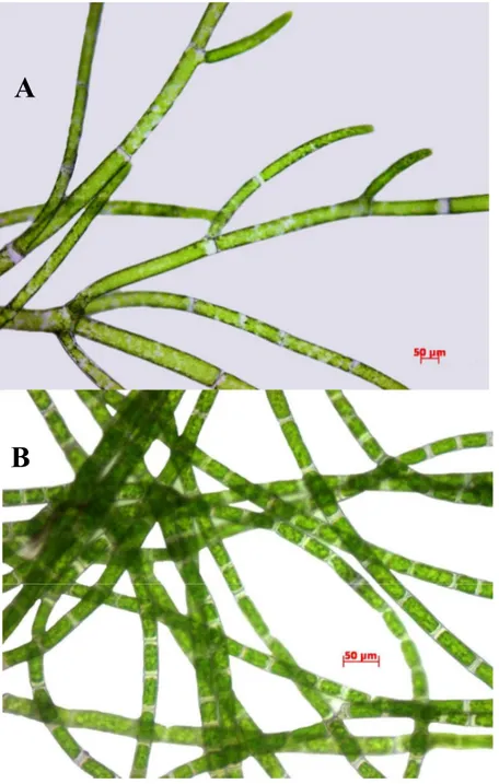
Cladophora glomerata Kiitzing and Microspora floccose (Vaucher) Thuret (Figure 1) were collected from three areas of Nan River, all located in Nan province, during the dry season (December 2009) when the algae were at their peak biomass (0-2 m in depth). Freshly collected algae were washed thoroughly in water to remove epiphytes, small invertebrates and extraneous matter. The samples were separated into two portions: one was used for morphological identification and the other was freeze-dried.
Preparation of Extracts and Preliminary Analysis
A 100-g portion of each freeze-dried macroalgal sample was extracted suceessively with 600 mL each of methanol, hexane and ethyl acetate at room temperature. Each extract was then clarified by centrifugation and the pellet was re-extracted twice with the same solvent. The supernatants were then pooled and filtered. The solvent was then removed from the filtrate by rotary evaporation and the dry crude extract was kept at 25°C and protected from light in a dessicator under an atmosphere of nitrogen gas until use.
Another 100-g sample of each freeze-dried alga was extracted with boiling deionised water for 1 hr and the water was removed by lyophilisation. The resulting crude extract was kept at 25°C and protected from light in a dessicator under an atmosphere of nitrogen gas until use.
All the extracts obtained were analysed by TLC on Kieselgel 60 F254 aluminum support
A
Figure 1. Cladophora glomerata Kützing (A) and Microspora floccosa (Vaucher) Thuret (B)
Determination of Total Phenolic Content (TPC)
The TPC was evaluated using Folin-Ciocalteu method as described previously [18, 19, 20] with modifications. Each extract (1.0 mL) was mixed with 1.0 mL of 2% Na2CO3 and 0.2 mL of
50% (v/v) Folin-Ciocalteu reagent was then added, mixed, allowed to stand at room temperature for 30 min. and then centrifuged. The absorbance of the supernatant was measured with a spectrophotometer at 750 nm. A calibration curve of gallic acid was prepared and the TPC were expressed as mg gallic acid equivalents (GAE)/g dry weight.
Free Radical Scavenging Activity by DPPH
The DPPH free-radical scavenging assay was performed according to established methods [20, 21] with some modifications. One mL of each extract in methanol at 100 g/mL was added to
%DPPH= {[Abscontrol –Abssample –Absblank)/Abscontrol} x 100, where DPPH (2.0 mL) + methanol (1.0
mL) was used for Abscontrol; DPPH (2.0 mL) + extract (1.0 mL) was used for Abssample; and
methanol (2.0 mL) and extract (1.0 mL) was used for Absblank. Ascorbic acid was used as positive
control.
Anticancer Assay against Human Oral Cavity Cell Lines (KB)
These experiments were based on the resazurin microplate assay (REMA) as described by Brien et al [22]. In brief, three KB cell lines (epidermoid carcinoma of oral cavity, ATCC CCL-17) at logarithmic growth phase were harvested and diluted to 7x10⁴ cells/mL in fresh medium. Five
L of test sample diluted in 5% DMSO and 45 L of cell suspension were successively added to a 384-well plate and incubated at 37°C in a 5% CO2 incubator. After 3 days of incubation, 12.5 L of
62.5 g/mL resazurin solution was added to each well and the plate was then incubated at 37°C for 4 hr. Fluorescence signals were measured using a SpectraMax M5 multi-detection microplate reader (Molecular Devices, USA) at excitation and emission wavelengths of 530 nm and 590 nm respectively. Per cent inhibition of cell growth was calculated with the following equation: % Inhibition = [1- (FUT/ FUC)] x 100, where FUT and FUC are the mean fluorescent intensity from
treated and untreated conditions respectively. Dose response curves were plotted from 6 concentrations of twofold serially diluted test compounds and the sample concentrations that inhibit cell growth by 50% (IC50) were derived using SOFTMax Pro software (Molecular Devices, USA).
Ellipticine and doxorubicin were used as positive controls and 0.5% DMSO as negative control. Cytotoxicity against Normal Cell Lines (Vero)
The cytotoxicity experiments were based on the green fluorescent protein (GFP)-expressing Vero cell lines [23]. The method was generated in-house by stably transfecting the African green monkey kidney cell lines (Vero, ATCC CCL-81) with the pEGFP-N1 plasmid (Clontech). The cell line was maintained in minimal essential medium supplemented with 10% heat-inactivated fetal bovine serum, 2 mM L-glutamine, 1 mM sodium pyruvate, 1.5 g/L sodium bicarbonate and 0.8 mg/mL geneticin at 37°C in a humidified incubator with 5% CO2.
The assay was carried out by adding 45 L of cell suspension at 3.3x10⁴ cells/mL to each
well of a 384-well plate containing 5 L of test compounds previously diluted in 0.5% DMSO. The plate was then incubated in a 37°C incubator with 5% CO2 for 4 days. Fluorescence signals were
measured with the SpectraMax M5 microplate reader in the bottom reading mode with excitation and emission wavelengths of 485 and 535 nm respectively. The fluorescence signal at day 4 was subtracted from the background fluorescence at day 0. The per cent inhibition was calculated with the following equation: % inhibition = [1-(FUT / FUC)] × 100, where FUT and FUC represent the
fluorescence units of cells treated with test compound and untreated cells respectively.
The IC50 values (extract concentrations resulting in a 50% inhibition) were derived from
RESULTS AND DISCUSSION
TLC analysis of the extracts obtained from all locations revealed distinct different chemical profiles between the two algae. However, the same algal extracts from different locations showed similar TLC profiles and the presence of the same major secondary metabolites.
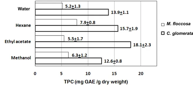
Phenolic compounds are known to act as antioxidants not only because of their ability to donate hydrogen atoms or electrons but also because of their stable radical intermediates [19], which prevent the oxidation of various ingredients, particularly fatty acids and oils. For example, minerals, polysaccharides and antioxidant properties were reported for macroalgae in the Noto Peninsula, Ishikawa, Japan [24]. The TPC of C. glomerata and M. floccosa extracts, when assayed at a concentration of 100 g/mL, was found to vary with the extraction method and type of macroalga (Figure 2). The order of extraction efficiency varied between the two algae (ethyl acetate > hexane > hot water > methanol for C. glomerata, compared to hexane > methanol > ethyl acetate > hot water for M. floccosa). However, for each solvent the TPC level was always higher (2 times for methanol and hexane, and 3.3 times for ethyl acetate) for C. glomerata than M. floccosa.
Figure 2. TPC of C. glomerata and M. floccosa extracts. The data are based on duplicates from three distinct areas of the Nan River. The results are expressed as means±SD (n=6).
Radical scavengers were evaluated in eachof the C. glomerata and M. floccosa extracts by their reactivity towards the stable free radical DPPH (Figure 3). Similar to the TPC, the radical scavenger level varied with extraction method and type of macroalga. Indeed, the order of scavenging activity was observed to be the same as that of TPC, with %DPPH scavenging activity being 1.8 times (in methanol) to 5.6 times (in ethyl acetate) higher for C. glomerata than M. floccosa. However, all %DPPH scavenging activities observed were significantly lower than that of the ascorbic acid positive control at the same concentration.
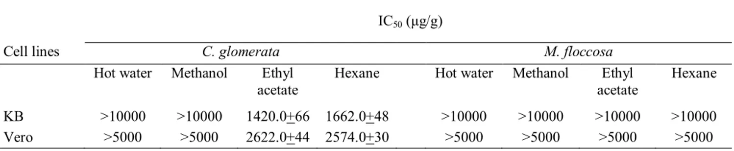
From the determination of the effect of C. glomerata and M. floccosa extracts on in vitro inhibition of the growth (metabolism) of the KB cell lines in tissue culture, a significant decrease in the total cellular metabolic (reductase) activity (assumed number of viable cells) compared to the negative control was observed for the hexane and ethyl acetate extracts but not for the more polar methanol and hot water extracts of C. glomerata. In contrast, none of the four solvent extracts from M. floccosa elicited any significant cytotoxicity against the KB cell lines. The IC50 of ethyl acetate
C. glomerata M. floccosa
These results suggest that C. glomerata contains certain useful biological compounds that have anticancer activity against KB cells and low cytotoxic against Vero cells. However, it is not yet known if this is a general cytotoxic activity towards any human cell lines or is indeed carcinoma-specific. Future research is needed to delineate the relative contribution of this pathway to cytotoxicity.
Figure 3. % DPPH radical scavenging activity of C. glomerata and M. floccosa extracts. The data are based on duplicates from three distinct areas of the Nan River. The results are expressed as means±SD (n=6).
Table 1. Cytotoxicity tests for C. glomerata and M. floccosa extracts against cell lines
IC50 (μg/g)
Cell lines C. glomerata M. floccosa
Hot water Methanol Ethyl
acetate Hexane Hot water Methanol acetate Ethyl Hexane KB >10000 >10000 1420.0+66 1662.0+48 >10000 >10000 >10000 >10000
Vero >5000 >5000 2622.0+44 2574.0+30 >5000 >5000 >5000 >5000
Note: The results are expressed as means±SD (n=6)
CONCLUSIONS
It has been shown that of the two freshwater green algae studied, i.e. Cladophora glomerata and Microspora floccosa, the former is a potential source of biologically active compounds that may be useful as therapeutic agents including an anticancer.
C. glomerata M. floccosa
ACKNOWLEDGEMENTS
This study is supported by the 90th Anniversary of Chulalongkorn University Fund (Ratchadaphiseksomphot Endowment Fund). The authors would like to thank Professor Yuwadee Peerapornpisal and Dr. Sorrachat Thiamdao from the Department of Biology, Faculty of Science, Chiang Mai University for identifying the macroalgae.
REFERENCES
1. O. Pulz and W. Gross, “Valuable products from biotechnology of microalgae”, Appl. Microbiol. Biotechnol., 2004, 65, 635-648.
2. I. Rodriguez-Meizoso, L. Jaime, S. Santoyo, F. J. Señoráns, A. Cifuentes and E. Ibáñez, “Subcritical water extraction and characterization of bioactive compounds from Haematococcus pluvialis microalga”, J. Pharm. Biomed. Anal., 2010, 51, 456-463.
3. T. Hoshino, T. Hayashi, K. Hayashi, J. Hamada, J. B. Lee and U. Sankawa, “An antivirally active sulfated polysaccharide from Sargassum horneri (Tunner) C. AGARDH”, Biol. Pharm. Bull., 1998, 21, 730-734.
4. T. Kuda, T. Kunii, H. Goto, T. Suzuki and T. Yano, “Varieties of antioxidant and antibacterial properties of Ecklonia stolonifera and Ecklonia kurome products harvested and processed in the Noto peninsula, Japan”, Food Chem., 2007, 103, 900-905.
5. S. J. Heo, E. J. Park, K. W. Lee and Y. J. Jeon, “Antioxidant activities of enzymatic extracts from brown seaweeds”, Bioresour. Technol., 2005, 96, 1613-1623.
6. Y. L. Chew, Y. Y. Lim, M. Omar and K. S. Khoo, “Antioxidant activity of three edible seaweeds from two areas in South East Asia”, LWT-Food Sci. Technol., 2008, 41, 1067-1072. 7. K. Iwai, “Antidiabetic and antioxidant effects of polyphenols in brown alga Ecklonia
stolonifera in genetically diabetic KK-A(y) mice”, Plant Foods Hum. Nutr., 2008, 63, 163-169.
8. T. Kuda, M. Tsunekawa, H. Goto and Y. Araki, “Antioxidant properties of four edible algae harvested in the Noto Peninsula, Japan”, J. Food Comp. Anal., 2005, 18, 625-633.
9. Y. V. Yuan and N. A. Walsh, “Antioxidant and antiproliferative activities of extracts from a variety of edible seaweeds”, Food Chem. Toxicol., 2006, 44, 1144-1150.
10. M. Zubia, D. Robledo and Y. Freile-Pelegrin, “Antioxidant activities in tropical marine macroalgae from the Yucatan Peninsula, Mexico”, J. Appl. Phycol., 2007, 19, 449-458.
11. Y. Peerapornpisal, I. Pongsirikul and D. Kanjanapothi, “Potential of freshwater macroalgae as food and medicine”, Final report submitted to Thailand Research Fund (TRF), 2005.
12. P. Fahprathanchai, K. Saenphet, Y. Peerapornpisal and S. Aritajat, “Toxicological evaluation of Cladophora glomerata Kützing and Microspora flocosa Thuret in albino rats”, SE Asian J. Trop. Med. Pub. Health, 2006, 37, 206-209.
13. Y. Peerapornpisal, D. Amornledpison, C. Rujjanawate, K. Ruangrita and D. Kanjanapothib, “Two endemic species of macroalgae in Nan River, northern Thailand, as therapeutic agents”, Sci. Asia, 2006, 32, 71-76.
14. M. Kuniyoshi, K. Yamada and T. Higa, “A biologically active diphenyl ether from the green alga Cladophora fascicularis”,Experientia, 1985, 41, 523-524.
composition of Cladophora vagabunda”, Phytochem., 1996, 42, 39-44.
18. H-B. Li, K-W. Cheng, C-C. Wong, K-W. Fan, F. Chen and Y. Jiang, “Evaluation of antioxidant capacity and total phenolic content of different fractions of selected microalgae”, Food Chem., 2007, 102, 771-776.
19. M. Hajimahmoodi, M. A. Faramarzi, N. Mohammadi, N. Soltani, M. R. Oveisi and N. Nafissi-Varcheh, “Evaluation of antioxidant properties and total phenolic contents of some strains of microalgae”, J. Appl. Phycol., 2010, 22, 43-50.
20. G. Miliauskas, P. R. Venskutonis and T. A. van Beek, “Screening of radical scavenging activity of some medicinal and aromatic plant extracts”, Food Chem., 2004, 85, 231-237. 21. B-G. Wang, W-W. Zhang, X-J. Duan and X-M. Li, “In vitro antioxidative activities of extract
and semi-purified fractions of the marine red alga, Rhodomela confervoides (Rhodomelaceae)”, Food Chem., 2009, 113, 1101-1105.
22. J. O′Brien, I. Wilson, T. Orton and F. Pognan, “Investigation of the Alamar Blue (resazurin) fluorescent dye for the assessment of mammalian cell cytotoxicity”, Eur. J. Biochem., 2000, 267, 5421-5426.
23. L. Hunt, M. Jordan, M. De Jesus and F. M. Wurm, “GFP-expressing mammalian cells for fast, sensitive, noninvasive cell growth assessment in a kinetic mode”, Biotechnol. Bioeng., 1999, 65, 201-205.
24. T. Kuda and T. Ikemori, “Minerals, polysaccharides and antioxidant properties of aqueous solutions obtained from macroalgal beach-casts in the Noto Peninsula, Ishikawa, Japan”, Food Chem., 2009, 112, 575-581.