Efectiveness of cyanoacrylate adhesive in rabbit aortorrhaphy
Efetividade do adesivo de cianoacrilato para aortarrafia em coelhos
Marcus Vinicius Henriques de Carvalho1
*
, Evaldo Marchi1, Mario Pantaroto1
Abstract
Background: Even when properly performed, arterial sutures are not always absolutely hemostatic. Tissue sealants and adhesives have become available that can be used to complete sutures, preventing hemorrhage problems. Objectives: To evaluate the efectiveness of cyanoacrylate adhesive for sealing aortotomies in rabbits in a coagulopathic state, by analyzing survival of the animals and the time taken to achieve hemostasis. Methods: Ten-mm long aortotomies were performed on the infrarenal aortas of 12 animals. In four animals, aortorrhaphy was conducted using four interrupted polypropylene sutures and in the remaining animals aortorrhaphy was performed using four or fewer polypropylene stitches, followed by application of a cyanoacrylate adhesive. Results: he four animals sutured without adhesive died within the postoperative period due to severe hemorrhage (3 animals) or intestinal ischemia. he eight animals to which the adhesive was applied exhibited no transoperative bleeding, but one animal died during the postoperative period. Using cyanoacrylate permitted more rapid procedures (p<0.004) and no local reactions were observed in macroscopic examination of tissues. Conclusions: he cyanoacrylate adhesive demonstrated efectiveness for sealing aortotomies, considering the survival outcomes, duration of procedures and absence of local reactions on macroscopic examination.
Keywords: tissue adhesives; aorta; surgery; animal experimentation.
Resumo
Contexto: Ainda quando feitas adequadamente, suturas arteriais não são absolutamente hemostáticas. Vedantes e adesivos teciduais estão disponíveis e podem ser usados para completar as suturas, prevenindo problemas hemorrágicos.
Objetivos: Avaliar a efetividade do adesivo de cianoacrilato em vedar aortotomias de coelhos, considerando a sobrevida dos animais e o tempo para hemostasia. Métodos: Em todos os doze animais, aortotomias de 10 mm de extensão foram feitas na aorta abdominal infrarrenal. Em quatro animais a aortorraia foi feita com quatro pontos separados de polipropileno, e no outro grupo, a aortorraia foi feita com quatro ou menos pontos separados e, depois, o adesivo de cianoacrilato foi aplicado. Resultados: Os quatro animais que receberam somente a sutura morreram no primeiro dia de pós-operatório devido à hemorragia intensa (três animais) e por isquemia intestinal. Nos oito animais nos quais o adesivo foi aplicado não houve sangramento transoperatório, mas um animal morreu no período pós-operatório. A utilização do cianoacrilato tornou o procedimento mais rápido (p < 0,004). Não foram observadas reações locais sob o ponto de vista macroscópico. Conclusões: O adesivo de cianoacrilato mostrou efetividade em vedar aortotomias, considerando o desfecho sobrevida e tempo para obter hemostasia e o fato de não ocorrerem reações locais sob o ponto de vista macroscópico.
Palavras-chave: adesivos teciduais; aorta; cirurgia; experimentação animal.
1Faculdade de Medicina de Jundiaí – FMJ, Department of Surgery, Jundiaí, SP, Brazil.
Financial support: None.
Conlicts of interest: No conlicts of interest declared concerning the publication of this article. Submitted: December 30, 2014. Accepted: April 10, 2015.
INTRODUCTION
Routine arterial closure using surgical sutures is effective in the great majority of cases and patients
have beneited from the technique for many years.
However, arterial suture is time-consuming, which is sometimes undesirable in cases such as carotid,
abdominal aorta and coronary graft sutures. Even
when properly performed, arterial sutures may fail
to achieve absolute hemostasis, most frequently
in patients on antiplatelet drugs or in patients with
coagulopathies who require surgery despite the presence of these conditions. Surgical sealants and
adhesives have recently become available and can be used to supplement sutures, preventing hemorrhagic
problems. There are two main types of surgical adhesive: biological adhesives based on ibrin and chemical adhesives based on cyanoacrylate. Adhesives
based on cyanoacrylate began to be used in 1960, but at that time there were concerns that cyanoacrylate
could lead to strong tissue reactions. However, in
modern formulations the product is safe and causes
almost imperceptible reactions.1,2 Nevertheless, there
is a need for further research into the effectiveness
of cyanoacrylate in vascular sutures.
This research was conducted to investigate
the effectiveness of cyanoacrylate adhesive in an experimental model of aortorrhaphies in rabbits, evaluating postoperative complications (hemorrhage, thrombus, death, gross macroscopic changes, and local
reactions) and the time required to perform aortorrhaphy using the gluing method. The coagulopathic state
was induced by administration of heparin, without
reversal using protamine.
MATERIAL AND METHODS
The study was conducted on 12 rabbits with an
average weight of 4,070 kg (3,500 to 4,450 kg), handled in accordance with rules published by Brazil’s National
Council for the Control of Animal Experimentation.3
The study was also approved by the Research Ethics Committee at the Institution. Animals were kept in individual cages and cared for in the FMJ vivarium.
On the day of the procedure, each animal was brought
to the FMJ Surgical Technique Laboratory and anesthetized with acepromazine (Acepran, Univet, S.
Paulo) and tiletamine+zolazepam (Zoletil, Virbac, S.
Paulo). After anesthesia, venous access was obtained
and the abdominal region was shaved and submitted to antisepsis and asepsis, followed by placement of sterile
surgical ields. All operations were performed by the same surgeon with constant asepsis. Frontal light and a surgical loupe with 2.5x magniication were used
in all operations, to facilitate careful arteriorrhaphy,
avoiding stenosis of the vessel.
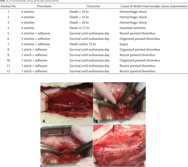
An intraperitoneal approach to the infrarenal
abdominal aorta was obtained by anterior median laparotomy and a segment of the vessel approximately
30 mm long was exposed. At this stage, heparin (100 mg/kg) (Hepamax-S, Blausiegel, S. Paulo)
was administered intravenously and after 2 minutes an infrarenal aorta segment was occluded with gentle compression by two vascular clamps (Codman Instruments, USA) to prevent bleeding. The heparin dose was not reversed with protamine in order to
create a state of coagulopathy. A 10-mm incision was
made in the anterior part of this aortic segment using
an ophthalmic scalpel. Up to this point, all animals were submitted to the same procedures. They were then assigned to one of two groups, as deined in Table 1. Briely, in the suture group aortorrhaphy was
performed using four interrupted sutures and in the suture + adhesive group aortorrhaphy was accomplished
using interrupted sutures + adhesive. The reason for
choosing interrupted sutures instead of continuous sutures was to make it possible to determine how large the distance between stitches could be and still
be sealed by the adhesive. To determine this limit, the
authors reduced the numbers of stitches by one stitch per case successively, because the adhesive applied after aortorrhaphy exhibited high sealing power and
reliability. As a result, animal No. 5 (the irst in the
sutures + adhesive group) received four stitches,
animal No. 6, three stitches, animal No. 7, two stitches
and animals 8 through 12 were treated with just one
stitch each. After removing the vascular clamps from
the aorta and checking for bleeding, intestinal loops were re-conducted to their original positions over the aorta, the abdominal wall was sutured, and the
animals were returned to the vivarium. The sequence
of procedures is illustrated in Figure 1. Postoperative analgesia was provided by adding dipyrone to the
animals’ water dispensers.
The tissue adhesive used was Omnex (Ethicon,
Johnson & Johnson, S. José dos Campos, SP, Brazil). The product was applied sparingly, using a partial drop to create a thin ilm over the suture line.
Statistical analysis
Fisher’s exact test was used to determine whether there were differences in the primary outcome (animal death) when those animals treated with sutures only and those treated with suture + adhesive
were compared. Student’s t test was used to assess differences between the time taken to perform
RESULTS
All four animals in which the aortotomy was closed
using four separate 7-0 polypropylene sutures without the adhesive suffered severe hemorrhages when the
distal vascular clamp itted to the aorta was opened.
Despite intense compression with sponges, it was necessary to clamp the aorta again and to perform a repeat aortorrhaphy with running 7-0 polypropylene
sutures in order to achieve hemostasis. All four animals had poor postoperative outcomes. Three of them
died the day after the operation due to complications of the severe hemorrhage that occurred when the aorta clamp was opened (before the supplementary
running sutures were performed). The fourth animal
died about 60 hours after surgery due to intestinal ischemia, undoubtedly caused by excessive handling
and long aortic cross-clamp time needed to stop the aorta bleeding (Table 1). The operations in which adhesive was applied progressed without hemorrhage or complications, but one of these animals also died
within approximately 72 hours after surgery. This was
probably due to sepsis, since it presented fever, tachycardia
and tachypnea. Although the death of animal No. 5
was considered to be caused by sepsis, this case was nonetheless recorded as an unsuccessful outcome in
the suture + adhesive group. No bleeding occurred
in any of the animals in which the cyanoacrylate
adhesive was used. As soon as the authors realized
that cyanoacrylate adhesive was highly effective for sealing, they reduced the number of stitches used to suture the aorta in the group in which glue was
applied after aortorrhaphy. As a result, the animals
in the suture + adhesive group had aortorrhaphy
Table 1.Procedures and animal outcome.
Animal No. Procedure Outcome Cause of death/macroscopic tissue examination
1 4 stitches Death < 24 hr Hemorrhagic shock
2 4 stitches Death < 24 hr Hemorrhagic shock
3 4 stitches Death < 36 hr Hemorrhagic shock
4 4 stitches Death in 72 hr Intestinal ischemia
5 4 stitches + adhesive Survival until euthanasia day Recent parietal thrombus
6 3 stitches + adhesive Survival until euthanasia day Organized parietal thrombus
7 2 stitches + adhesive Death within 72 hr Sepsis
8 1 stitch + adhesive Survival until euthanasia day Organized parietal thrombus
9 1 stitch + adhesive Survival until euthanasia day Recent parietal thrombus
10 1 stitch + adhesive Survival until euthanasia day Organized parietal thrombus
11 1 stitch + adhesive Survival until euthanasia day Recent parietal thrombus
12 1 stitch + adhesive Survival until euthanasia day Recent parietal thrombus
conducted using four stitches (animal No. 5), three stitches (animal No. 6), two stitches (animal No. 7)
or just one stitch in the center of the aortotomy
(animals 8 through 12). Thus, although the last ive
animals submitted to arteriorrhaphy with the use of
adhesive received only one stitch equidistant from
the edges (resulting in an interval between the suture and the edges of approximately 5 mm), no bleeding
occurred.
The cyanoacrylate adhesive demonstrated eficacy
in all eight animals in which it was used for sealing
aortotomies. These aortotomies were of the same
dimensions as those performed in animals in which
a suture technique employing four separated stitches
without adhesive resulted in hemorrhage of such
intensity that it led to the animals’ deaths.
Table 1 lists the procedures and results obtained.
For statistical analysis, the animals were divided into two groups as follows: a suture group, comprising four animals in which aortorrhaphy was performed using sutures only, and a suture + adhesive group, in which the animals received four, three, two or one
sutures in combination with tissue adhesive. The reason why the suture group was limited to
just four animals was that all of these animals had poor outcomes due to severe hemorrhage and so that the authors were certain that it was unnecessary to continue to use this approach that would lead to the
unnecessary deaths of more animals. The primary
outcome (animal death) was compared across groups, including the animal in the suture + adhesive group
that had probably died from sepsis.
Analysis of the endpoint (death) showed that technique including application of cyanoacrylate produced better outcomes (p<0.010). Analysis of the
time needed to perform aortorrhaphy using only suture
(average time 39.7 minutes) and suture + adhesive (average time 4.31 minutes) also showed that using cyanoacrylate enabled quicker procedures (p<0.004),
since aortorrhaphy using continuous sutures in a
conined area was time consuming.
One month after the procedure, the surviving animals (seven) were euthanized with lethal doses of anesthetic and the segment of the aorta where the procedure had
been conducted was examined. Segments of aorta
extending from the infrarenal area to the bifurcation of the iliac arteries were withdrawn and submitted to anatomic pathology investigation, which revealed
only parietal thrombus with an inlammatory iniltrate.
During dissection of the aorta segment submitted to the procedure using suture + adhesive, no evidence of adhesions or major macroscopically observable local
reactions was detected, but residues of cyanoacrylate
adhesive were observed over the aorta.
The frequency of occurrence of intraluminal
parietal thrombus in the animals in which adhesive was used could not be compared to the rate in animals in which aortorrhaphy was conducted using sutures
only because none of the second group survived.
DISCUSSION
Bleeding along the suture line is a frequent problem
that usually occurs in operations on blood vessels and which can prolong operating times and increase
morbidity and mortality. Routine actions taken to
avoid undesirable bleeding include gentle compression over the suture line, reversal of the effect of heparin
injections and administration of oxidized cellulose.
However, the introduction of tissue adhesives has provided a new approach for cases of mild to severe
hemorrhage along the suture lines.4
There are two main types of tissue adhesives.
Fibrin glue has been used for more than 30 years to promote hemostasis in several parts of the human
body.3 This adhesive is effective in certain situations,
but since it is prepared with a concentrate of plasma from several blood donors it involves the potential for
transmission of viral disease. Additionally, it has been shown that ibrin glue provides insuficient adhesion
force to support anastomosis without suture and is
not recommended for vascular sutures.5
When cyanoacrylate was irst used as a surgical
adhesive in 1940 it provoked a strong local reaction, but recent new formulations have exhibited low toxicity
and limited local reactions.2 This was achieved by
addition of an organic chemical group to the main chain of the chemical formula, increasing its absorption
capacity.2 Cyanoacrylate glue has exhibited high
effectiveness, quick ixation and strong enough tensile force to maintain tissues closed.1,6-8 Cyanoacrylate
has also exhibited greater strength than ibrin glue
for maintaining tissues together5 and for controlling
bleeding from needle holes.2,4 This adhesive has the
advantage of not being dependent on the patient’s coagulation status and it can also be used in cases in
which coagulopathies are present.9-11 There are different
subtypes of this class of products and for the present study Omnex (2-octyl-cyanoacrylate and butyl-lacoyl-cyanoacrylate) was chosen, which is formulated to biodegrade in a slow and safe manner over 36 months so that only small amounts of degradation products
such formaldehyde are produced at any given time.12
and is indicated for vascular sealing.12 It is available
for immediate use in storage containers with integrated
applicators, but should be applied in a ‘dry ield’. The present study investigated the distance between
two separate suture stitches that could be effectively
sealed by the cyanoacrylate glue. The importance of
this information is that if cyanoacrylate can provoke complete sealing even when larger distances are left
between suture stitches, it should allow for quicker suture procedures and shorter duration tissue ischemia.
In this study the cyanoacrylate adhesive proved to be an effective sealant for rabbit aortotomies in all cases, even when 10-mm aortotomies were closed with just
a single suture stitch equidistant from the edges, with distances as long as 5 mm left free of sutures. Additionally,
all of the animals in which only sutures were applied (without the use of adhesives or of other hemostatic substances) suffered major hemorrhaging from the spaces between sutures, showing that the adhesive is
truly effective. Furthermore, the cyanoacrylate glue
provided effective tensile strength, since these animals had systolic blood pressures of 90 to 130 mmHg and
diastolic pressures of 60 to 90 mmHg.13 This degree of
effectiveness has prompted research to test the tissue adhesive in animal models of vascular anastomoses
without using any sutures at all.1
The secondary objectives of this study were to observe
macroscopically whether the cyanoacrylate adhesive produced a severe local reaction, important vessel wall changes, or severe adhesions to adjacent intestinal
loops. In the present study we observed no remarkable
degenerative changes to the aorta wall or adhesions to surrounding structures; but 1 month after the procedure there were still vestiges of the adhesive that had been
applied to the vessel surface. An unexpected inding
was observation of parietal nonocclusive thrombus in all aorta segments that had been subjected to the
procedures. Since aortorrhaphy was performed by a
trained surgeon using 7-0 polypropylene sutures and
working with frontal lighting and a 2.5x magniication
loupe, the reason for thrombus formation should not
be attributed to technical laws. Two other possibilities should be considered, i.e., the low heparin doses
and the fact that part of the adhesive drains into the
vessel between the sutures. One published study has
shown that cyanoacrylate glue can reach the vascular
intima sheath and be thrombogenic.14 In view of this,
further studies are needed to determine whether this
adhesive penetrates the lumen of the vessels. It is also
possible, however, that the thrombus could be due to
the low heparin doses used in this study (100 mg/kg).
Heparin dosages vary between different studies,
with Saba et al.,1 for instance, using 100 mg/kg and
Barbalinardo et al.8 using double this dose.
In conclusion, this study shows that cyanoacrylate glue promotes efficient sealing in rabbit aortas (mean blood pressure 70 to 90 mmHg) even when the distance between sutures is as long as 5 mm,
and requires a shorter time to achieve hemostasis. Gross tissue reactions were nonsigniicant and there
was no adhesion between the adhesive applied over
the arteriotomy and the intestinal loops. However,
nonocclusive parietal thrombus was found in all aorta
segments treated with the adhesive.
REFERENCES
1. Saba D, Yilmaz M, Yavuz H, et al. Sutureless vascular anastomoses by N-butyl-2 cyanoacrylate adhesive: an experimental animal study. Eur Surg Res. 2007;39(4):239-44. http://dx.doi.org/10.1159/000101893. PMid:17446710.
2. Ellman PI, Brett Reece T, Maxey TS, et al. Evaluation of an absorbable cyanoacrylate adhesive as a suture line sealant. J Surg Res. 2005;125(2):161-7. http://dx.doi.org/10.1016/j.jss.2004.12.014. PMid:15854669.
3. Brasil. Ministério da Ciência, Tecnologia e Inovação. Conselho Nacional de Controle de Experimentação Animal – CONCEA. [site on the internet]. Brasília: Ministério da Ciência, Tecnologia e Inovação; 2014. [cited 2014 may 05]. http://www.mct.gov.br/ index.php/content/view/310553.html.
4. Spotnitz WD, Burks S. State-of-the-art review: Hemostats, sealants, and adhesives II: Update as well as how and when to use the components of the surgical toolbox. Clin Appl Thromb Hemost. 2010;16(5):497-514. http://dx.doi.org/10.1177/1076029610363589. PMid:20699255.
5. Jackson MR, Gillespie DL, Longenecker EG, et al. Hemostatic efficacy of fibrin sealant (human) on expanded poly-tetrafluoroethylene carotid patch angioplasty: a randomized clinical trial. J Vasc Surg. 1999;30(3):461-6. http://dx.doi.org/10.1016/S0741-5214(99)70073-X. PMid:10477639.
6. Ludsen AB, Heyman ER, Closure Medical Surgical Sealant Study Group. Prospective randomized study evaluating an absorbable cyanoacrylate for use in vascular reconstruction. J Vasc Surg. 2006;4:1002-9.
7. Shapiro AJ, Dinsmore RC, North JH Jr. Tensile strength of wound closure with cyanoacrylate glue. Am Surg. 2001;67(11):1113-5. PMid:11730233.
8. Barbalinardo RJ, Citrin P, Franco CD, Hobson RW 2nd. A comparison of isobutyl 2-cyanoacrylate glue, fibrin adhesive, and oxidized regenerated cellulose for control of needle hole bleeding from polytetrafluoroethylene vascular prostheses. J Vasc Surg. 1986;4(3):220-3. http://dx.doi.org/10.1016/0741-5214(86)90189-8. PMid:2427751.
9. Sileshi B, Achneck HE, Lawson JH. Management of surgical hemostasis: topical agents. Vascular. 2008;16(Suppl 1):S22-8. PMid:18544302.
10. Achneck HE, Sileshi B, Jamiolkowski RM, Albala DM, Shapiro ML, Lawson JH. A comprehensive review of topical hemostatic agents: efficacy and recommendations for use. Ann Surg. 2010;251(2):217-28. PMid:20010084.
2009;157(1):e15-21. http://dx.doi.org/10.1016/j.jss.2009.01.034. PMid:19439320.
12. Spotnitz WD, Burks S. Hemostats, sealants, and adhesives III: a new update as well as cost and regulatory considerations for components of the surgical toolbox. Transfusion. 2012;52(10):2243-55. http:// dx.doi.org/10.1111/j.1537-2995.2012.03707.x. PMid:22612730.
13. Marques RG, Caetano CER. Parâmetros fisiológicos em animais de pequeno e médio porte. In: Marques RG, editor. Técnica operatória e cirurgia experimental. Rio de Janeiro: Guanabara Koogan; 2005. p. 821-23.
14. Scheetz WL, Matsumoto T. Cyanoacrylate tissue adhesive: thrombogenic effect. Am Surg. 1970;36(7):418-22. PMid:4915721.
*
Correspondence
Marcus Vinicius Henriques de Carvalho Faculdade de Medicina de Jundiaí – FMJ Rua Francisco Telles, 250 CEP 13202-550 – Jundiaí (SP), Brazil Tel.: +55 (11) 4587-1095 E-mail: [email protected]
Author information
MVHC - MD, PhD. Cardiovascular surgeon; associate professor at Faculdade de Medicina de Jundiaí. EM - MD, PhD. horacic surgeon; associate professor at Faculdade de Medicina de Jundiaí. MP - MD. General surgeon; assistant professor at Faculdade de Medicina de Jundiaí.
Author contributions
Conception and design: MVHC Analysis and interpretation: MVHC, EM Data collection: MVHC, EM, MP Writing the article: MVHC Critical revision of the article: EM Final approval of the article*: MVHC, EM, MP Statistical analysis: MVHC, EM Overall responsibility: MVHC