Comparison of Bite Force with Locking Plates
versus Non-Locking Plates in the Treatment of
Mandibular Fractures: A Meta-Analysis
José Cristiano Ramos Glória
1Ighor Andrade Fernandes
1Esmeralda Maria da Silveira
2Glaciele Maria de Souza
1Ricardo Lopes Rocha
2Endi Lanza Galvão
3Saulo Gabriel Moreira Falci
11Department of Oral and Maxillofacial Surgery, Universidade Federal
dos Vales do Jequitinhonha e Mucuri, Diamantina, MG, Brazil
2Department of Dentistry, Universidade Federal dos Vales do
Jequitinhonha e Mucuri, Diamantina, MG, Brazil
3Research Department, Centro de Pesquisas Rene Rachou, Belo
Horizonte, MG, Brazil
Int Arch Otorhinolaryngol 2018;22:181–189.
Address for correspondence Ighor Andrade Fernandes, DDs,
Departmento de Cirurgia Oral e Maxilofacial, Universidade Federal dos Vales do Jequitinhonha e Mucuri, Rua da Glória 187, Diamantina, Minas Gerais, 39100-000, Brazil (e-mail: [email protected]).
Introduction
Mandibular fractures represent a high percentage of all facial fractures.1–3The most common causes of these injuries are
interpersonal violence, falls, lesions practicing sports, and accidents at work and traffic accidents.4The consequences of mandibular fractures can include malocclusion, temporo-mandibular joint syndrome, and poor mastication,5leading to a decrease in quality of life.6
The mandibular fracturefixation by metallic plates was first described by Michelet in 1973,7and was subsequently improved by Champy in 1975,8 who described the ideal places in the mandible to apply plates that could resist to torsional forces during osteosynthesis. The standard plate system works when the heads of the screws compress the plate to the bone to maintain stability, preventing changes in the alignment of the fragments and maintaining occlusal stability.9 On the other hand, the locking plate system Keywords
►
bite force
►
mandibular fracture
►
fracture
fi
xation
►
bone plates
►
internal
fi
xators
Abstract
Introduction
Mandibular fractures represent a high percentage of all facial fractures,
and the bite force is a fundamental parameter to measure the actual mandibular
function and, subsequently, the masticatory ef
fi
ciency and quality of life.
Objectives
The purpose of the present systematic review was to verify if there is any
difference in the bite forces of patients with mandibular fractures
fi
xed by locking or
non-locking plates, testing the null hypothesis of no difference in this parameter.
Data Synthesis
A systematic review of the literature was conducted using four
databases (PubMed, Virtual Health Library, Web of Science and Science Direct) without
restrictions as to publication date or language. We found 3,039 abstracts, and selected
4 articles for this review.
Conclusion
The overall results show better performance in bite force for the locking
plates when compared with the non-locking plates in the incisor region (mean
deviation [MD]: 1.18; 95% con
fi
dence interval [95%CI]: 0.13
–
2.23), right molar region
(MD: 4.71; 95%CI: 0.63
–
8.79) and left molar region (MD: 10.34; 95%CI: 4.55
–
16.13).
Although the results of this study indicated a better bite force result with the locking
plates, there is still no suf
fi
cient evidence to support this information safely.
received April 1, 2017 accepted May 9, 2017 published online July 14, 2017
DOI https://doi.org/ 10.1055/s-0037-1604056. ISSN 1809-9777.
Copyright © 2018 by Thieme Revinter Publicações Ltda, Rio de Janeiro, Brazil
attaches the plates to the bone by locking the screws to both the bone and the plate, which maintains the stability of the system.10Therefore, treatments with locking plates lead to fewer changes in occlusal relationships, fewer screw-loosen-ing events, less problems with bone vascularization, greater stability between fragments, and easier placement of the plates when compared with treatments with non-locking systems.11,12
Some characteristics of the locking plate systems are recognized as disadvantages, such as the fact that they are more expensive than the standard systems, and need a specific apparatus in order to be applied in the bone. Thus, for each method and instrument launched by new studies, it is necessary to evaluate the actual advantages and disadvan-tages, so they may be properly applied in clinical situations. According to a recent meta-analysis,13there are no signifi -cant differences in the postoperative complication rates with the use of locking plate systems when compared with the use of standard plate systems in the management of mandibular fractures, but this study did not assess the bite force mea-surements. Bite force is a fundamental parameter to measure the actual mandibular function and, subsequently, the mas-ticatory efficiency and quality of life.
The purpose of the present systematic review was to verify if there is any difference in the bite force of patients with mandibular fracturesfixed by locking or non-locking plates, testing the null hypothesis of no difference in this parameter.
Review of the Literature
The present study was conducted according to theCochrane Handbook for Systematic Reviews of Interventions (apud Chrcanovic, 2014),13and the Preferred Reporting Items for Systematic Reviews and Meta-Analyses (PRISMA) statement using the Review manager (RevMan) software, version 5.3 (The Cochrane Collaboration, Copenhagen). It was registered in the International Prospective Register of Systematic Reviews (PROSPERO; CRD42016051937).
We conducted an electronic search to identify relevant literature published since November 11, 2016, in the PubMed, the Virtual Health Library (VHL), the Web of Science and the Science Direct databases. The search was performed using four different combinations of terms in all databases, following these stages: stage 1–(mandibular fracture) AND osteosynthesisAND (bite force); stage 2–(mandibular frac-ture) AND osteosynthesis AND (occlusal force); stage 3– (mandibular fracture) ANDosteosynthesisAND (masticatory force); and stage 4–(mandibular fracture) AND (locking plate ORnon-locking plateORnon-locking plateORstandard plate ORconventional plate).
Furthermore, we also searched the gray literature (Google Scholar), published theses (http://bancodeteses.capes.gov. br/banco-teses), and the reference lists of all studies identi-fied as relevant reviews for possible additional studies. We also searched for relevant ongoing clinical trials in the Clinical Trials Registry (http://www.clinicaltrials.gov).
The included studies must have reported the criteria for bite force measurement of the patients and the follow-up
measurements. The exclusion criteria were: textbooks, case reports, technical reports, review papers, opinion articles, cover letters, in vitro studies, animal studies, and papers without available abstracts. After independently reading paper titles and abstracts, the reviewers checked the results of the selected papers. Disagreements were resolved by consensus. The same process was performed when reading the full text for the final inclusion of the papers in the systematic review.
After the database search, 3,039 studies were identified. After removing duplicates, 1,697 studies remained. After screening titles and abstracts, we retrieved 25 full-text studies. The reference lists of the selected papers and a hand search revealed one additional relevant paper. A total of 4 studies involving 130 patients4,14–16evaluated bite force and were
included in the qualitative synthesis (►Fig. 1). One study was mentioned in two separate papers14,16that reported the same statistical data. These two articles were thus considered a single paper for the meta-analysis. One study presented the results through graphs, and it was not possible to extract the adequate statistical data for it.15The author was contacted by e-mail, but we could not obtain an answer. Therefore, only two studies4,14were included in the meta-analyses.
The relevant characteristics of the included studies are presented in►Table 1. All studies evaluated and compared the bite force of patients treated with locking and non-locking plates for the osteosynthesis of the mandibular fractures. The follow-up periods ranged from 1 to 12 weeks. The number of fractures ranged from 31 to 34. Out of the four studies, three used the very own patients as controls by measuring the bite force preoperatively. All plates systems were 2.0 mm (►Table 1). The results were checked by reviewers after data collection to ensure accuracy.
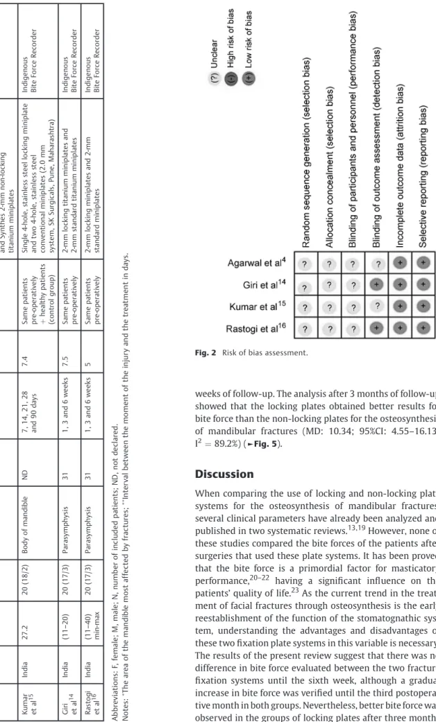
The quality of the included studies was independently assessed by the same reviewers using a specific protocol developed by Higgins & Green (2011,apudChrcanovic, 2014) 13for the assessment of the risk of bias. The classification of the potential risk of bias for each study was based on the following criteria: sequence generation; allocation sequence concealment; blinding of participants, personnel, and out-come assessors; incomplete outout-come data; and selective outcome reporting. Each study was submitted to analysis, and the criteria were rated as exhibiting low, high, or unclear (no information or uncertain) risk of bias. The disagreements between reviewers were resolved by consensus for thefinal classification. Our assessment is presented in►Fig. 2.
The meta-analysis was performed using the R software (R Foundation for Statistical Computing, Vienna, Austria), ver-sion 3.3.1. The packages“meta”and“metafor”were used to perform the statistician calculation and the forest plots. The heterogeneity of the results of the studies was assessed using the I-squared (I2) test. Values above 25% and 50% were considered indicatives of moderate and high heterogeneity respectively. As heterogeneity was present (I2>0) in all tests, the random effect model was used to perform the meta-analysis.17,18
by comparing the baseline mean values at the end of the study for each group.
All studies evaluated the bite forces by the same method, using the indigenous Bite Force Recorder, which consists of four strain gauges mounted on steel bars, forming a Wheat-stone bridge. Load changes in the steel bar produced a measurable voltage change across the four strain gauges. All measurements were made with the patient seated with the head upright, in an unsupported natural head position, and looking forward. The patients were instructed to bite on the pads of bite force gauge to the maximal level. The results were analyzed in the follow-ups at 1, 3, 6, and 12 weeks, observing the mean of the bite force measurements in the incisor, left molar, and right molar regions (►Table 2).4,14–16 The results of the meta-analysis showed that when the bite force in the incisor region was evaluated, the patients treated with locking plates showed no difference in bite force when compared with the patients treated with non-locking plates in thefirst (mean deviation [MD] :0.84; 95% confi
-dence interval [95%CI]: -0.62–2.30), third (MD: 0.91; 95%CI: -0.29–2.10), and sixth (MD: 1.05; 95%CI: -1.11–3.20) weeks. However, when evaluating the pooled bite force including 3 months of follow-up, the locking plates obtained better results than the non-locking plates for the osteosynthesis of mandibular fractures (MD: 1.18; 95%CI: 0.13–2.23; I2¼57.2%) (
►Fig. 3).
weeks of follow-up. The analysis after 3 months of follow-up showed that the locking plates obtained better results for bite force than the non-locking plates for the osteosynthesis of mandibular fractures (MD: 10.34; 95%CI: 4.55–16.13; I2¼89.2%) (
►Fig. 5).
Discussion
When comparing the use of locking and non-locking plate systems for the osteosynthesis of mandibular fractures, several clinical parameters have already been analyzed and published in two systematic reviews.13,19However, none of these studies compared the bite forces of the patients after surgeries that used these plate systems. It has been proved that the bite force is a primordial factor for masticatory performance,20–22 having a significant influence on the
patients’quality of life.23As the current trend in the treat-ment of facial fractures through osteosynthesis is the early reestablishment of the function of the stomatognathic sys-tem, understanding the advantages and disadvantages of these twofixation plate systems in this variable is necessary. The results of the present review suggest that there was no difference in bite force evaluated between the two fracture fixation systems until the sixth week, although a gradual increase in bite force was verified until the third postopera-tive month in both groups. Nevertheless, better bite force was observed in the groups of locking plates after three months
Table 1 C h ar ac te ri st ics of stu d ies in cl ud e d in th e sy st e ma ti c rev ie w Stu d y Co un tr y Me a n Ag e (yea rs) N (M /F ) S it e of m an d ib ul a r fr a ct u re Nu m be r of fr ac tur es Fo ll ow -u p In te rva l (m ea n) Co nt rol fo r b it e fo rc e Typ e of p la te s us ed B it e fo rc e m eas ur em en ts me th o d Ag ar wa l et al 4 In d ia (16 – 30 ) 2 0 (19 /1) P ar asy m ph ys is 34 1 , 3 , 6 w ee k s an d 3 m o nt h s 8 .7 S am e p at ien ts p re -o pe rat ive ly Syn th e s (We st Ch e st e r, P en n sy lva n ia , US ) 2 -mm lo cki n g tit an iu m mi n ip lat es an d Sy nt he s 2 -m m n on -l o cki ng tit a ni u m m in ipl at e s In di g e no us B it e Fo rce Reco rd e r Kum a r et al 15 In d ia 2 7 .2 2 0 (18 /2) B od y o f m an d ib le ND 7 , 1 4, 2 1 , 28 a nd 9 0 day s 7 .4 S am e p at ien ts p re -o pe rat ive ly þ h e a lth y p at ie nt s (c on tr ol gr ou p) Si ng le 4-h ol e, st ai n le ss st e el lo ck in g mi ni p la te an d two 4 -ho le, st a in les s st ee l co nve n ti on a l mi n ip la tes (2. 0 mm sy st e m, S K S ur g ic a ls , Pu ne, M ah a ras h tr a) In di g e no us B it e Fo rce Reco rd e r Gi ri et al 14 In d ia (11 – 20 ) 2 0 (17 /3) P ar asy m ph ys is 31 1 , 3 an d 6 we ek s 7 .5 S am e p at ien ts p re -o pe rat ive ly 2 -m m lo ck in g tit a ni u m m in ip la te s an d 2 -m m st an da rd tit an iu m mi n ip lat e s In di g e no us B it e Fo rce Reco rd e r Ras to gi et al 16 In d ia (11 – 40 ) mi n-ma x 2 0 (17 /3) P ar asy m ph ys is 31 1 , 3 an d 6 we ek s 5 S am e p at ien ts p re -o pe rat ive ly 2 -m m lo ck in g mi n ip la tes a nd 2-m m st a nd ar d m in ip la te s In di g e no us B it e Fo rce Reco rd e r Ab b rev ia ti on s: F, fe ma le ; M , ma le ; N, nu mb er of inc lud e d p at ien ts ; ND , n ot de cl a red . No te s: Th e a rea o f the m an d ib le m os t a ffe ct ed by fr ac tur e s; In ter va l b et w ee n th e mo m en t of th e in ju ry an d th e tr eat m en t in day s.
of follow-up. These results enable us to imply that the locking plate system promotes the reestablishment of masticatory functions earlier than the non-locking plate system. This may be due to the locking plate system’s ability to join the fragments of the fracture without leaving gaps.12In the non-locking plates system, the existence of these gaps enables the formation of bony callus between the fragments of the fracture, delaying the repair process and thus reducing the effectiveness of this system.24
Four articles met the inclusion criteria for this study.4,14–16
Two of these articles presented identical results;14,16 there-fore, the most recent article was excluded from the meta-analysis.16Another study included in this review expressed the results in graphs, so it was not included in the meta-analysis due to the impossibility of extracting raw data such as mean and standard deviation.15 Despite the possibility of performing a meta-analysis with only two studies,25this is a limitation of this study. The lack of studies is justified by the
fact the other studies that investigated bite force comparing locking and non-locking plate systems were laboratory studies usingfinite element analysis,26ex vivo human27or animal28 parts, or a combination of methods,29and they failed to meet the inclusion criteria adopted in this review.
In order to evaluate the bite force, one could use a dynamometer,30a bite fork,31or the indigenous Bite Force Recorder, which was used in all the studies included in this review.4,14–16
In addition to the diversity of methods for measuring bite force, there is also no established standard anatomical sites used for this purpose.30,31 In this systematic review, all studies performed the measurement of bite force in the incisor, left molar and right molar regions, in agreement with the studies by Gupta and coleagues (2012) and Kshir-sagar and colleagues (2011).32,33
To avoid possible biases and facilitate the comparison among future studies, a single measurement of bite force Table 2 Data extracted from the included studies (mean and standard deviation of bite forces)
Follow-up 1stweek
Author, year Incisor region: MeanSD Left molar region: MeanSD Right molar region: MeanSD
Locking Non-locking Locking Non-locking Locking Non-locking
Agarwal et al4 2.471.74 0.900.91 14.66.30 4.733.74 8.395.72 5.553.68
Giri et al14 5.541.67 5.461.22 16.922.87 15.544.26 16.603.45 16.104.46
Kumar et al15 ND ND ND ND ND ND
Rastogi et al16 5.541.67 5.461.22 16.922.87 15.544.26 16.603.45 16.104.46
Follow-up 3rdweek
Author, year Incisor region: MeanSD Left molar region: MeanSD Right molar region: MeanSD
Locking Non-locking Locking Non-locking Locking Non-locking
Agarwal et al4 4.222.45 2.611.75 27.7911.85 12.235.94 20.529.58 15.269.69
Giri et al14 7.261.96 6.851.59 19.832.19 18.004.97 19.52.17 19.344.99
Kumar et al15 ND ND ND ND ND ND
Rastogi et al16 7.261.96 6.851.59 19.832.19 18.004.97 19.52.17 19.344.99
Follow-up 6thweek
Author, year Incisor region: MeanSD Left molar region: MeanSD Right molar region: MeanSD
Locking Non-locking Locking Non-locking Locking Non-locking
Agarwal et al4 7.934.94 5.172.41 43.2713.80 18.996.08 37.9210.44 22.737.40
Giri et al14 8.831.97 8.491.39 22.692.09 21.205.70 23.643.02 23.085.99
Kumar et al15 ND ND ND ND ND ND
Rastogi et al16 8.831.97 8.491.39 22.692.09 21.205.70 23.643.02 23.085.99
Follow-up 3rdmonth
Author, year Incisor region: MeanSD Left molar region: MeanSD Right molar region: MeanSD
Locking Non-locking Locking Non-locking Locking Non-locking
Agarwal et al4 16.769.34 6.952.02 63.0721.30 28.697.42 58.4320.25 31.589.58
Giri et al14 ND ND ND ND ND ND
Kumar et al15 ND ND ND ND ND ND
Rastogi et al16 ND ND ND ND ND ND
Fig. 3 Forest plot for the bite forces in the incisor region. Abbreviations: 95%CI, 95% confidence interval; MD, mean deviation; SD, standard deviation.
would enable a better evaluation of the performance of the plate systems, since there is no significant difference in the bite force between the fractured side and the non-fractured side30because, in the immediate postoperative period, there is a reduction in this force caused by protective neuromus-cular mechanisms. This single measurement could further help control the bias of measuring the bite forces of one edentulous side and another toothed side. In addition, it is suggested that the maximum bite force would be obtained after at least three attempts, since the first attempt at measuring should be considered a stage of adaptation to the technique by the patient.34 Some clinical parameters such as infection, malocclusion, hardware failure, wound dehiscence, paresthesia and pain may decrease the bite force of patients treated after mandibular fractures.13,19,30 Although it is difficult to establish what would be a short follow-up period following osteosynthesis surgeries, these factors may have contributed to the fact that in the follow-ups of only a few weeks after surgery there were no sig-nificant differences in the means of the bite forces in the regions evaluated when comparing thefixation plates sys-tems. This suggests that the success of the treatment is more related to bone quality, fracture site and surgical technique than to the type of plate system.13,19This is a limitation that must be recognized in this review. Although the studies state that most of the treated fractures have occurred in the symphysis and mandibular parasymphysis regions, standar-dization cannot be achieved due to the peculiarities of each type of fracture. Other limiting factors that may influence the
results of this review are the size of the plates and the size of the screws used in each type of fracture.35Since there was some standardization during data collection for the bite force variable in the studies included in this meta-analysis, the high heterogeneity present in this review can be attributed to the variability of osteosynthesis techniques due to the peculiarity of each type of mandibular fracture treated.
The literature points to a higher financial cost of the locking system in relation to the non-locking system.36 However, it is possible that this cost difference is compen-sated by the increase in bite force in the third month of follow-up, and by the decrease in postoperative complica-tions associated with the locking system.11,12 Cost-effective-ness studies are needed to compare the costs of each fracture fixation system with its respective impact on clinical out-comes, and to identify the best alternative for the treatment. The current review presents a summary of the effect of the measures regarding the bite force of patients treated with locking and non-locking plates, which may contribute to the validity of future economic analyzes.
Final Comments
results. For a better consistency of the presented evidences, it is suggested that more clinical trials should be performed to evaluate the bite force, measuring the whole mouth in a single time, or even using electromyographic analysis to avoid possible biases. In addition, greater standardization of fracture site, and size of plates and screws used would increase the homogeneity of the results. Using the patient as his or her own control is also a way to standardize the analysis of the results in a more reliable way, which was not done by one of the four included studies.15Once statis-tical significance was found only at the 3-month follow-up, a longer follow-up period should be considered in the next experiments, considering a reduced interval between the measurements in order to accurately estimate the moment of return to functional normality.
Although the results of this study indicated a better bite force result with the locking plates, there is still no sufficient evidence to support this information safely.
References
1 Almahdi HM, Higzi MA. Maxillofacial fractures among Sudanese children at Khartoum Dental Teaching Hospital. BMC Res Notes 2016;9:120
2 Ribeiro Ribeiro AL, da Silva Gillet LC, de Vasconcelos HG, de Castro Rodrigues L, de Jesus Viana Pinheiro J, de Melo Alves-Junior S. Facial Fractures: Large Epidemiologic Survey in Northern Brazil Reveals Some Unique Characteristics. J Oral Maxillofac Surg 2016; 74(12):2480.e1–2480.e12
3 Singaram M, G SV, Udhayakumar RK. Prevalence, pattern, etiol-ogy, and management of maxillofacial trauma in a developing country: a retrospective study. J Korean Assoc Oral Maxillofac Surg 2016;42(04):174–181
4 Agarwal M, Mohammad S, Singh RK, Singh V. Prospective rando-mized clinical trial comparing bite force in 2-mm locking plates versus 2-mm standard plates in treatment of mandibular frac-tures. J Oral Maxillofac Surg 2011;69(07):1995–2000
5 Nabil Y. Evaluation of the effect of different mandibular fractures on the temporomandibular joint using magnetic resonance ima-ging: five years of follow-up. Int J Oral Maxillofac Surg 2016; 45(11):1495–1499
6 Conforte JJ, Alves CP, Sánchez MdelP, Ponzoni D. Impact of trauma and surgical treatment on the quality of life of patients with facial fractures. Int J Oral Maxillofac Surg 2016;45(05):575–581 7 Champy M, Loddé JP, Schmitt R, Jaeger JH, Muster D. Mandibular
osteosynthesis by miniature screwed plates via a buccal ap-proach. J Maxillofac Surg 1978;6(01):14–21
8 Champy M, Wilk A, Schnebelen JM. [Tretment of mandibular fractures by means of osteosynthesis without intermaxillary immobilization according to F.X. Michelet’s technic]. Zahn Mund Kieferheilkd Zentralbl 1975;63(04):339–341
9 Haug RH, Street CC, Goltz M. Does plate adaptation affect stabi-lity? A biomechanical comparison of locking and nonlocking plates. J Oral Maxillofac Surg 2002;60(11):1319–1326
10 Alpert B, Gutwald R, Schmelzeisen R. New innovations in cranio-maxillofacialfixation: the 2.0 lock system. Keio J Med 2003;52 (02):120–127
11 Collins CP, Pirinjian-Leonard G, Tolas A, Alcalde R. A prospective randomized clinical trial comparing 2.0-mm locking plates to 2.0-mm standard plates in treatment of mandible fractures. J Oral Maxillofac Surg 2004;62(11):1392–1395
12 Gutwald R, Alpert B, Schmelzeisen R. Principle and stability of locking plates. Keio J Med 2003;52(01):21–24
13 Chrcanovic BR. Locking versus non-locking platefixation in the management of mandibular fractures: a meta-analysis. Int J Oral Maxillofac Surg 2014;43(10):1243–1250
14 Giri KY, Sahu P, Rastogi S, et al. Bite Force Evaluation of Conven-tional Plating System Versus Locking Plating System for Mandib-ular Fracture. J Maxillofac Oral Surg 2015;14(04):972–978 15 Kumar S, Gattumeedhi SR, Sankhla B, Garg A, Ingle E, Dagli N.
Comparative evaluation of bite forces in patients after treatment of mandibular fractures with miniplate osteosynthesis and inter-nal locking miniplate osteosynthesis. J Int Soc Prev Community Dent 2014;4(Suppl 1):S26–S31
16 Rastogi S, Reddy MP, Swarup AG, Swarup D, Choudhury R. Assessment of Bite Force in Patients Treated with 2.0-mm Tradi-tional Miniplates versus 2.0-mm Locking Plates for Mandibular Fracture. Craniomaxillofac Trauma Reconstr 2016;9(01):62–68 17 Borenstein M, Hedges LV, Higgins J, Rothstein HR. Random-Effects
Model. Introduction to Meta-analysis 2009:69–75
18 Higgins JP, Thompson SG. Quantifying heterogeneity in a meta-analysis. Stat Med 2002;21(11):1539–1558
19 Zhan S, Jiang Y, Cheng Z, Ye J. A meta-analysis comparing the 2.0-mm locking plate system with the 2.0-mm nonlocking plate system in treatment of mandible fractures. J Craniofac Surg 2014; 25(06):2094–2097
20 Hatch JP, Shinkai RS, Sakai S, Rugh JD, Paunovich ED. Determinants of masticatory performance in dentate adults. Arch Oral Biol 2001;46(07):641–648
21 Koc D, Dogan A, Bek B. Bite force and influential factors on bite force measurements: a literature review. Eur J Dent 2010;4(02): 223–232
22 Okiyama S, Ikebe K, Nokubi T. Association between masticatory performance and maximal occlusal force in young men. J Oral Rehabil 2003;30(03):278–282
23 Said MM, Otomaru T, Aimaijiang Y, Li N, Taniguchi H. Association Between Masticatory Function and Oral Health-Related Quality of Life in Partial Maxillectomy Patients. Int J Prosthodont 2016; 29(06):561–564
24 Henderson CE, Lujan T, Bottlang M, Fitzpatrick DC, Madey SM, Marsh JL. Stabilization of distal femur fractures with intramedul-lary nails and locking plates: differences in callus formation. Iowa Orthop J 2010;30:61–68
25 Shrier I. Cochrane Reviews: new blocks on the kids. Br J Sports Med 2003;37(06):473–474
26 Bujtár P, Simonovics J, Váradi K, Sándor GK, Avery CM. The biomechanical aspects of reconstruction for segmental defects of the mandible: afinite element study to assess the optimisation of plate and screw factors. J Craniomaxillofac Surg 2014;42(06): 855–862
27 Grohmann I, Raith S, Kesting M, et al. Experimental biomechanical study of the primary stability of different osteosynthesis systems for mandibular reconstruction with an iliac crest graft. Br J Oral Maxillofac Surg 2013;51(08):942–947
28 Miller EI, Acquaviva AE, Eisenmann DJ, Stone RT, Kraus KH. Perpendicular pull-out force of locking versus non-locking plates in thin cortical bone using a canine mandibular ramus model. Vet Surg 2011;40(07):870–874
29 Goulart DR, Kemmoku DT, Noritomi PY, de Moraes M. Develop-ment of a Titanium Plate for Mandibular Angle Fractures with a Bone Defect in the Lower Border: Finite Element Analysis and Mechanical Test. J Oral Maxillofac Res 2015;6(03):e5
30 Pepato AO, Palinkas M, Regalo SC, et al. Effect of surgical treat-ment of mandibular fracture: electromyographic analysis, bite force, and mandibular mobility. J Craniofac Surg 2014;25(05): 1714–1720
32 Gupta A, Singh V, Mohammad S. Bite force evaluation of man-dibular fractures treated with microplates and miniplates. J Oral Maxillofac Surg 2012;70(08):1903–1908
33 Kshirsagar R, Jaggi N, Halli R. Bite force measurement in man-dibular parasymphyseal fractures: a preliminary clinical study. Craniomaxillofac Trauma Reconstr 2011;4(04):241–244 34 Araújo SCCSd, Vieira MM, Gasparotto CA, Bommarito S. Bite Force
Analysis in Different Types of Angle Malocclusions. Rev CEFAC 2014;16(05):1567–1578
35 de Medeiros RC, Sigua EA, Navarro P, Olate S, Albergaria Barbosa JR. In Vitro Mechanical Analysis of Different Techniques of Inter-nal Fixation of Combined Mandibular Angle and Body Fractures. J Oral Maxillofac Surg 2016;74(04):778–785