The use o f filte r pape r plasticize d with
po lyvinyl alco ho l-glutaralde hyde in
ELISA
1Laboratório de Imunopatologia Keizo Asami, Departamentos de 2Medicina Tropical
and 3Bioquímica, Universidade Federal de Pernambuco, 4Centro de Pesquisas Aggeu Magalhães, FIO CRUZ, and
5Fundação de Hematologia e Hemoterapia de Pernambuco, Recife, PE, Brasil 6Departamento de Q uímica, Universidade Federal de São Carlos, São Carlos,
SP, Brasil G.H.T.S. Barbosa1,
E.M. Santana1,2,
A.M.P. Almeida4,
A.M. Araujo5,
O . Fatibello-Filho6 and
L.B. Carvalho Jr.1,3
Abstract
F1-antigen purified from Yersinia pestis was covalently linked to 5-mm diameter filter paper discs plasticized with polyvinyl alcohol-glutaraldehyde. These discs were used both for ELISA and dot-ELISA for the detection of anti-F1 IgG in rabbits. The best conditions were achieved using 1.25 µg of F1 antigen/disc, 3% w/v skim milk in PBS as blocking agent, anti-IgG peroxidase conjugate diluted 12,000 times, and serum from rabbits immunized or not against Y. pestis, diluted 6,400 times. The absorbance values obtained from the com-parative study between this procedure and conventional ELISA were not significantly different but the low cost of the reagents employed in ELISA using the filter paper discs plasticized with polyvinyl alcohol-glutaraldehyde makes this method economically attractive.
Co rre spo nde nce
L.B. Carvalho Jr.
Laboratório de Imunopatologia Keizo Asami, UFPE 50670-420 Recife, PE Brasil
Fax: + 55-81-271-8485 E-mail: lbcj@ npd.ufpe.br
Publication supported by FAPESP.
Received June 21, 1999 Accepted March 10, 2000
Ke y words
·Filter paper
·Polyvinyl alcohol
·Glutaraldehyde
·ELISA
·Dot-ELISA
·Plague
Intro ductio n
Plague, an acute tropical disease caused by Yersinia pestis, is still an important public health issue in developing countries (1). Hu-man cases of plague, which had virtually disappeared in Madagascar after the 1930’s, reappeared in 1990 with more than 200 con-firmed or presumptive cases reported each year since then (2). From 1980 to 1993, 763 cases of plague were recorded in Brazil (3). This disease is unlikely to disappear due to the wide range of mammalian hosts and their at-tendant fleas (4). On the other hand, the pres-ence of significant levels of specific anti-F1A antibodies among rodents and wild or domes-tic carnivores (dogs and cats) indicates that all Brazilian plague foci remain active in spite
of the absence of human cases (5).
The Centro de Pesquisas Aggeu Maga-lhães/FIOCRUZ is responsible for monitor-ing the occurrence of new plague cases in Northeastern Brazil. The serologic diagnosis is made by the passive hemagglutination as-say (PHA), as recommended by the World Health Organization Committee specialized in plague.
Detection of anti-F1 IgG by ELISA has also been used for sero-epidemiological stud-ies in Madagascar (7). Modern approaches have been reported such as the use of a fiber optic biosensor (8,9) and a PCR-based proce-dure (10). In our laboratories, different meth-ods for the serologic diagnosis of this patholo-gy have been proposed (10-14). Dacron plates and polyvinyl alcohol-glutaraldehyde discs have been used in these procedures.
In the present study, filter paper plasti-cized with polyvinyl alcohol-glutaraldehyde was investigated as support for the serologic diagnosis of plague.
Mate rial and Me thods
Synthe sis of filte r pape r plasticize d with
polyvinyl alcohol-glutaralde hyde
Polyvinyl alcohol (0.5 g; Reagen, Rio de Janeiro, RJ, Brazil) was dissolved in 5 ml of deionized water by heating. After cooling at 25oC, glutaraldehyde (1.25 ml of 25% w/v
solution; Sigma Chemical Co., St. Louis, MO, USA) and H2SO4 (1 ml of a 0.3 M solution)
were successively added under stirring. This mixture (about 7.3 ml) was then poured onto a Petri dish (9 cm in diameter) and 1 sheet of filter paper (ADVANTEC Toyo Roshi Inter-national, Inc., Dublin, CA, USA), measuring 5.5 cm in diameter, grade 2, was immersed in the mixture on the Petri dish. The soaked filter paper was allowed to dry at 25oC lying on the
top of a beaker for 2-3 h. The entire procedure was carried out under a hood.
Antige n and se ra
F1 antigen (4 mg/ml) from Y. pestis was obtained by the method of Baker et al. (15). Sera from rabbits immunized or not against
Y. pestis and 67 µM phosphate-buffered sa-line (PBS), pH 7.2, containing 1 M NaCl were used as control. Goat anti-rabbit IgG labeled with horseradish peroxidase was purchased from Sigma.
Antige n immobilization
The plasticized filter paper was cut into small discs using a paper punch (5 mm in diameter). The discs were then introduced into microwells of tissue culture plates con-taining 100 µl of F1 antigen, diluted to 1:320 (1.25 µg) and incubated at 25oC for 1 h.
These antigen/plasticized discs were then washed twice with PBS containing 0.05% w/v Tween 20 (Sigma), blocked with skim milk (3% w/v in PBS; Molico milk, Nestlé) at 4oC
for 12 h and washed once with PBS/Tween.
ELISA
The antigen/plasticized discs were incu-bated with 100 µl of serum diluted 1:6,400 in PBS at 37oC for 30 min. After washing five
times with PBS/Tween, the antigen-antibody-disc complex was incubated with 100 µl of goat anti-rabbit IgG conjugated with horse-radish peroxidase, diluted 12,000 times in PBS, at 37oC for 30 min. Five washings with
PBS/Tween were then carried out. The sub-strate solution (100 µl), consisting of 0.325% (w/v) orthophenylenediamine dihydrochlo-ride (OPD; Sigma) and 0.085% v/v H2O2,
prepared in 0.3 M Tris-citrate buffer, pH 6.0, was added and incubated at room tempera-ture (28oC) for 15 min in the dark. The
reaction was stopped with 2.5 M H2SO4 (25
µl), the discs were removed and the plates read in an ELISA reader (Bio-rad) at 492 nm.
O ptimization procedure
The procedure was standardized accord-ing to previous studies on the best antigen concentration, blocking conditions, conju-gate titration, and sera.
protein (F1 antigen) immobilization.
Blocking conditions.The conditions used were as described by Araujo et al. (13).
Conjugate tritation. Goat anti-rabbit IgG conjugated with horseradish peroxidase was diluted from 1:1,500 to 1:24,000 in PBS.
Serum titration. Positive sera were di-luted from 1:400 to 1:51,200 in PBS and ELISA was carried out using the best condi-tions for blocking, conjugate dilution and antigen concentration.
Re sults and D iscussion
Several polymers have been investigated as an alternative solid phase to be used in immunoassays. Lehtonen and Viljanen (16), studying the concentration of antigen in ELISA for the detection of chicken anti-bovine serum albumin, used three polymers as solid phase and two immobilization pro-cedures: polystyrene and nylon (adsorption) and cyanogen bromide-activated paper (co-valent link). There was a noticeable release of the adsorbed antigen during the assay from both polystyrene (30%) and nylon (60%). Desorption of antigen from activated paper during the assay was lower than with polystyrene or nylon. Bode et al. (17), study-ing the detection of antigen of four adenovi-ruses and anti-adenoviadenovi-ruses, reported the sensitivity of the membrane discs (nitrocel-lulose) to be 9-10-fold higher than in a con-ventional ELISA using polystyrene solid phase supports.
De Saeger and Van Peteghem (18) pro-posed a dipstick enzyme immunoassay for rapid antigen detection on a membrane precoated with rabbit anti-mouse immuno-globulins and monoclonal anti-T-2 toxin an-tibodies for the detection of Fusarium T-2 toxin in wheat.
In the present study, ELISA was based on the covalent link of F1 antigen on discs of filter paper plasticized with polyvinyl alco-hol-glutaraldehyde.
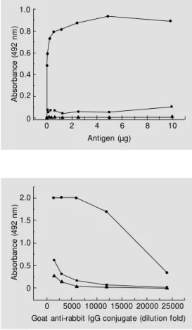
Figure 1 shows the relationship between
the concentration of F1 antigen on the pro-posed matrix and the results obtained by ELISA. A typical hyperbolic Michaelis-Menten curve (19) obtained due to the satu-ration of antigen-antibody complex revealed that the minimum amount of fixed antigen to produce maximum absorbance is equal to 1.25 µg protein. Figure 2 shows the relation-ship between goat anti-rabbit IgG conjugate dilution and ELISA performance. The 1:12,000 dilution can be used as the best conjugate concentration. The titration of immunized and non-immunized serum against F1 antigen, illustrated in Figure 3, shows that a 1:6,200 dilution would be rec-ommended since the highest F factor value was estimated at this concentration (F @ 12). A remarkable difference in absorbance was observed between immunized and non-im-munized sera, while values close to zero were detected for the control (PBS).
A
b
s
o
rb
a
n
c
e
(
4
9
2
n
m
)
1.0
0.8
0.6
0.4
0.2
0
0 2 4 6 8 10
Antigen (µg)
Figure 1 - Relationship betw een ant igen im m obilized (F1) on discs of filter paper plasticized w ith polyvinyl alcohol-glutaralde-hyde and ELISA results. Discs containing F1 antigen (100 µl) w it h prot ein concent rat ions ranging from 0.08 to 10 µg w ere incubated w ith 100 µl of immu-nized serum dilut ed 1:6,400 (circles), non-immunized serum (squares) and PBS (triangles). Goat anti-rabbit IgG conjugate (100 µl) diluted 12,000 times and substrate solution (100 µl), in-cluding OPD and H2O2, w ere
used. The discs w ere removed and the absorbance of the su-pernatant at 492 nm w as deter-mined w ith an ELISA reader.
A
b
s
o
rb
a
n
c
e
(
4
9
2
n
m
)2.0
1.0
0 1.5
0.5
0 5000 10000 15000 20000 25000 Goat anti-rabbit IgG conjugate (dilution fold)
A comparative study between this method and conventional ELISA based on antigen adsorption on polyvinyl chloride plates is shown in Figure 4. In this experiment, sera were collected from immunized rabbits (N = 8) and non-immunized rabbits (N = 9). Firstly, regarding the sera from immunized rabbits, the mean values (± SD) of 0.667 ± 0.264 and 0.536 ± 0.326 were calculated for the ELISA absorbance values using plasticized filter paper and conventional procedures, respec-tively. The independent t-test applied to both mean values at the 0.05 level showed no significant difference (t = 0.875 and P = 0.396). Also, a linear correlation was ob-served when plasticized filter paper values were plotted against those obtained by the conventional procedure (r = 0.986 and P = 0.00037). For the sera collected from non-immunized rabbits (N = 9) mean values of 0.127 ± 0.103 and 0.065 ± 0.045 were esti-mated for the plasticized filter paper and conventional ELISA procedures, respec-tively. The independent t-test between these values was not significant (t = 1.652 and P = 0.118). A linear correlation was also ob-served between the two procedures (r = 0.952 and P = 0.00007). In summary, ELISA per-formed on this basis is comparable to that performed by the conventional procedure.
The present results indicate that filter paper plasticized with polyvinyl alcohol-glu-taraldehyde can be an alternative support for immunoassays. It is easy to synthesize and the antigen is covalently linked, therefore avoiding false-negative results due to antigen leaching. The reagents are of low cost and easy acquisition; ELISA and dot-ELISA can be simultaneously performed either in the laboratory or in the field. Small amounts of antigen (1.25 µg) and conjugate (dilution 1:12,000) were employed in the present model (experimental plague). High sensitiv-ity was also achieved (serum dilution up to 1:51,200 still revealed differences between immunized and non-immunized serum, with an F factor higher than 5).
A b s o rb a n c e ( 4 9 2 n m ) 1.4 1.2 1.0 0.8 0.6 0.4 0.2 0.0 Non-immune PVC Non-immune plasticized filter paper Immune PVC Immune plasticized filter paper
Figure 4 - Comparison of the ab-sorbance values obtained by conventional ELISA based on polyvinyl chloride (PVC) and ELISA based on filter paper plas-ticized w ith polyvinyl alcohol-glu-taraldehyde. Sera from immu-nized (N = 8) and non-immu-nized (N = 9) rabbits w ere ana-lyzed for the presence of anti-F1 antigen IgG. The absorbance re-ported is the mean of tw o deter-minations. The arrow s indicate the positions of the mean val-ues. A b s o rb a n c e ( 4 9 2 n m ) 2.0 1.5 1.0 0.5 0.0 A b s o rb a n c e r a ti o (i m m u n iz e d /n o n -i m m u n iz e d s e ru m ) 12 10 8 6 4
0 1000020000 30000 40000 50000 60000 Serum (dilution fold)
0 10000 20000 30000 40000 50000 60000
Serum (dilution fold)
Figure 3 - Titration of serum from rabbits immunized or not against F1 antigen by ELISA using filter paper plasticized w ith polyvinyl alcohol-glutaraldehyde. Sera (100 µl) from immunized rabbit (circles), non-immunized rabbit (squares) and PBS (triangles) w ere diluted from 1:400 to 1:51,200 in PBS and ELISA w as carried out according to the best conditions for antigen (1.25 µg/disc) and conjugate concentration (diluted 1:12,000). Insert, The ratio (factor F) betw een the absorbance values recorded for the immunized and non-immunized serum w as calculated for each dilution.
The antigen/plasticized discs used for this serum titration were impregnated by the col-ored products released during ELISA devel-opment (OPD oxidation by peroxidase). The color intensity was directly proportional to absorbance and remained unaltered for about two days. Thus, one can set up a dot-ELISA based on this antigen/plasticized discs.
!
!
Re fe re nce s
1. Butler T (1989). The Black Death past and present. 1. Plague in the 1980s. Transac-tions of the Royal Society of Tropical
M edicine and Hygiene, 83: 458-460.
2. Chant eau S, Rat sif asoam anana L, Rasoam anana B, Rahalison L, Randriambelosoa J, Roux J & Rabeson D (1998). Plague, a reemerging disease in M adagascar. Emerging Infectious
Dis-eases, 4: 101-104
3. Vieira JBF, Almeida AM P & Almeida CR (1994). Epidemiologia e controle da peste no Brasil. Revista da Sociedade Brasileira
de M edicina Tropical, 27 (Suppl III): 51-58.
4. Perry RD & Fetherston JD (1997). Yersinia
pestis - etiologic agent of plague. Clinical
M icrobiology Review s, 10: 35-66.
5. Almeida AM , Leal NC, de Carvalho FG, Dantas Sobrinho J & de Almeida CR (1995). Plague surveillance in Brazil: 1983-1992. Revista do Instituto de M edicina
Tropical de São Paulo, 37: 511-516.
6. Almeida AM & Ferreira LC (1992). Evalua-tion of three serological tests for the de-tection of human plague in northeast Bra-zil. M emórias do Instituto Osw aldo Cruz, 87: 87-92.
7. Leroy F (1997). Etude séro(-)épidémio-logique de la peste humaine à M adagas-car. Annales de Biologie Clinique, 55: 332-336.
8. Cao LK, Anderson GP, Ligler FS & Ezzell J
(1995). Detection of Yersinia pestis frac-tion 1 antigen w ith a fiber optic biosensor.
Journal of Clinical M icrobiology, 33:
336-341.
9. Anderson GP, King KD, Cao LK, Jacoby M , Ligler FS & Ezzell J (1998). Quantifying serum antiplague antibody w ith a fiber-optic biosensor. Clinical and Diagnostic
Laboratory Immunology, 5: 609-612.
10. Leal NC, Abath FG, Alves LC & de Almeida AM (1996). A simple PCR-based proce-dure for plague diagnosis. Revista do Instituto de M edicina Tropical de São
Paulo, 38: 371-373.
11. M ontenegro SM L, De Alm eida AM P, Carvalho AB & Carvalho Jr LB (1991). The use of Dacron plates for dot enzyme-linked immunosorbent assay (dot-ELISA).
M emórias do Instituto Osw aldo Cruz, 86:
461-465.
12. M ont enegro SM L, Alm eida AM P & Carvalho Jr LB (1993). Standardization of the dot enzyme-linked immunosorbent assay (dot -ELISA) f or experim ent al plague. M emórias do Instituto Osw aldo Cruz, 88: 119-123.
13. Araujo AM , Petribú ATS, Barbosa GHTS, Diniz JRP, Almeida AM P, Azevedo WM , M alagueño E & Carvalho Jr LB (1996). The use of polyvinyl alcohol glutaralde-hyde as solid-phase in ELISA for plague.
M emórias do Instituto Osw aldo Cruz, 91:
195-198.
14. Carvalho Jr LB, Araujo AM , Almeida AM P & Azevedo WM (1996). The use of polyvi-nyl alcohol glutaraldehyde antigen coated discs for laser induced fluorescence de-tection of plague. Sensors and Actuators, B 35-36: 1-4.
15. Baker EE, Sommer H, Foster LE, M eyer E & M eyer KF (1952). Studies on immuniza-tion against plague. I. The isolaimmuniza-tion and characterization of the soluble antigen of
Pasteurella pestis. Journal of
Immunol-ogy, 68: 131-140.
16. Lehtonen OP & Viljanen M KJ (1980). Antigen attachment in ELISA. Journal of
Immunological M ethods, 34: 61-70.
17. Bode L, Beutin L & Köhler HJ (1984). Nitrocellulose-enzyme-linked immunosor-bent assay (NC-ELISA) - a sensitive tech-nique for the rapid visual detection of both viral antigens and antibodies. Journal of
Virological M ethods, 8: 111-121.
18. De Saeger S & Van Peteghem C (1996). Dipstick enzyme immunoassay to detect Fusarium T-2 toxin in w heat. Applied and
Environmental M icrobiology, 62:
1880-1884.