Trabeculectomy and optic nerve
head topography
1Department of Ophthalmology and Visual Science,
Yale University, New Haven, CT, USA
2Departamento de Oftalmologia, Universidade Federal de São Paulo,
São Paulo, SP, Brasil
3Jules Stein Eye Institute, UCLA, Los Angeles, CA, USA
A. Paranhos Jr.1,2,
M.C. Lima1,
S. Salim1,
J. Caprioli3 and
M.B. Shields1
Abstract
The objective of the present study was to evaluate changes in optic nerve head parameters, measured by confocal laser tomography, before and after trabeculectomy in order to identify outcome measures for the management of glaucoma. The optic nerve head of 22 eyes (22 patients) was analyzed by confocal laser tomography with the Heidel-berg retinal tomogram (HRT) before and after trabeculectomy. The median time between the first HRT and surgery was 4.6 months (mean: 7.7 ± 8.3) and the median time between surgery and the second HRT was 10.8 months (mean: 12.0 ± 6.8). The patients were divided into two groups, i.e., those with the highest (group A) and lowest (group B) intraocular pressure (IOP) change after surgery. Differences in the 12 standard topographic parameters before and after surgery for each group were evaluated by the Wilcoxon signed rank test and the differences in these parameters between the two groups were com-pared by the Mann-Whitney rank sum test. Multiple regression anal-ysis was used to evaluate the influence of the change in IOP (∆IOP and ∆IOP%) and the changes in the other parameters. There were
signifi-cant differences in the HRT measures before and after surgery in group A only for cup volume. In group B, no parameter was statistically different. The changes in group A were not significantly different than those in group B for any parameter (P > 0.004, Bonferroni correction for multiple comparisons). ∆IOP and ∆IOP% had a statistically
sig-nificant effect on ∆ cup disk area, ∆ cup volume and ∆ mean cup depth.
Changes in cup shape size were influenced significantly only by
∆IOP. Some optic disc parameters measured by HRT presented a
significant improvement after filtering surgery, depending on the amount of IOP reduction. Long-term studies are needed to determine the usefulness of these findings as outcome measures in the manage-ment of glaucoma.
Correspondence
A. Paranhos Jr.
Departamento de Oftalmologia UNIFESP
Rua Botucatu, 824 04023-062 São Paulo, SP Brasil
Fax: +55-11-5573-4002
E-mail: [email protected] Research supported in part by an unrestricted grant from the Connecticut Lions Eye Research Foundation and Research to Prevent Blindness.
Publication supported by FAPESP.
Received November 23, 2004 Accepted September 15, 2005
Key words •Optic nerve head
•Confocal laser tomography •Intraocular pressure •Trabeculectomy •Glaucoma
Introduction
The traditional outcome measure for ad-equacy of intraocular pressure (IOP) reduc-tion with any medical or surgical
inadequacy of the therapy is recognized. Although most glaucomatous damage is irreversible, some optic nerve head and vi-sual field improvement following IOP re-duction have been reported. The improve-ment has been associated with the amount of pressure reduction (1-3). Spaeth and associ-ates (4,5) have suggested that improvement in the appearance of the optic disc and/or visual field is an important early indicator of the long-term adequacy of glaucoma man-agement. With the advent of technologies to more precisely and quantitatively measure changes in optic disc topography, investiga-tors are beginning to take a closer look at the influence of lowering the IOP on disc mor-phology (6,7).
The purpose of the present study was to quantitatively analyze changes in optic nerve head topography, as measured by confocal laser tomography before and after trabecu-lectomy, and by attempting to correlate the changes with the degree of IOP reduction.
Material and Methods
This is a retrospective study of patients who had undergone confocal laser tomogra-phy with the Heidelberg retinal tomogram (HRT, Software IR1-V2.01, Heidelberg En-gineering, Heidelberg, Germany) before and after trabeculectomy. Only one eye of each patient was included in the study. If both eyes of one patient were eligible, data from the eye with the highest IOP reduction after surgery were used for analysis. Patients were excluded if HRT studies were thought to be unreliable or if they had other optic nerve disease in addition to glaucoma.
IOP in mmHg was measured with a Gold-mann tonometer, and the absolute IOP change (∆IOP) and the percent IOP reduction (∆IOP%) before and after surgery were based
on measurements obtained at the time of the HRT studies.
The principles and technique of confocal laser tomography with the HRT have been
previously described (8,9). The disc margin was defined by the contour line of the optic nerve head, which was drawn by the techni-cian. For each image, the scanning depth was chosen by the operator, based on the depth of the optic disc cup. The mean of at least three images was used for the analyses of each exam.
A reference plane was defined as 0.05 mm posterior to the mean retinal surface at the disc margin near the papillo-macular bundle. Structures posterior to the plane were assigned to the optic nerve head cup, whereas structures anterior to the reference plan were regarded as the neuroretinal rim. The HRT stereometric parameters used in the analyses are listed in Table 1.
The patients were divided into two equal groups according to the amount of ∆IOP or ∆IOP% using the median as the cut-off point.
Both divisions gave coincident subgroups hence group A had higher ∆IOP and ∆IOP%
while group B had lower ∆IOP and ∆IOP%. Patient age and cup/disc area ratio before surgery and ∆IOP, ∆IOP% and the changes
in HRT parameters after surgery were com-pared between the two groups using the Mann-Whitney rank sum test. The changes in HRT parameters before and after surgery for each group were evaluated by the Wil-coxon signed rank test. Non-parametric tests were used because of the high variability of all parameters in both groups.
Multiple regression analysis was also used with a backward stepwise regression strat-egy. In this analysis, the dependent variables were the differences of all stereometric pa-rameters before and after surgery. The inde-pendent variables were ∆IOP, ∆IOP%, the
patient’s age at the time of the surgery, and the cup/disc area ratio before surgery and the time between surgery and the last HRT exam. The variance inflation factor was used to evaluate colinearity of independent variables. Homoscedasticity and normality tests were also performed.
Depart-ment of Ophthalmology, Yale University, New Haven, CT, USA, with the approval of the Yale Hospital Ethics Committee.
Results
Twenty-two eyes of 22 patients were included in this study. There were 15 (68.2%) women. The ages ranged from 33 to 73 years (mean: 60.3 ± 11.0). Fourteen patients (63.3%) had chronic open-angle glaucoma, 3 (16.6%) had pigmentary glaucoma, 3 (16.6%) had the pseudoexfoliation syndrome, and 2 (9.1%) had low tension glaucoma. The median cup/disc area ratio before surgery, as measured by the HRT, was 0.52 (mean: 0.52 ± 0.21). The median time between the first HRT and surgery was 4.65 months (mean: 7.74 ± 8.30) and the median time between surgery and the second HRT was 10.8 months (mean: 12.0 ± 6.85). In group A, the eleven patients had a median ∆IOP of 11 mmHg
(mean: 13.45 ± 6.14) and a median ∆IOP%
of 52.4% (mean: 52.21 ± 15.46), while the corresponding values for group B were 3 mmHg (2.36 ± 2.69) and 26.7% (18.50 ± 16.66), respectively. These differences be-tween the two groups were statistically sig-nificant (P < 0.0001). There were no signifi-cant differences between the two groups regarding the time of the HRT exam before or after surgery (P = 0.249 and 0.247,
re-spectively), or the age or cup/disc area ratio (P = 0.9215 and 0.0878, respectively).
The measures of each HRT parameter before and after surgery and the compari-sons between the changes in the two groups are listed in Table 1. There were significant differences between the HRT measures made before and after surgery in group A only for cup volume. In group B, none of the param-eters was statistically different. The changes in group A were not significantly greater than those in group B for any parameter (P > 0.004, Bonferroni correction for multiple comparisons).
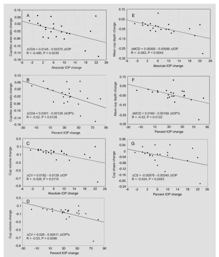
The stepwise regression analysis excluded age and cup/disc area ratio before surgery and the time between surgery and the last HRT exam as independent variables due to lack of a significant correlation with any change in HRT (P > 0.05). Because ∆IOP% and ∆IOP have a high colinearity, as indicated by the variance inflation factor (value of 4), the two variables were considered in different simple linear re-gression models. The results demonstrated that ∆IOP and ∆IOP% have a statistically
significant influence on ∆ cup disc area,
∆ cup volume and ∆ mean cup depth. The
change in cup shape measurements was sig-nificantly influenced only by ∆IOP (Figure 1A-G). Linear regression analysis was con-sidered to be appropriate by the normality and homoscedasticity tests (P > 0.05).
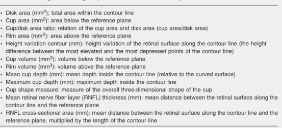
Table 1. Heidelberg retinal tomogram parameters included in the analysis.
·
Disk area (mm2): total area within the contour line·
Cup area (mm2): area below the reference plane·
Cup/disk area ratio: relation of the cup area and disk area (cup area/disk area)·
Rim area (mm2): area above the reference plane·
Height variation contour (mm): height variation of the retinal surface along the contour line (the height difference between the most elevated and the most depressed points of the contour line)·
Cup volume (mm3): volume below the reference plane·
Rim volume (mm3): volume above the reference plane·
Mean cup depth (mm): mean depth inside the contour line (relative to the curved surface)·
Maximum cup depth (mm): maximum depth inside the contour line·
Cup shape measure: measure of the overall three-dimensional shape of the cup·
Mean retinal nerve fiber layer (RNFL) thickness (mm): mean distance between the retinal surface along the contour line and the reference planeDiscussion
An improvement in the appearance of the optic nerve head after reduction of IOP has been documented in children with glaucoma (10) and also in adults (1,2,11). Improve-ment of the optic disc has been reported in adults after both surgical and medical reduc-tion of IOP and in a variety of glaucomas with different techniques used to measure the changes (12).
Experimental studies suggest that appar-ent improvemappar-ent in optic disc morphology may be due to compliance of the lamina cribrosa in response to changes in IOP. Levy and Crapps (13) demonstrated movement of the lamina in response to IOP change in an experimental model using human cadaver eyes in which platinum wires were placed in the lamina to allow radiographic observa-tion of the displacement. Coleman et al. (14) also observed displacement of the optic nerve head in monkeys’ eyes with a change in IOP and noted that this was more prominent in ocular hypertensive than normotensive ani-mals, although there was considerable vari-ability within each group. Shirakashi et al. (15) reported that the extent of cupping re-versal after IOP reduction in primates with experimental glaucoma may decrease with progressive glaucomatous damage. Bur-goyne et al. (12) also demonstrated reduced compliance of the optic disc in monkeys with chronic experimental glaucoma.
The variable compliance of the optic nerve head to changes in IOP may relate to ultrastructural differences in the lamina cribrosa. Hernandes (16) showed that elastin in the human lamina has ultrastructural dif-ferences in normal eyes as compared to those with glaucoma, and that this difference is influenced by age in normal eyes and by whether the glaucoma is early or advanced. In the early stages of chronic open-angle glaucoma, there appears to be new forma-tion of elastin, while in the later stages, an abnormal enzymatic activity appears to
en-hance degradation or impaired maturation of the newly formed material. In chronic open-angle glaucoma, Hernandes (16) also found a decrease in collagen content, proliferation of basement membranes, and a change in elastic fibers with loss of the tubular struc-ture.
Because of this apparent difference in the characteristics of the lamina cribrosa with age and the stage of glaucoma, we per-formed multiple regression analysis with the age of the patient and the cup/disc area ratio before surgery, using the latter as a param-eter of the glaucoma severity. We did not find a significant correlation with either vari-able, although this could be due to an insuf-ficient sample size to adequately represent different ages and levels of nerve damage. Shin et al. (17) and Topouzis et al. (18) also failed to find a significant association be-tween reversal of cupping and age.
Another possible explanation for changes in the optic disc following IOP reduction with filtering surgery is edema of the nerve head and peripapillary retina, which has been de-scribed by Kawasaki and Purvin (19). Since this most likely occurs in the early postopera-tive period, it probably does not explain the disc changes in the present study, since the postoperative image analyses were performed an average of one year after surgery.
Whatever the mechanism of change in the optic disc following IOP reduction may be, it could offer a different approach to the management of glaucoma. Spaeth et al. (4,5) noted that these changes obviously do not indicate that the disc has returned to normal in most cases, nor that they occur in every patient. However, the authors suggested that, if the ophthalmologist does not find im-provement in the optic disc on careful ex-amination after lowering the IOP, further lowering of the pressure should be consid-ered.
suffi-ciently accurate to detect a subtle improve-ment in all cases. The newer technologies of image analysis may improve our ability to detect these subtle changes. Shin et al. (17) reported that in 13 eyes with IOP reduction greater than 5 mmHg, 10 (77%) had im-provement in at least one optic disc param-eter when analyzed by computerized stereo-photogrammetry with the Rodenstock nerve head analyzer (Munich, Germany), while only 5 eyes (38%) showed qualitative evi-dence of optic disc cup reversal with stereo photography. Lesk et al. (20) also found that the HRT was more sensitive in detecting changes in disc cupping than was the case with stereo photography.
The HRT, which is based on the principle of confocal laser tomography, appears to be a good tool to detect subtle improvement in the optic nerve since it has been shown to have good reproducibility (8,9), has been used to demonstrate progression and im-provement of the optic disc cupping with changes in IOP (21), and may be able to discriminate between persons with normal optic nerve heads and those with early glau-comatous damage (22).
Using HRT technology in the present study, we found statistical improvement af-ter IOP reduction in optic disc parameaf-ters related only to cup volume in group A, which had relatively greater IOP reduction, and none in group B, which had less IOP reduc-tion. Other topographic parameters improved in group A, but did not reach statistical significance, probably because of lack of power to detect these differences. The sample size is to small to find significant differences for most of the HRT parameters and larger studies or meta-analyses are needed to clarify the real influence of the IOP on HRT param-eters. Nevertheless, the findings of some outliers with big differences in HRT param-eters after surgery should be a concern in clinical practice.
We have confirmed and extended the observations of other investigators. Irak et
al. (6) used the HRT to evaluate changes in the optic nerve head in 49 patients after trabeculectomy and found improvement in the same parameters as in the present study, as well as in rim area, rim volume and height variation contour. With the exception of rim area, these latter parameters also improved in our study, although the change was not statistically significant. Other investigators have also reported improvement in the optic disc parameters, as measured with the HRT, after lowering the IOP (7,20,23).
Topouzis et al. (18) suggested that the improvement in disc morphology following IOP reduction is time-limited, since the changes at 2 weeks after surgery did not persist 4 and 8 months later, with the excep-tion of cup shape, which was still improved by the fourth month. However, Shin et al. (17) reported that the time after IOP reduc-tion did not correlate with changes in the optic disc, and Raitta et al. (23) found statis-tically significant improvement in cup vol-ume and mean cup depth after a mean fol-low-up of 3.7 months, which remained sig-nificant for cup volume at 12.1 months. In the latter study, cup/disc area ratio and mean height contour, which were not significantly changed at the first follow-up, were signifi-cantly improved in the later exam. In our study, the longer follow-up between surgery and the second HRT of 12.0 ± 6.85 months suggests some degree of stability in the pa-rameters that improved after IOP reduction, as also does the nonsignificant correlation with the improvement of any parameter and the time of follow-up. However, the lack of shorter-term follow-up is a limitation of the present study, and further studies are needed to clarify the stability of improvement in optic disc parameters following a sustained reduction of IOP.
In the present study, regression analysis showed that ∆IOP and ∆IOP% can
although some have found a less consistent correlation (18,24).
The most important result presented here is that, although the correlation with the
∆IOP and ∆IOP% and optic disc changes is
statistically significant, it probably has no influence on clinical evaluation of the disc and the damage progression as most of all parameters did not change significantly.
The important question that remains to be answered is whether improvement in one or more disc parameters in association with
a particular degree of IOP reduction is a reliable indicator that the new pressure level, assuming it remains constant, will be ad-equate to prevent further glaucomatous op-tic neuropathy. The same question must also be applied to visual function tests. Large population, long-term, prospective trials with both functional and morphologic tests after IOP reduction are needed to determine whether these outcome measures can im-prove our ability to manage glaucoma.
References
1. Pederson JE & Herschler J (1982). Reversal of glaucomatous cup-ping in adults. Archives of Ophthalmology, 100: 426-431.
2. Greenidge KC, Spaeth GL & Traverso CE (1985). Change in ap-pearance of the optic disc associated with lowering of intraocular pressure. Ophthalmology, 92: 897-903.
3. Tsai CS, Shin DH, Wan JY et al. (1991). Visual field global indices in patients with reversal of glaucomatous cupping after intraocular pressure reduction. Ophthalmology, 98: 1412-1419.
4. Spaeth GL, Fellman RL, Starita RL et al. (1985). A new manage-ment system for glaucoma based on improvemanage-ment of the appear-ance of the optic disc or visual field. Transactions of the American Ophthalmological Society, 83: 268-284.
5. Spaeth GL (1988). A new management system for glaucoma based on improvement of the appearance of the optic disc or visual field. Fortschritte der Ophthalmologie, 85: 614-619.
6. Irak I, Zangwill L, Garden V et al. (1996). Change in optic disk topography after trabeculectomy. American Journal of Ophthalmol-ogy, 122: 690-695.
7. Park KH, Kim DM & Youn DH (1997). Short-term change of optic nerve head topography after trabeculectomy in adult glaucoma pa-tients as measured by Heidelberg retina tomograph. Korean Journal of Ophthalmology, 11: 1-6.
8. Weinreb RN, Lusky M, Bartsch DU et al. (1993). Effect of repetitive imaging on topographic measurements of the optic nerve head. Archives of Ophthalmology, 111: 636-638.
9. Rohrschneider K, Burk ROW, Kruse FE et al. (1994). Reproducibility of the optic nerve head topography with a new laser topographic scanning device. Ophthalmology, 101: 1044-1049.
10. Quigley HA (1982). Childhood glaucoma. Results with trabeculotomy and study of reversible cupping. Ophthalmology, 89: 219-226. 11. Spaeth GL (1989). Reversibility of optic disc cupping. Archives of
Ophthalmology, 107: 1583-1584.
12. Burgoyne CF, Quigley HA, Thompson HW et al. (1995). Early changes in optic disc compliance and surface position in experimen-tal glaucoma. Ophthalmology, 102: 1800-1809.
13. Levy N & Crapps EE (1984). Displacement of optic nerve head in response to short-term intraocular pressure elevation in human eyes. Archives of Ophthalmology, 102: 782-786.
14. Coleman AL, Quigley HA, Vitale S et al. (1991). Displacement of the optic nerve head by acute changes in intraocular pressure in mon-key eyes. Ophthalmology, 98: 35-40.
15. Shirakashi M, Nanba K & Iwata K (1992). Changes in reversal cupping in experimental glaucoma. Ophthalmology, 99: 1104-1110. 16. Hernandes MR (1992). Ultrastructural immunocytochemical analy-sis of elastin in the human lamina cribrosa. Investigative Ophthal-mology and Visual Science, 33: 2891-2903.
17. Shin DH, Bielik M, Hong JY et al. (1989). Reversal of glaucomatous optic disc cupping in adult patients. Archives of Ophthalmology, 107: 1599-1603.
18. Topouzis F, Peng F, Kotas-Neumann R et al. (1999). Longitudinal changes in optic disc topography of adult patients after trabeculec-tomy. Ophthalmology, 106: 1147-1151.
19. Kawasaki A & Purvin V (1998). Unilateral optic disc edema following trabeculectomy. Journal of Neuro-Ophthalmology, 18: 121-123. 20. Lesk MR, Spaeth GL, Azuara-Blanco A et al. (1999). Reversal of
optic disc cupping after glaucoma surgery analyzed with a scanning laser tomograph. Ophthalmology, 106: 1013-1018.
21. Azura-Blanco A, Harris A, Cantor LB et al. (1998). Effects of short term increase of intraocular pressure on optic disc cupping. British Journal of Ophthalmology, 82: 880-883.
22. Uchida H, Brigatti L & Caprioli J (1996). Detection of structural damage from glaucoma with confocal laser image analysis. Investi-gative Ophthalmology and Visual Science, 37: 2393-2401. 23. Raitta C, Tomita G, Vesti E et al. (1996). Optic disc topography
before and after trabeculectomy in advanced glaucoma. Ophthalmic Surgery and Lasers, 27: 349-354.