ABSTRACT
Purpose: To determine the relationship between anterior segment and optic nerve head (ONH) parameters.
Methods: Two hundred healthy subjects were enrolled in this study. The par-ticipants underwent measurement of anterior segment parameters with a Pen-tacam Scheimpflug system and imaging of the optic disc with confocal scanning laser ophthalmoscopy. Pearson’s correlation coefficient was calculated to assess the associations between ONH and anterior segment parameters. Multivariate regression analyses controlling for age and disc area as confounding factors were also conducted.
Results: There were significant negative relationships between optic disc area and corneal thickness (p=0.03, r=-0.217) and volume (p=0.017, r=-0.239). Corneal refractive power was significantly correlated with cup area, rim area, rim volume, and cup/disc ratio (p<0.05 for all). An increase of 1 diopter in anterior corneal refractive power corresponded to a decrease of 0.022 in cup/disc ratio. Rim volume was negatively correlated with anterior chamber depth and anterior chamber volume (p<0.05 for both). A 1-mm3 increase in anterior chamber depth corresponded to a 0.154-mm3 decrease in rim volume. Mean and maximum cup depth measurements were not associated with any of the anterior segment parameters.
Conclusions: In nonglaucomatous eyes, the dimensions and shape of the ONH may be related to anterior segment morphology. Therefore, it may be helpful to take anterior segment parameters into consideration in evaluating the OHN.
Keywords: Anterior eye segment, Cornea; Glaucoma; Optic disc
RESUMO
Objetivo: Determinar a relação entre os parâmetros do segmento anterior e da cabeça do nervo óptico (CNO).
Métodos: Duzentos indivíduos saudáveis foram incluídos neste estudo. Os parti-cipantes foram submetidos à medição dos parâmetros do segmento anterior com o sistema Pentacam Scheimpflug e à obtenção de imagens de disco óptico com oftalmoscopia a laser de varredura confocal. O coeficiente de correlação de Pearson foi calculado para avaliar as associações entre CNO e os parâmetros do segmento anterior. Realizaram-se também análises de regressão multivariada que controlam a idade e a área do disco como factores de confusão.
Resultados: Houve uma relação negativa estatisticamente significativa entre a espessura da córnea, o volume da córnea e a área do disco óptico (p=0,03, r=-0,217 e p=0,017, r=-0,239, respectivamente). As medições do poder de refração da córnea mostraram correlações significativas com a área da escavação, a área da rima, o volume da rima (RV) e a relação escavação/disco (C/D). (P<0,05 para todos). Um aumento de dioptria no poder de refração corneana anterior corresponde a uma diminuição de 0,022 em C/D. O RV mostrou correlação negativa com a profundidade da câmara anterior (PCA) e volume da câmara anterior. (P<0,05 para ambos). O aumento de 1 mm3 na PCA corresponde a uma diminuição de 0,154 mm3 no RV. As
médias e as medidas máximas de profundidade de rima não mostraram associação com nenhum dos parâmetros do segmento anterior.
Conclusões: Em olhos não glaucomatosos, as dimensões e forma da CNO podem estar relacionadas com a morfologia do segmento anterior. Portanto, pode ser útil levar em consideração os parâmetros do segmento anterior na avaliação da CNO.
Descritores: Segmento anterior do olho; Cornea; Glaucoma; Disco óptico
Submitted for publication: October 7, 2016 Accepted for publication: March 19, 2017
1 Faculty of Medicine, Hacettepe University, Ankara, Turkey. 2 Ulucanlar Eye Research Hospital, Ankara, Turkey.
Funding: No specific financial support was available for this study.
Disclosure of potential conflicts of interest: None of the authors have any potential conflict of interests to disclose.
Corresponding author: Ali Bulent Cankaya. Hacettepe University. Faculty of Medicine - Sıhhiye -
Ankara - 06100 - Turkey - E-mail: [email protected], [email protected]
Approved by the following research ethics committee: Institutional Review Board of the Ankara Numune Research and Education Hospital (# E-14-315).
INTRODUCTION
Increasing attention is being focused on the impact of corneal parameters as potential determinants of the risk of glaucoma. Many studies have investigated the question of whether corneal thickness and/or biomechanical properties have an influence on glaucoma. The Ocular Hypertensive Treatment Study (OHTS) and the Early Manifest Glaucoma Treatment Study (EMGT ) demonstra-ted a link between central corneal thickness (CCT ) and the risk of glaucoma(1,2).
A morphological link is possible between aspects of the front of the eye and optic nerve head (ONH) parameters. Owing to the ana-tomic continuity of the cornea, sclera, and optic disc lamina, it can be hypothesized that corneal parameters may relect parameters of the ONH, such as optic disc area (DA) and deformability of the lamina
cribrosa. For example, in a study analyzing anterior segment structu-res in eyes with an optic disc pit, a signiicantly smaller back radius of corneal curvature was found in eyes with an optic disc pit(3). Since the corneal stroma, corneal endothelium, and optic nerve lamina cribrosa diferentiate from the neural crest, they are embryologically related to one another(4).
Many reports have suggested that the risk of glaucoma is re-lated in some way to optic disc parameters, particularly optic disc size and cup/disc ratio (C/D)(5). For example, it has been suggested that large discs may be susceptible to glaucomatous damage at low levels of intraocular pressure (IOP)(6). In support of this hypothesis, the size of the ONH has been reported to be larger in patients with normotensive glaucoma than in those with primary open angle glaucoma (POAG)(7). Although disc size was not associated with the
Relationship between anterior segment and optic nerve head parameters
in healthy subjects
Relações entre os parâmetros do segmento anterior e da cabeça do nervo óptico em indivíduos saudáveis
development of POAG in OHTS participants(8), the OHTS suggested that vertical and horizontal C/D was a good predictor of the onset of POAG(1).
Although the relationships among CCT, corneal biomechanics, and ONH parameters have been examined, there is little informa-tion on the relainforma-tionships between other corneal parameters (such as corneal volume and corneal refractive power) and the OHN in healthy subjects. Moreover, it is also unclear whether anterior cham-ber parameters (depth and volume) are significantly correlated with ONH parameters.
The aim of this study was to determine whether there is a rela-tionship between anterior segment morphology and the dimensions and shape of the ONH. To the best of our knowledge, no study has evaluated the correlations between anterior segment parameters measured by the Scheimplug imaging system and optic disc para-meters measured by confocal scanning laser ophthalmoscopy.
METHODS
This research was carried out in accordance with the Declara-tion of Helsinki and after approval from the InstituDeclara-tional Review Board of the Ankara Numune Research and Education Hospital. All participants provided informed consent for participation in this study. The subjects were between 20 and 60 years of age and were prospectively recruited among patients presenting to the outpatient clinic of Ulucanlar Eye Research and Education Hospital.
Individuals with a history of vascular, circulatory (diabetes melli-tus, hypertension, or hypotension), autoimmune, or neurological di-seases (including migraine), neoplasia, or chronic medication usage were excluded. The exclusion criteria included history of glaucoma, uveitis, ocular surgery, or trauma; any corneal scarring pathology, such as infection or trauma, and any previous history of corneal or intraocular surgery. The participants were not using topical eye drops. We limited the contribution of each patient to the right eye to avoid interdependency.
The inclusion criteria were best corrected visual acuity ≥20/20, spherical equivalent between -1.0 and +1.0 diopters (D) with cylin-der power <1 D, IOP<22 mmHg, open anterior chamber angle, and normal-appearing ONH. Normal-appearing ONH was deined as in-tact neuroretinal rim without splinter hemorrhage, notches, localized pallor, or asymmetry of the cupping <0.2 between the eyes, taking into account disc size. Participants with an optic disc abnormality (such as drusen or tilted disc) were excluded.
Eligible subjects were consecutively included and received a comprehensive ocular examination, including medical history, manifest refraction, IOP measurement by Goldmann applanation tonometry, gonioscopy, and slit lamp and fundus examination. The subjects underwent disc imaging with a confocal scanning laser ophthalmoscope (Heidelberg Retina Tomograph III (HRT III); Heidel-berg Engineering, Dossenheim, Germany). Subjects whose HRT III images had a standard deviation <50 were included in the inal analysis. The same examiner performed all measurements. The details of this technique have been described elsewhere(8). The parameters obtained from HRT III hardware were DA (mm2), cup area (CA) (mm2), rim area (RA) (mm2), cup volume (CV) (mm3), rim volume (RV) (mm3), cup to disc area ratio (C/D area), linear cup to disc ratio (linear C/D), and mean and maximum cup depth (CD) (mm).
Anterior segment parameters were obtained automatically by the Pentacam Scheimplug system (Oculus Gmbh, Wetzlar, HE, Ger-many) with a quality score >95. Pentacam software constructs the three-dimensional image of the anterior segment and calculates the anterior chamber parameters. This imaging provides measurements of anterior chamber depth (ACD) and anterior chamber volume (ACV). Corneal thickness at the apex point (regarded as the CCT) and corneal volume values were also recorded. The amount of anterior and posterior corneal refractive power (the dioptric power of the posterior corneal surface is negative) within a span of 3.0 mm from the centre was also measured.
Axial length measurements were recorded with an 11-MHz probe of an A-mode ultrasound device (Cine Scan; Quantel Medical SA, Clermont-Ferrand, France). Measurements were performed 5 min after instillation of one drop of proparacaine hydrochloride 0.5% (Alcaine; Alcon Laboratories, Hemel Hempstead, UK).
The data were evaluated by the SPSS program version 22.0 (IBM Corp.) The data obtained with both devices were analyzed descriptively. Normality of the variables was checked by the Kol-mo gorov-Smirnov test. The distribution of the main variables was normal. Age and optic DA have a known impact on anterior seg-ment and ONH parameters, respectively. We carried out a partial correlation analysis to investigate the relationship between anterior segment and optic nerve head parameters, controlling for age and optic DA. In addition, we used multiple linear regression analysis to examine the associations of the anterior segment and ONH para-meters. All models were adjusted for age and optic DA.
RESULTS
This clinical observational study included 200 eyes of 200 Cau-casian individuals (82 men and 118 women). The mean age was 25.8 ± 10.5 years (range, 20-60 years), and the mean refractive error was -0.14 ± 0.28 D as spherical equivalent (range, -1.50 to +1.25 D). The mean axial length was 22.85 ± 0.49 mm (range, 22.06-23.49 mm). The mean IOP was 15.6 ± 3.66 mmHg (range, 8-22 mmHg). The an-terior segment and optic disc parameters are summarized in tables 1 and 2, respectively.
The study found statistically signiicant negative correlations between CCT and DA (p=0.002, r=-0.217) and between corneal volu-me and DA (p=0.001, r=-0.239). Signiicant negative correlations were found between ACV and DA (p=0.034, r=-0.150) and between ACD and DA (p=0.028, r=-0.156).
Partial correlation analysis to investigate the relationships among anterior segment and optic nerve head parameters, controlling for age and optic DA, showed that corneal refractive power (both anterior and posterior) correlated well with most ONH parameters except CV and CD (Table 3). Pearson’s partial correlation coeicient showed no statistically signiicant correlations between CCT and ONH parameters.
Signiicant correlations between anterior or posterior corneal refractive power and CA, RA, RV, C/D area, and linear C/D were also apparent after multiple regression analysis. For example, a 1-D in-crease in anterior corneal refractive power corresponds to a 0.034-mm3 increase in RV. Correlations between anterior and posterior corneal refractive power and RV were shown on the basis of the scatterplot distribution (Figures 1 and 2).
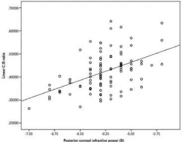
CV and CD measurements showed signiicant relationships with none of the corneal parameters. As seen in table 3, none of the ante-rior segment parameters except refractive power of the cornea (both anterior and posterior) correlated with C/D (C/D area and linear C/D). In multivariate regression models, a 1-D increase in anterior corneal refractive power was associated with a 0.022 decrease in linear C/D (Figure 3). Posterior corneal refractive power was also strongly corre-lated with linear C/D (Figure 4).
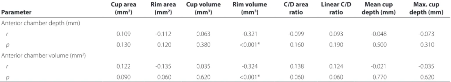
Pearson’s partial correlation coeicient showed a statistically significant negative correlation between ACD and DA (p=0.001, r=-0.228). ACV was also negatively correlated with DA (p=0.001, r=-0.230). Table 4 shows the partial correlation coeicient between ACD measurements and ONH parameters. Only RV showed signi-icant associations with depth (p<0.001, r=-0.321) and volume (p<0.001, r=-0.324) of the anterior chamber. We also found signii-cant negative correlations of ACV and ACD with RV in multivariable re gression analysis. According to our analyses, a 0.1-mm increase in ACD corresponds to a 0.015-mm3 decrease in RV (Figure 5).
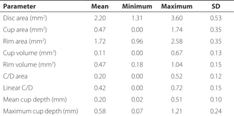
Table 1. Optic nerve parameters of the participants
Parameter Mean Minimum Maximum SD
Disc area (mm2) 2.20 1.31 3.60 0.53
Cup area (mm2) 0.47 0.00 1.74 0.35
Rim area (mm2) 1.72 0.96 2.58 0.35
Cup volume (mm3) 0.11 0.00 0.67 0.13
Rim volume (mm3) 0.47 0.18 1.04 0.15
C/D area 0.20 0.00 0.52 0.12
Linear C/D 0.42 0.00 0.72 0.15
Mean cup depth (mm) 0.20 0.02 0.51 0.10
Maximum cup depth (mm) 0.58 0.07 1.21 0.24
Table 2. Anterior segment parameters of the participants
Parameter Mean Minimum Maximum SD
Anterior corneal refractive power (D) 043.16 0-39.90 0-47.50 01.45 Posterior corneal refractive power (D) 0 -6.27 00-5.70 00-7.00 00.25
Central corneal thickness (μm) 555.20 -460.00 -671.00 33.10
Corneal volume (mm3) 060.85 0 53.10 0-72.20 03.43 Anterior chamber volume (mm3) 162.84 0 87.00 -278.00 40.23
Anterior chamber depth (mm) 002.89 00-2.07 -003.85 00.37
Table 3. Partial correlation coeicients between corneal and optic nerve head parameters after eliminating the inluence of age and optic disc area of the participants
Parameter
Cup area (mm2)
Rim area (mm2)
Cup volume (mm3)
Rim volume (mm3)
C/D area ratio
Linear C/D ratio
Mean cup depth (mm)
Max cup depth (mm)
Anterior corneal refractive power (D)
r -0.242 0.240* -0.028 -0.347* <-0.238* -0.244* -0.035 -0.017
p <0.001* -0.001* -0.690 -<0.001* -0.001* <0.001* -0.620 -0.810
Posterior corneal refractive power (D)
r -0.225 -0.223* *0.048 -0.357* -<0.222* -0.251* -0.034 -0.037
p <0.001* -0.002* -0.500 -<0.001* -0.002* <0.001* -0.630 -0.600
Corneal thickness (μm)
r -0.049 -0.051* -0.036 <--0.100* -0.002* --0.049* -0.006 -0.033
p -0.490 -0.470* -0.610 -0.160* -<0.940* -0.490* -0.940 -0.640
Corneal volume (mm3)
r -0.050 0.048* -0.015 <-0.074* -0.090* --0.112* -0.001 -0.054
p -0.480 -0.500* 0.840 -0.300* <-0.210* -0.120* -0.990 -0.450
*= statistically signiicant.
DISCUSSION
The thickness and biomechanical properties of the cornea have been of interest as important risk factors for the development of glaucomatous optic nerve damage(1,8-10). In view of the potential clinical importance of ONH parameters for glaucomatous optic neu-ropathy, we undertook the current study to assess the relationship between anterior segment and ONH parameters in healthy, non-glaucomatous subjects.
Some studies reported no correlation between optic disc para-meters and corneal measurements or optic disc parapara-meters and ACD(11). However, in the present study we have demonstrated a sta-tistically significant negative correlation between corneal thickness and optic DA. Therefore, it can be concluded that eyes with thicker
corneas have smaller discs. Moreover, DA was negatively associated with corneal volume. The association between corneal parameters and optic disc morphology can be explained by the embryologic relationship between the OHN and the cornea, since both the cor-neal stroma and the optic disc lamina cribrosa differentiate from the neural crest(4). Moreover, DA was negatively associated with corneal volume. We also found negative correlations among ACD, ACV, and DA and found that eyes with deeper anterior chambers had smaller ONHs.
Several studies have yielded inconsistent results on the rela-tionship between corneal characteristics and optic disc parameters. In our previous study in which CCT was measured by ultrasonic pachymeter in healthy eyes, we noted an inverse correlation between CCT and optic DA(12). Moreover, we found negative correlations between CCT and RA as well as RV, and RNFL cross-sectional area. Pakravan et al. reported a similar negative relationship in POAG pa-tients(13). Insull et al. also observed an inverse relationship between optic DA and corneal thickness in a combined group of glaucoma patients and healthy subjects; however, this relationship was not significant when the results were evaluated separately for the glaucoma patients and the control subjects(14). Moreover, such in -ver se correlations could not be conirmed in some studies. In a po pulation-based study, no significant relationships between CCT and optic disc parameters were observed(15). Bourne et al. reported no association between DA and CCT(11). In contrast, a recent study revealed statistically significant positive correlations between CCT and DA, RA, and RV in normal subjects (16).
Optic disc size is an important parameter in the susceptibility of axonal damage in glaucoma. In the Blue Mountains Eye Study, eyes with glaucoma were found to have larger optic discs than nonglau-comatous eyes and those with ocular hypertension(17). It has been hypo-thesized that the strength of the lamina cribrosa might be correlated with optic DA. In large discs, a higher lamina cribrosa pore-to-disc area ratio and thinner connective tissue support provide less tissue support. Conversely, when disc size is decreased, the pore-to-disc area ratio also decreases, providing greater tissue support against glaucomatous damage(18,19).
Figure 1. Correlation between anterior corneal refractive power and rim volume.
Figure 2. Correlation between posterior corneal refractive power and rim volume.
Figure 3. Correlation between anterior corneal refractive power and linear C/D.
Figure 4. Correlation between posterior corneal refractive power and linear C/D.
segment parameters, was associated with the increased deformation of the optic nerve surface during transient elevations of IOP(22).
It has been reported that increased vertical and horizontal C/D ratio is a risk factor for the development of POAG(1). In our present study, we found a signiicant relationship between corneal refractive power and C/D. Statistically signiicant negative correlations were observed between anterior corneal refractive power and linear C/D and C/D area ratios (i.e., the latter the cornea, the larger the C/D ratio). Posterior corneal refractive power, which is negative, was positively correlated with C/D. Among the anterior segment parameters, only corneal volume had a signiicant correlation with C/D.
The possibility of a correlation between optic nerve shape and corneal curvature has also been suggested. A previous study re-ported that an abnormally shaped optic nerve was strongly related to corneal astigmatism(23). Our results are in good agreement with the results of the study by Jonas and Königsreuther, in which large optic discs were found in eyes with flat corneas(24). Similarly, a clinical observational study noted large optic discs with low keratometric readings (diopters)(25). Kim et al. also reported that eyes with large, flat corneas had large C/D(26). Nevertheless, no study has assessed the relationship between corneal refractive power and the risk of glaucoma. Further studies are needed to clarify whether corneal refractive power is a risk factor for glaucoma.
In a population-based study in which the anterior segment OCT and optic disc photographs were used, shallow ACD was associated with a small optic disc(27). Interestingly, we found statistically signii-cantly smaller optic discs in eyes with deep anterior chambers. Mo-reover, we found signiicant negative correlations between anterior chamber parameters and optic disc rim parameters.
Our study showed that corneal volume was signiicantly corre-lated with almost all ONH parameters except RV. Simirlaly, RV was signiicantly correlated with almost all anterior segment parameters (except corneal volume). However, the interpretation of these corre-lations is unclear.
Because this was a hospital-based study, the question arises of how much the results can be generalized. Moreover, the cross-sec-tional design of the study and its modest sample size may afect the validity of our conclusions and their signiicance. Nevertheless, our indings may be helpful in understanding the relationship between corneal and optic disc parameters.
Table 4. Partial correlation coeicients between anterior chamber and optic nerve head parameters after eliminating inluence of age and optic disc area of the participants
Parameter
Cup area (mm2)
Rim area (mm2)
Cup volume (mm3)
Rim volume (mm3)
C/D area ratio
Linear C/D ratio
Mean cup depth (mm)
Max. cup depth (mm)
Anterior chamber depth (mm)
r 0.109 -0.112 0.063 -0.321 -0.099 0.093 -0.048 -0.073
p 0.130 -0.120 0.380 <0.001* -0.160 0.190 -0.500 -0.310
Anterior chamber volume (mm3)
r 0.122 -0.135 0.035 -0.324 -0.138 0.124 -0.021 -0.035
p 0.090 -0.060 0.620 <0.001* -0.060 0.060 -0.770 -0.620
*= statistically signiicant.
Figure 5. Correlation between anterior chamber depth and rim volume.
cornea, may be associated with some important ONH parameters, such as DA, RA, and C/D. These results may support the hypothesis that there is a structural relationship between the cornea and the optic disc. Our indings also show that the relationship between the cornea and glaucomatous optic neuropathy could be more complex than the role of CCT itself in glaucoma.
ACKNOWLEDGMENT
The authors appreciate the help of statistician Sevilay Karahan for her valuable advice in the statistical analysis.
REFERENCES
1. Gordon MO, Beiser JA, Brandt JD, Heuer DK, Higginbotham EJ, Johnson CA, et al. The ocular hypertension treatment study: baseline factors that predict the onset of primary open-angle glaucoma. Arch Ophthalmol. 2002;120(6):714-20; discussion 829-30.
2. Leske MC, Heijl A, Hyman L, Bengtsson B, Dong L, Yang Z; EMGT Group. Predictors of long-term progression in the early manifest glaucoma trial. Ophthalmology. 2007; 114(11):1965-72.
3. Goktas A, Goktas S. Common embryological links to the optic pit and anterior seg-ment structures. Optom Vis Sci. 2010;87(8):585-7.
4. Sellheyer K, Spitznas M. Development of the human sclera. A morphological study. Graefes Arch Clin Exp Ophthalmol. 1988;226(1):89-100.
5. Hofmann EM, Zangwill LM, Crowston JG, Weinreb RN. Optic disk size and glaucoma. Surv Ophthalmol. 2007;52(1):32-49. Review.
6. Burk RO, Rohrschneider K, Noack H, Völcker HE. Are large optic nerve heads suscep-tible to glaucomatous damage at normal intraocular pressure? A three-dimen sional study by laser scanning tomography. Graefes Arch Clin Exp Ophthalmol. 1992;230(6): 552-60.
7. Kiriyama N, Ando A, Fukui C, Nambu H, Nishikawa M, Terauchi H, et al. A comparison of optic disc topographic parameters in patients with primary open angle glaucoma, normal tension glaucoma, and ocular hypertension. Graefes Arch Clin Exp Ophthal-mol. 2003;241(7):541-5.
8. Zangwill LM, Weinreb RN, Beiser JA, Berry CC, Cioi GA, Coleman AL, et al. Baseline topographic optic disc measurements are associated with the development of pri-mary open-angle glaucoma: the Confocal Scanning Laser Ophthalmoscopy Ancillary Study to the Ocular Hypertension Treatment Study. Arch Ophthalmol. 2005;123(9): 1188-97.
9. Jonas JB, Stroux A, Velten I, Juenemann A, Martus P, Budde WM. Central corneal thickness correlated with glaucoma damage and rate of progression. Invest Ophthalmol Vis Sci. 2005;46(4):1269-74.
10. Medeiros FA, Meira-Freitas D, Lisboa R, Kuang TM, Zangwill LM, Weinreb RN. Corneal hysteresis as a risk factor for glaucoma progression: a prospective longitudinal study. Ophthalmology. 2013;120(8):1533-40.
11. Bourne RR, Foster PJ, Bunce C, Peto T, Hitchings RA, Khaw PT, et al. The morphology of the optic nerve head in the Singaporean Chinese population (the Tanjong Pagar study): part 2-Biometric and systemic associations. Br J Ophthalmol. 2008;92(3):310-4. 12. Cankaya AB, Elgin U, Batman A, Acaroglu G. Relationship between central corneal
thickness and parameters of optic nerve head topography in healthy subjects. Eur J Ophthalmol. 2008;18(1):32-8.
13. Pakravan M, Parsa A, Sanagou M, Parsa CF. Central corneal thickness and correlation to optic disc size: a potential link for susceptibility to glaucoma. Br J Ophthalmol. 2007;91(1):26-8.
14. Insull E, Nicholas S, Ang GS, Poostchi A, Chan K, Wells A. Optic disc area and correla-tion with central corneal thickness, corneal hysteresis and ocular pulse amplitude in glaucoma patients and controls. Clin Experiment Ophthalmol. 2010;38(9):839-44. 15. Wu RY, Zheng YF, Wong TY, Cheung CY, Loon SC, Chauhan BC, et al. Relationship of
central corneal thickness with optic disc parameters: the Singapore Malay Eye Study. Invest Ophthalmol Vis Sci. 2011;52(3):1320-4.
16. Vergados A, Papaconstantinou D, Diagourtas A, Theodossiadis PG, Vergados I, Geor-galas I. Correlation between optic nerve head parameters, RNFL, and CCT in patients with bilateral pseudoexfoliation using HRT-III. Semin Ophthalmol. 2015;30(1):44-52. 17. Healey PR, Mitchell P. Optic disk size in open-angle glaucoma: the Blue Mountains
eye study. Am J Ophthalmol. 1999;128(4):515-7.
18. Jonas JB, Mardin CY, Schlotzer-Schrehardt U, Naumann GO. Morphometry of the human lamina cribrosa surface. Invest Ophthalmol Vis Sci. 1991;32(2):401-5. 19. Bellezza AJ, Hart RT, Burgoyne CF. The optic nerve head as a biomechanical structure:
initial inite element modeling. Invest Ophthalmol Vis Sci. 2000;41(10):2991-3000. 20. Ren R, Li B, Gao F, Li L, Xu X, Wang N, et al. Central corneal thickness, lamina cribrosa
and peripapillary scleral histomorphometry in nonglaucomatous Chinese eyes. Graefes Arch Clin Exp Ophthalmol. 2010;248(11):1579-85.
21. Jonas JB, Holbach L. Central corneal thickness and thickness of the lamina cribrosa in human eyes. Invest Ophthalmol Vis Sci. 2005;46(4):1275-9.
22. Wells AP, Garway-Heath DF, Poostchi A, Wong T, Chan KC, Sachdev N. Corneal hys-teresis but not corneal thickness correlates with optic nerve surface compliance in glaucoma patients. Invest Ophthalmol Vis Sci. 2008;49(8):3380-6.
23. Jonas JB, Kling F, Grundler AE. Optic disc shape, corneal astigmatism, and amblyopia. Ophthalmology. 1997;104(11):1934-7.
24. Jonas JB, Königsreuther KA. Macrodiscs with lat and large corneas. German J Oph-thalmol. 1994;3(3):179-81.
25. Jonas JB, Stroux A, Martus P, Budde W. Keratometry, optic disc dimensions, and degree and progression of glaucomatous optic nerve damage. J Glaucoma. 2006; 15(3):206-12.
26. Kim JM, Park KH, Kim SH, Kang JH, Cho SW. The relationship between the cornea and the optic disc. Eye (Lond). 2010;24(11):1653-7.