Lo w fre que ncy o f
p5 3 m utatio ns in
ce rvical carcino m as am o ng Brazilian
wo m e n
1Departamento de Bioquímica, Instituto de Q uímica,
Universidade de São Paulo, São Paulo, SP, Brasil
2Instituto Ludwig de Pesquisa sobre o Câncer, São Paulo, SP, Brasil N.A. Pinheiro1,2
and L.L. Villa2
Abstract
Human papillomavirus (HPV) infections of the high-risk types are strongly linked to the development of cervical carcinoma. The HPV oncoproteins E6 and E7 are thought to play a crucial role in this process through their interactions with the p53 protein and the retino-blastoma susceptibility gene product pRb, respectively. E6 binds to p53 protein promoting its degradation. This is considered to contri-bute to the oncogenesis of HPV-associated anogenital cancer. On the other hand, in HPV-negative cervical carcinoma, p53 mutations are thought to have a role in the transformation process. A total of 122 HPV-positive cervical carcinoma tissue samples were evaluated for the presence of mutations in exons 5-8 of the p53 gene by single-stranded conformation polymorphism analysis and DNA sequencing. Only four missense point mutations were detected. These findings suggest that other mechanisms independent of p53 inactivation may play a role in the genesis of cervical carcinomas.
Co rre spo nde nce
L.L. Villa
Instituto Ludwig de Pesquisa sobre o Câncer
Rua Prof. Antonio Prudente, 109 4º andar
01509-010 São Paulo, SP Brasil
Fax: + 55-11-270-7001 E-mail: llvilla@ node1.com.br
N.A. Pinheiro was the recipient of a fellowship from CNPq. Publication supported by FAPESP.
Received August 21, 2000 Accepted April 2, 2001
Ke y wo rds
·Single-stranded conformation polymorphism
·SSCP
·Suppressor gene ·HPV
·PCR
·DNA sequencing
Intro ductio n
Carcinoma of the uterine cervix is the third most frequent of the female genital malignancies. In recent years, human papil-lomavirus (HPV) has been identified as the etiological agent involved in the pathogen-esis of this cancer (1,2). Among more than 70 HPV types reported to date, types HPV 16 and 18 are the most prevalent and are found in more than 90% of primary cervical carci-nomas in different geographic regions of the world (3,4). These high-risk HPVs encode two transforming gene products, E6 and E7,
whose proteins bind to p53 and pRb,
respec-tively (5-7). The high-risk HPV E6 oncopro-tein targets p53 degradation through a
ubiq-uitin-dependent proteolysis system (6,8). p53 is also functionally inactivated by interac-tion with SV40 TAg, and E1B of adenovirus type 5 (Ad5E1B) (9).
The p53 protein is a nuclear phosphopro-tein whose function is classified as a tumor suppressor (11) and has the properties of a transcriptional activator (12). The ability of p53 to bind to specific DNA sequences and to activate transcription indicates that this pro-tein plays an important role in the regulation of cell proliferation. This gene is frequently mu-tated in nearly all types of human cancers (13). The majority of the studies involving the p53 gene examined only exons 5-8 in the central DNA-binding region, where the most com-monly identified alterations of the p53 gene are single base pair substitutions (13,14).
Several studies have examined the status of p53 in cervical carcinomas and recent data have suggested that it may have an important role (12,15). Sequencing of p53
DNA from cervical carcinoma tissue and
cell lines revealed wild-type p53 in HPV-positive tissues, whereas the mutated form was demonstrated only in HPV-negative tis-sues (15). Additional studies demonstrated
that p53mutations occur at higher
frequen-cies in HPV-negative cervical carcinoma cell lines, but occur rarely in HPV-positive lines (7,16). These findings led to the suggestion that inactivation of p53 function, either by mutation or by interaction with the HPV E6 gene product, is central to carcinogenesis in the cervix (17). The p53 mutants identified in HPV-positive anogenital cancers exhibit increased resistance to HPV E6-directed deg-radation, suggesting that mutation of p53 may play a role in the progression of HPV-positive cervical cancer (18).
To further understand the role of this tumor suppressor gene in HPV-associated neoplasia we performed an analysis of p53 gene alterations in a large series of cervical carcinomas from Brazil.
Mate rial and Me tho ds
Samples, DNA extraction and HPV DNA typing
Cervical carcinoma tissues were obtained
during surgery from patients admitted to the Napoleão Laureano Hospital, João Pes-soa, PB, Brazil, a high-risk area for this neoplasia. The institutions Ethics Commit-tee approved the study and all patients gave their written consent. High molecular weight DNA was extracted from the tissue samples as previously described (19). Most of these tumors were classified histologically as squa-mous cell carcinomas. HPV DNA sequences were evaluated by both Southern blot analy-sis and PCR using generic primers MY09 and MY11, which amplify a 450-bp frag-ment of the L1 gene from the genital HPV types, followed by dot-blot hybridization and restriction fragment length polymorphism, allowing the detection of more than 40 HPV types (20,21).
PCR-SSCP analysis o f p53
Genomic DNA isolated from HPV-nega-tive and HPV-posiHPV-nega-tive cervical carcinomas was amplified by PCR for each of exon 5, 6, 7 and 8 so-called hot spots for p53 gene mutations. Single-stranded conforma-tion polymorphism (SSCP) analysis of p53 mutations has been previously described (22-25).
The oligonucleotide primers employed flanked each exon and were obtained based on genomic sequences deposited in Genbank: for exon 5, sense: 5'-TACTCCCCTGCCCTC AACAAG-3' and antisense: 5'-CACCATCG CTATCTGAGCAGCG-3'; for exon 6, sense: 5'-CAGGGCTGGTTTCCCAGGGTCC CCA-3' and antisense: 5'-CAGGCGGCTCA TAGGGCA-3'; for exon 7, sense: 5'-GTGT TATCTCCTAGGTTGGC-3' and antisense: 5'-CAAGTGGCTCCTGACCTGGA-3'; for exon 8, sense: 5'-AGTGGTAATCTAC TGGGACGC-3' and antisense: 5'-TATC TCCATCCAGTGGTTTC-3'. These primer pairs for exons 5, 6, 7 and 8 amplify products of 184, 110, 113 and 137 bp, respectively.
DNA (200 ng) was subjected to PCR
deoxynucle-otide triphosphates (0.2 mM), 10 mM
Tris-HCl, pH 8.3, 50 mM KCl, 1.5 mM MgCl2,
0.5 U Taq DNA polymerase (Cenbiot, Porto
Alegre, RS, Brazil), 1 µM of each primer, and the following cycling profile: for exons
5 and 8, after heating for 5 min at 93o
C, 35
cycles of 5 min at 93o
C, 30 s at 58o
C and 2
min at 72o
C; for exons 6 and 7 heating for 5
min at 94o
C, and 32 cycles of 5 min at 94o
C,
1 min at 63o
C and 7 min at 72o
C. A 4-µl aliquot of the PCR products was diluted with a loading solution (95% formamide contain-ing 0.05% xylene cyanol, 0.05% bromophe-nol blue and 20 mM EDTA), denatured at 95o
C for 10 min and then applied to 5% nondenaturing polyacrylamide gel contain-ing either 5 or 10% glycerol. Electrophoresis was performed at 3 and 6 watts for the 5 and 10% glycerol gels, respectively. The gel was dried on filter paper and exposed to X-ray
film at 37o
C for 12 h with an intensifying screen.
We used DNAs extracted from the fol-lowing tumors, previously shown to contain mutations in the p53 gene as positive
con-trols: a gastric carcinoma with a G®A (codon
157) change in exon 5, an HPV-negative
penile carcinoma with an A®G (codon 213)
change in exon 6, a penile carcinoma with
two changes (C®A and C®T in codons 247
and 248, respectively) in exon 7, and C33, an HPV-negative cervical carcinoma cell line
that harbors a C®T change in codon 273 of
exon 8.
p53 se que ncing
PCR-amplified DNA fragments with al-tered mobility as determined by SSCP-PCR analysis were cloned with the SureClone Ligation Kit (Pharmacia Biotech, Uppsala, Sweden) and the recombinant plasmids se-quenced in an ALF Express DNA Se-quencer (Pharmacia Biotech). Two different bacterial clones with the mutated or wild-type alleles were sequenced for each tumor sample.
Re sults
Analysis o f the p53 ge ne by SSCP-PCR
HPV type distribution in these 122 cervi-cal carcinomas was as follows: HPV 16 was the most prevalent type (79/122, 64.7%), followed by HPV 18 (6/122, 4.9%), HPV 31 (1/122, 0.81%), HPV 33 (2/122, 1.6%), and HPV 45 (2/122, 1.6%). We observed a high frequency of multiple infections: 11 tumors (9.0%) contained HPV 16 and 18, and an even larger number of samples, 21/122 (17.21%), showed multiple infections with HPV 16 and other types.
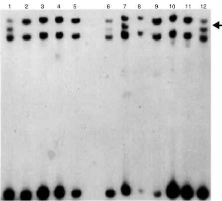
DNA extracted from the tumors was sub-jected to PCR-mediated amplification of ex-ons 5-8, which cover the coding region that encompasses most of the described muta-tions of the p53 gene. The amplified frag-ments for each exon were analyzed by SSCP. A representative example is shown in Figure 1. From this analysis, we inferred p53 muta-tions in only 4 out of the 122 tumors analyzed.
1 2 3 4 5 6 7 8 9 10 11 12
Figure 1. PCR-SSCP of p53exon 6 in cervical carcinoma. Four samples present band shifts
Se que ncing o f p53 ge ne e xo ns 5-8
Sequencing was performed on both the sense and antisense strands of the cloned exon 5, 6 and 8 fragments obtained from the HPV-positive cervical carcinomas. One specimen showed a single nucleotide change
at codon 175 (GCG®GCA), which does not
lead to amino acid substitution. On the other hand, two samples showed mutations at
codon 213 (GAC®GGC), which results in
an Asp to Gly change. Another nucleotide
alteration was found in codon 277 (ACA®A
AA), causing a Thr-Lys substitution.
D iscussio n
Almost all of human cervical carcinomas harbor HPV DNA sequences, and the viral E6 and E7 oncoproteins are generally ex-pressed within these tumors (21). Evidence that the HPV E6 oncoprotein can bind p53 protein and promote its degradation suggests one mechanism by which the HPV viruses could mediate transformation (26,27). The presence of an HPV sequence in a cell could represent the loss of the p53 function result-ing from either a deletion or a mutation. If the abrogation of p53 function is really criti-cal to cervicriti-cal carcinogenesis, then either HPV infection or p53 gene mutation could fulfill this requirement. However, discrep-ancies are observed when comparing p53 data obtained from cell lines and clinical samples. It has been reported that
HPV-negative cell lines contain p53point
muta-tions, whereas in HPV-positive cell lines, p53 is always of the wild type, suggesting that expression of E6 would mimic p53 mu-tations in the latter (6,26,28). Data from several studies employing cervical carcino-ma samples have failed to substantiate this, pointing to a small percentage of p53 muta-tions in this neoplasm, which occur irrespec-tive of the HPV status of the tumor (4,14, 29,30).
Interaction of E6 from high-risk HPVs
and mutant forms of p53 may be rare in vivo
and complex to study in vitro. Some p53
mutants may present an E6-resistant pheno-type, either by reduced affinity for E6, or by being less prone to proteolysis, and may accumulate even upon HPV 16 E6 expres-sion. This apparent contradiction may be explained by E6 and mutated p53 not being present in the same cells, which is unlikely since it has been shown that E6 expression is required for progression of HPV-related tu-mors. Another explanation could be that E6
expression in vivo is not sufficient to
elimi-nate the p53 protein or that in these tumors p53 complexes with cellular proteins hin-dering E6 access to it. These data indicate that E6-p53 interaction should not be con-sidered the single mechanism of HPV-medi-ated transformation.
We have screened for p53 gene muta-tions 122 HPV-positive cervical carcinomas by SSCP analysis and DNA sequencing of exons 5-8, which have been reported to be the most common sites of mutations in this gene (13). Codon 213 was affected in 2 of the 4 p53-mutated samples, confirming this position as a hot spot in tumors from dif-ferent anatomical sites (13). Of the 4 point mutations detected, 3 corresponded to mis-sense mutations that may have implications for protein conformation. Moreover, these amino acid substitutions map to a region important for p53 DNA-binding activity, im-plying an eventual loss of function. How-ever, functional studies are needed to con-firm this implication. In fact, it was recently reported that cervical carcinoma cell lines containing transcriptionally active HPV dis-play normal p53 transactivating function,
including cell cycle arrest at G1 upon
cervical cancers. Kim et al. (33) showed that 2 out of 136 (1.5%) tumors demonstrated SSCP band shifts. One sample (positive for HPV 18) had a nonsense mutation of codon 101 in exon 4 from AAA to TAA transver-sion. Another (HPV positive for the L1 con-sensus primer set) showed a point mutation involving codon 179 in exon 5 changing CAT to CGT transition. Three specimens negative for HPV did not contain p53 gene mutations. In another study 64 cases of pri-mary cervix cancers were analyzed with a
screening of the p53gene mutations in exons
5 through 9 of this gene. SSCP analysis showed mobility shifts in 8 cases (6 in HPV-positive cases and 2 in HPV-negatives cases) and sequence analysis confirmed the results of SSCP (34).
Nakagawa et al. (35) analyzed mutation of the p53 gene in 45 women with cervical carcinomas. p53 mutations were analyzed by PCR-based SSCP and DNA sequencing techniques. Point mutation of the p53 gene was detected in 5 of 46 (11%) cervical carci-nomas, 1 of 17 (8%) samples associated with high-risk HPVs (HPV 16 and HPV 18), and 4 of 27 samples (15%) with intermediate risk HPVs, whereas no mutation was found in 2 HPV-negative cases.
Levi et al. (36) analyzed the presence of HPV DNA in a series of 84 paraffin-embed-ded penile carcinomas. They also investigated the presence of p53 mutations in these tumors by immunohistochemistry, SSCP and DNA sequencing. These data indicate that subsets of penile carcinomas are etiologically related to HPV and that an overlapping subset may rise from mutational events in the p53 gene.
The p53 gene regions examined in the
present study represent only a fraction of this gene. However, the vast majority of known mutations identified in different primary tu-mors and cell lines clustered between amino acid residues 130 and 290 (11). This is a region where the DNA sequence is highly conserved among several different species (37). Although the frequency and distribution of these
muta-tions may differ among cancers from different tissue types, p53 mutations in cervical carci-noma are localized within this region (38). Therefore, it is relatively safe to assume that we probably would have detected some p53 mutations within exons 5-8. However, one cannot exclude that some mutations are lo-cated outside the regions of the p53 gene examined by us and others (14,32,39,40). Kurvinen et al. (40) determined the state of the p53 gene in 20 genital precancer lesions and carcinomas. Exons 5-9 of the p53 gene were analyzed by SSCP-PCR, and no muta-tions were detected in any of the specimens, including the 3 HPV-negative cases.
Coexistence of HPV DNA and a mutated form of p53 may suggest that these cells were mutated at the p53 locus and then became infected with HPV. Alternatively, the infection was an initial event and a point mutation on the p53 gene provided an addi-tional growth advantage to the cells. In fact, Crook et al. (41) indicated that mutations within the p53 gene in an HPV-positive pri-mary cancer might confer a growth advan-tage and contribute to the acquisition of metastatic potential in these cells.
The present results may account to the existence of other tumor suppressor genes whose inactivation or loss of function is important for cervical carcinogenesis. This is compatible with the fact that the frequency of p53 mutation reported in these tumors is low when compared with other cancers. Our results confirm that the occurrence of so-matic mutations in the hot spot region of the p53 gene is indeed very low in HPV-positive cervical carcinoma.
Ackno wle dgm e nts
Re fe re nce s
1. Villa LL (1997). Papillomaviruses and
cer-vical cancer. Advances in Cancer
Re-search, 71: 321-341.
2. Zur Hausen H(1996). Papillomavirus
in-fections - a major cause of human
can-cers. Biochimica et Biophysica Acta, 1288:
F55-F78.
3. Bosch FX, M anos M M , M uñoz N, Sher-man M , Jansen AM , Peto J, SchiffSher-man M H, M oreno V, Kurman R, Shah KV & International Biological Study on Cervical Cancer (IBSCC) St udy Group (1995). Prevalence of human papillomavirus in cervical cancer: a w orldw ide perspective.
Journal of the National Cancer Institute, 87: 796-802.
4. Borresen AL, Helland A, Nesland J, Holm R, Trope C & Kaern J (1992).
Papillomavi-ruses, p53, and cervical cancer.Lancet,
339: 1350-1351.
5. Dyson N, How ley PM , M ünger K & Harlow E (1989). The human papillomavi-rus-16 E7 oncoprotein is able to bind to
the retinoblastoma gene product.
Sci-ence,243: 934-937.
6. Scheffner M , M unger K, Byrne JC & How ley PM (1991). The state of the p53 and retinoblastoma genes in human
cervi-cal carcinoma cell lines. Proceedings of
the National Academy of Sciences, USA, 88: 5523-5527.
7. Wrede D, Tidy JA, Crook T, Lane D & Vousden KH (1994). Expression of RB and p53 proteins in positive and HPV-negative cervical carcinoma cell lines. M o-lecular Carcinogenesis, 4: 171-175. 8. Huibregtse JM , Scheffner M & How ley
PM (1994). AP directs the HPV E6-dependent inactivation of p53 and is rep-resentative of a family of structurally and
functionally related proteins. Cold Spring
Harbor Symposia on Quantitative Biology, LIX: 237-245.
9. Lane DP & Craw ford LV(1979). T antigen
is bound to a host protein in
SV40-trans-formed cells. Nature, 278:261-263.
10. Reich NC, Oren M & Levine AJ (1983). Tw o mechanisms regulate the levels of a
cellular tumor antigen, p53. M olecular and
Cellular Biology, 3: 2143-2150.
11. Levine AJ, M omand J & Finlay CA (1991).
The p53 tumour suppressor gene. Nature,
351: 453-456.
12. Funk WD, Pak DT, Karas RH, Wright WE & Shay JW (1992). Transcriptionally active DNA-binding site for human p53 protein
complexes. M olecular and Cellular
Biol-ogy, 12: 2866-2871.
13. Hollstein M , Sidransky D, Vogelstein B &
Harris CC (1991). p53 mutations in human
cancers. Science, 253: 49-53.
14. Busby-Earle RM C, Steel CM , Williams ARW, Cohen B & Bird CC (1992). Papillo-maviruses, p53 and cervical carcinoma.
Lancet, 339: 1350.
15. Crook T, Wrede D & Vousden KH (1991). p53 point mutation in HPV negative
hu-man cervical carcinoma cell lines.
Onco-gene, 6: 873-875.
16. Srivastava S, Tong YA, Devadas K, Zou ZQ, Chen Y, Pirollo KF & Chang EH(1992). The status of the p53 gene in human papilloma virus positive or negative
cervi-cal carcinoma cell lines. Carcinogenesis,
13: 1273-1275.
17. Park DJ, Wilczynski SP, Paquette RL, M iller CW & Koeffler HP (1994). p53 mu-tations in HPV-negative cervical carcino-ma. Oncogene, 9: 205-210.
18. Crook T & Vousden KH(1992). Properties
of p53 mutations detected in primary and secondary cervical cancers suggest mechanisms of metastasis and
involve-m ent of environinvolve-m ent al carcinogens.
EM BO Journal, 11: 3935-3940.
19. Krieg P, Antmann E & Sauer G (1983). The simultaneous extraction of high molecu-lar w eight DNA and of RNA from solid
tumours. Analytical Biochemistry, 134:
288-294.
20. Bauer HM , Yi Ting M S, Chambers JC, Tashiro CJ, Chimera J, Reingold A & M anos M M (1991). Genital human papil-lomavirus infection in female university students as determined by PCR-based
methods. Journal of the American M
edi-cal Association,265: 23-30.
21. Walboom ers JM , Jacobs M V, M anos M M , Bosch FX, Kummer JA, Shah KV, Snijders PJ, Peto J, M eijer CJ & M unoz N (1999). Human papillomavirus is a neces-sary cause of invasive cervical cancer w orldw ide. Journal of Pathology, 189: 12-19.
22. Hayashi K, Orita M , Suzuki Y & Sekiya T (1989). Use of labeled primers in poly-merase chain reaction (LP-PCR) for a rapid
detection of the product. Nucleic Acids
Research, 17: 3605.
23. Kishimoto Y, M urakami Y, Shiraishi M ,
Hayashi K & SekiyaT (1992). Aberrations
of the p53 tumor suppressor gene in
hu-man non-small cell carcinomas of the
lung. Cancer Research, 52: 4799-4804.
24. M urakami Y, Hayashi K & Sekiya T (1991). Detection of aberrations of the p53 alleles
and the gene transcript in human tumor cell lines by single-strand conformation
polymorphism analysis. Cancer Research,
51: 3356-3361.
25. Orita M , Iw ahana H, Kanazaw a H, Hayashi K & Sekiya T (1989). Detection of poly-morphisms of human DNA by gel electro-phoresis as single-strand conformation
polymorphisms. Proceedings of the
Na-tional Academy of Sciences, USA, 86: 2766-2770.
26. Scheffner M , Werness BA, Huibregtse
JM , Levine AJ & How ley PM(1990). The
E6 oncoprotein encoded by human papil-lomavirus types 16 and 18 promotes the
degradation of p53. Cell, 63: 1129-1136.
27. Werness BA, Levine AJ & How ley PM (1990). Association of human
papillomavi-rus types 16 and 18. Science, 248: 76-79.
28. Liang XH, Volkman M , Klein R, Herman B & Lockett SJ(1993). Co-localization of the tumor-suppressor protein p53 and human papillomavirus E6 protein in human
cervi-cal carcinoma cell lines. Oncogene, 8:
2645-2652.
29. Busby-Earle RM , Steel CM , Williams AR,
Cohen B & Bird CC(1994). p53 mutations
in cervical carcinogenesis - low frequency and lack of correlation w ith human
papil-lomavirus status. British Journal of
Can-cer, 69: 732-737.
30. Kong G, Choo B & Chong KY (1993). Ab-sence of mutation in the p53 and the retinoblastoma susceptibility genes in
pri-mary cervical carcinomas. Virology, 193:
1042-1046.
31. Helland A, Holm R, Kristensen G, Kaern J, Karesen F, Trope C, Nesland JM &
Bfrresen A-L (1993). Genetic alterations
of the TP53 gene, p53 protein expression and HPV infection in primary cervical
car-cinomas. Journal of Pathology, 171:
105-114.
32. Choo KB, Chong KY, Liew L-N & Hsu Cheng W (1993). Unregulated and basal transcriptional activities of the regulatory sequence of the type 18 human
papillo-mavirus genome in transgenic mice.
Jour-nal of Virology, 188: 378-383.
33. Kim JW, Cho YH, Lee CG, Kim JH, Kim HK, Kin EJ, Ham KT & Namkoong SE (1997). Human papillomavirus infection and TP53 gene mutation in primary
cervi-cal carcinoma. Acta Oncologica, 36:
295-300.
34. Kim KH & Kim YS (1995). Role of human
papillomavirus and p53 tumor suppressor
M edical Journal, 36: 412-425.
35. Nakagaw a S, Yoshikaw a H, Jimbo H, Onda T, Yasugi T, M atsumoto K, Kino N, Kaw ana K, Kozuka T, Nakagaw a K, Aoki M
& Taketani Y (1999). Elderly Japanese
w omen w ith cervical carcinoma show higher proportions of both intermediate-risk human papillomavirus types and p53
mutations.British Journal of Cancer, 79:
1139-1144.
36. Levi JE, Rahal P, Sarkis AS & Villa LL (1998). Human papillomavirus DNA and
p53 status in penile carcinomas.
Interna-tional Journal of Cancer, 76: 779-783.
37. Soussi T, Caron de Fromentel C & M ay P (1990). Structural aspects of the p53
pro-tein in relation to gene evolution.
Onco-gene, 5: 945-952.
38. Pao CC, Kao SM , Tang GC, Lee KSIJ & Ruan S (1993). Human papillomavirus and cervical carcinoma in China and Taiw an.
Lancet, 342: 937.
39. Chen TM , Chen CA, Hsieh Cy, Chang Dy, Chen Yh & Defendi V (1992). The state of p53 in primary human cervical carcinomas and its effects in human
papillomavirus-immortalized human cervical cells.
Onco-gene, 8: 1511-1518.
40. Kurvinen K, Tervahauta A, Syrjanen S, Chang F & Syrjanen K (1994). The state of
the p53 gene in human papillomavirus
(HPV)-positive and HPV-negative genital precancer lesions and carcinomas as de-termined by single-strand conformation polymorphism analysis and sequencing.
Anticancer Research, 14: 177-181. 41. Crook T, Wrede D, Tidy JA, M ason WP,
Evans DJ & Vousden KH (1992). Clonal p53 mutation in primary cervical cancer: association w ith