Performance of TcI/TcVI/TcII Chagas-Flow
ATE-IgG2a for universal and genotype-specific
serodiagnosis of
Trypanosoma cruzi
infection
Glaucia Diniz Alessio1, Fernanda Fortes de Arau´jo2, Denise Fonseca Coˆrtes1, Policarpo Ademar Sales Ju´nior3, Daniela Cristina Lima2, Matheus de Souza Gomes4, Laurence
Rodrigues do Amaral4, Marcelo Antoˆnio Pascoal Xavier5, Andre´a Teixeira-Carvalho2, Olindo Assis Martins-Filho2*, Marta de Lana1
1Laborato´rio de Doenc¸a de Chagas, Nu´cleo de Pesquisas em Ciências Biolo´gicas (NUPEB), Instituto de Ciências Exatas e Biolo´gicas (ICEB), Universidade Federal de Ouro Preto (UFOP), Ouro Preto, Minas Gerais, Brazil,2Grupo Integrado de Pesquisas em Biomarcadores, Centro de Pesquisas Rene´ Rachou (CPqRR-FIOCRUZ/ MG), Belo Horizonte, Minas Gerais, Brazil,3Grupo de Genoˆmica Funcional e Proteoˆmica deLeishmania sppeTrypanosoma cruzi, Centro de Pesquisas Rene´ Rachou (CPqRR-FIOCRUZ/ MG), Belo Horizonte, Minas Gerais, Brazil,4Laborato´rio de Bioinforma´tica e Ana´lises Moleculares, Universidade Federal de Uberlaˆndia, INGEB/FACOM, Campus Patos de Minas, Patos de Minas, Minas Gerais, Brazil,5Grupo de Pesquisas Clı´nicas e Polı´ticas Pu´blicas em Doenc¸as Infecciosas e Parasita´rias, Centro de Pesquisas Rene´ Rachou (CPqRR-FIOCRUZ/ MG), Belo Horizonte, Minas Gerais, Brazil
*oamfilho@gmail.com
Abstract
DistinctTrypanosoma cruzigenotypes have been considered relevant for patient manage-ment and therapeutic response of Chagas disease. However, typing strategies for geno-type-specific serodiagnosis of Chagas disease are still unavailable and requires
standardization for practical application. In this study, an innovative TcI/TcVI/TcII Chagas Flow ATE-IgG2a technique was developed with applicability for universal and genotype-specific diagnosis ofT.cruziinfection. For this purpose, the reactivity of serum samples (percentage of positive fluorescent parasites-PPFP) obtained from mice chronically infected with TcI/Colombiana, TcVI/CL or TcII/Y strain as well as non-infected controls were deter-mined using amastigote-AMA, trypomastigote-TRYPO and epimastigote-EPI in parallel batches of TcI, TcVI and TcII target antigens. Data demonstrated that “α-TcII-TRYPO/ 1:500, cut-off/PPFP = 20%” presented an excellent performance for universal diagnosis of
T.cruziinfection (AUC = 1.0, Se and Sp = 100%). The combined set of attributes “α -TcI-TRYPO/1:4,000, cut-off/PPFP = 50%”, “α-TcII-AMA/1:1,000, cut-off/PPFP = 40%” and “α -TcVI-EPI/1:1,000, cut-off/PPFP = 45%” showed good performance to segregate infections with TcI/Colombiana, TcVI/CL or TcII/Y strain. Overall, hosts infected with TcI/Colombiana and TcII/Y strains displayed opposite patterns of reactivity with “α-TcI TRYPO” and “α-TcII AMA”. Hosts infected with TcVI/CL strain showed a typical interweaved distribution pattern. The method presented a good performance for genotype-specific diagnosis, with global accuracy of 69% when the population/prototype scenario include TcI, TcVI and TcII infec-tions and 94% when comprise only TcI and TcII infecinfec-tions. This study also proposes a receiver operating reactivity panel, providing a feasible tool to classify serum samples from a1111111111 a1111111111 a1111111111 a1111111111 a1111111111 OPEN ACCESS
Citation:Alessio GD, de Arau´jo FF, Coˆrtes DF, Sales Ju´nior PA, Lima DC, Gomes MdS, et al. (2017) Performance of TcI/TcVI/TcII Chagas-Flow ATE-IgG2a for universal and genotype-specific serodiagnosis ofTrypanosoma cruziinfection. PLoS Negl Trop Dis 11(3): e0005444.https://doi. org/10.1371/journal.pntd.0005444
Editor:Carlos A. Buscaglia, Instituto de Investigaciones Biotecnolo´gicas, ARGENTINA
Received:December 6, 2016
Accepted:March 1, 2017
Published:March 23, 2017
Copyright:©2017 Alessio et al. This is an open access article distributed under the terms of the
Creative Commons Attribution License, which permits unrestricted use, distribution, and reproduction in any medium, provided the original author and source are credited.
Data Availability Statement:All relevant data are within the paper and its Supporting Information files.
Funding:This work was supported by European Community’s Seventh Framework Program No. 602773- Project KINDRED). Fundac¸ão de Amparo
hosts infected with distinctT.cruzigenotypes, supporting the potential of this method for universal and genotype-specific diagnosis ofT.cruziinfection.
Author summary
Chagas disease remains a significant public health issue infecting 6–7 million people worldwide. The factors influencing the clinical heterogeneity of Chagas disease have not been elucidated, although it has been suggested that different clinical outcome may be associated with the genetic diversity ofT.cruziisolates. Moreover, differences in therapeu-tic response of distinctT.cruzigenotypes have been also reported. Typing strategies for genotype-specific diagnosis of Chagas disease to identify theT.cruzidiscrete typing units (DTU) have already been developed, including biochemical and molecular methods, how-ever the techniques have limitations. The majority of these methods can not directly be performed in biological and clinical samples. In addition, it has been proposed that para-site isolates from blood may not necessarily represent the full set of strains current in the individual as some strains can be confined to tissues. The improvement of genotype-spe-cific serology to identify theT.cruziDTU(s) present in a given host may provide a useful tool for clinical studies. In the present investigation, we developed an innovative TcI/ TcVI/TcII Chagas Flow ATE-IgG2a technique with applicability for universal and geno-type-specific diagnosis ofT.cruziinfection that may contribute to add future insights for genotype-specific diagnosis of Chagas disease.
Introduction
Trypanosoma cruzi, the etiological agent of Chagas disease [1] infects 6–7 million people worldwide, mainly in Latin America causing serious consequences for public health and national economies [2]. Geographical variations in the prevalence of clinical forms and mor-bidity of Chagas disease in different countries have been recorded [3]. Although the factors underlying the clinical heterogeneity of Chagas disease are still not completely understood, it has been suggested that different clinical outcome may be associated with the genetic diversity ofT.cruziisolates observed in the Americas [4]. Moreover, differences in therapeutic response of distinctT.cruzigenotypes have been also reported previously in mice infection [5–8].
Typing strategies for genotype-specific diagnosis of Chagas disease to identify the sixT.
cruzidiscrete typing units (DTU), named TcI, TcII, TcIII, TcIV, TcV and TcVI [9] have already been developed, including biochemical and molecular methods [4]. However, none of these methods allows a full resolution when used individually and a combinatory three-marker sequential typing strategy is usually required to confirm theT.cruzigenotype [10–12]. Straightforward, genotyping methods to identify theT.cruziDTUs are currently available, but research is still required to optimize sensitivity and simplify methods so that they can be easily applied in clinical laboratories. In fact, molecular methods require a measurable parasite load to directly identifyT.cruziDTUs in samples. Because of this, the approaches used forT.cruzi
genotyping requires parasite isolation by hemoculture/xenoculture followed by in vitro growth that may lead to clonal selection [13–16].
A feasible solution to overcome these problems is the design and development of genotype-specific serology to provide a current/historical profile ofT.cruziDTUs infecting an individual Fundac¸ão Oswaldo Cruz (FIOCRUZ) and
Universidade Federal de Ouro Preto. DCL received fellowship from CNPq/PIBITI program. DFC received a post-doc fellowship from FAPEMIG PDJ program. GDA received PhD research fellowship form CAPES. OAMF, ATC and MdL received financial support from CNPq PQ Fellowship program. FFdA received financial support from CAPES BJT Fellowship program. The authors thank the program for technological development in tools for health- PDTIS-FIOCRUZ for the use of its facilities. The funders had no role in study design, data collection and analysis, decision to publish, or preparation of the manuscript.
patient [17–20]. Moreover, genotypic-specific serodiagnosis has the potential to predict thera-peutic response and provide insights upon re-activation episodes.
Recently, a flow cytometry-based assay, named Chagas-Flow ATE (Amastigote, Trypomas-tigote and EpimasTrypomas-tigote), has been developed for simultaneous measurement of anti-amasti-gote, anti-trypomastigote and anti-epimastigote antibodies displaying high performance for the diagnosis and post-therapeutic monitoring of Chagas disease [21]. Aiming at optimizing the Chagas-Flow ATE for universal and genotypic-specific diagnosis ofT.cruziinfection, the present study proposed the development of modified Chagas-Flow ATE, using parallel batches of distinctT.cruzigenotypes as target antigens. StandardT.cruzistrains, representative of three major genotypes (TcI, TcII and TcVI) were used to setup the Chagas-Flow ATE-IgG2a methodology.
High-dimensional data handling were applied to select the sets attributes (“target-antigen/ serum dilution/cut-off”) applicable for universal and genotypic-specific diagnosis ofT.cruzi -infection. A receiver operating reactivity panel was proposed as a feasible tool to identify hosts infected with distinctT.cruzigenotypes. The results demonstrated the high-quality perfor-mance of TcI/TcVI/TcII Chagas-Flow ATE-IgG2a for universal and genotype-specific diagno-sis ofT.cruziinfection.
Methods
Ethics statement
All animals included in this study were maintained at the Animal Science Center of the Uni-versidade Federal de Ouro Preto, Ouro Preto, MG, Brazil, in strict accordance with the Brazil-ian College of Animal Experimentation Guidelines for ethical conduct in use of animals in research. Efforts were performed to reduce animal suffering. The study protocols were approved by the Ethics Committee on Animal Experimentation of the Federal University of Ouro Preto (Protocol approval numbers #2013/48 from December, 6th, 2013 for the experi-mental infection and collected blood by ocular plexus puncture in mice).
Trypanosoma cruzi
strains
StandardT.cruzistrains, representative of three major genotypes [9], involved in the domes-tic cycle of Chagas disease in Brazil, were used to setup the TcI/TcVI/TcII Chagas-Flow ATE-IgG2a methodology for the serodiagnosis ofT.cruziinfection. The Colombiana, acro-nyms “COL” (TcI) [22], CL (TcVI) [23] and Y (TcII)T.cruzistrains were used in this study [24]. All isolates were obtained from theT.cruzicryobank at Grupo de Genoˆmica Funcional e Proteoˆmica deLeishmania sppeTrypanosoma cruzi, Centro de Pesquisas Rene´ Rachou (CPqRR-FIOCRUZ/ MG). TheT.cruzistrains were maintained by consecutivein vivo
passages in Swiss female mice. Blood samples obtained from infected mice were used for experimental infection as well as for preparation of target antigens (amastigote-AMA, trypomastigote-TRYPO and epimastigote-EPI) used on each TcI/TcVI/TcII Chagas-Flow ATE-IgG2a platform.
Experimental infection with TcI, TcVI and TcII
T
.
cruzi
genotypes
The infection was confirmed in allT.cruzi-infected mice, by positivity at fresh blood exami-nation performed at day 7, 10 or 15 post-infection. The serum samples used for the TcI/TcVI/ TcII Chagas-Flow ATE-IgG2a serology were prepared from whole blood samples collected by ocular plexus puncture. Samples were collected from non-infected controls andT.cruzi -infected mice (day 90 and day180 post-infection) were inactivated at 56˚C for 30 min and stored at -20˚C until use. Considering the animal mortality during the experimental timeline, the final number of animals/group were (TcI/Colombiana strain, n = 29, TcVI/CL strain, n = 29, TcII/Y strain, n = 35 and NI, n = 10).
Preparation of AMA, TRYPO, EPI (ATE) target antigens from TcI, TcVI
and TcII
T
.
cruzi
genotypes
The amastigote/trypomastigote/epimastigote forms of TcI, TcVI and TcIIT.cruzigenotypes were obtained as described previously by Alessioet al. (2014) [21]. Enriched trypomastigotes (TRYPO) and amastigote (AMA) preparations were obtained from desynchronizedin vitro tis-sue cultures (L929 cell-line) harvested at day 4–6 and 8–15 post-inoculation, respectively. Epi-mastigote forms were obtained at log-phase growth of axenic culture in liver infusion tryptose medium [25]. Live amastigotes and trypomastigotes as well as fixed epimastigotes forms were stained with fluorescein isothiocyanate (FITC) as described by Alessioet al. (2014) [21]. Briefly, AMA/TRYPO mix and EPI suspensions (1x10⁷parasites/mL) were stained with FITC (100μg/mL for TcI/Colombiana strain and 200μg/mL for TcVI/CL strain and TcII/Y strain) for 30 min at 37˚C. After staining, AMA/TRYPO mix were kept at 37˚C for 60min and EPI preparation stored at 4˚C for 24h prior to use. The three FITC-labeled parasite preparations were mixed accordingly to obtain an equivalent proportion of AMA (33%), TRYPO (33%) and EPI (33%) in the final ATE-parasite Mix Platforms, monitored by flow cytometry checking performed prior use. The FITC-labeling approach led to a differential staining phenomenon previously described by Alessioet al., (2014) [21] that allowed the segregation of AMA, TRYPO and EPI organisms in distinct clusters, based on the FITC (Fluorescence 1- FL1)vs
Forward Scatter (FSC) dot plot distribution (Fig 1).
TcI/TcVI/TcII Chagas-Flow ATE-IgG2a serodiagnosis of
T
.
cruzi
infection
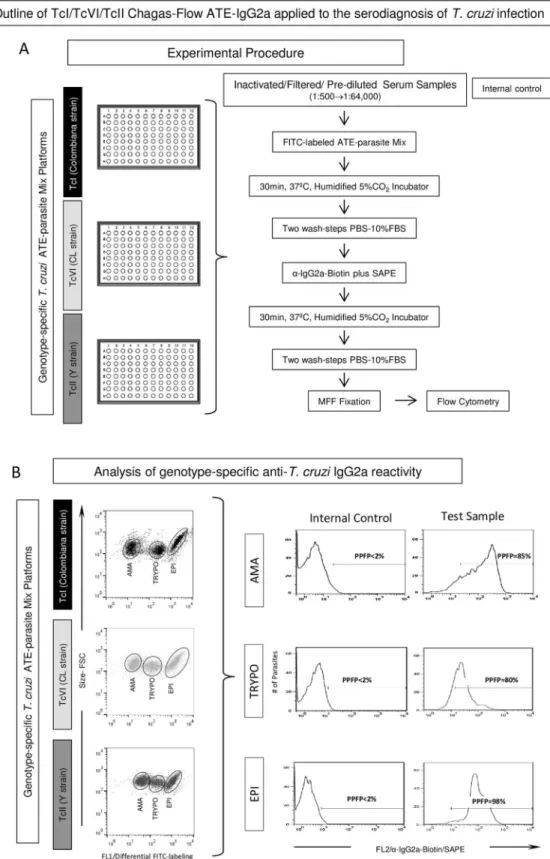
An outline of the TcI/TcVI/TcII Chagas-Flow ATE-IgG2a applied to the serodiagnosis ofT.
cruziinfection is provided in theFig 1. The method comprises two steps referred as: i) Experi-mental Procedure (Fig 1A) and ii) Analysis of genotype-specific anti-T.cruziIgG2a reactivity (Fig 1B).
Experimental procedure. The TcI/TcVI/TcII Chagas-Flow ATE-IgG2a serodiagnosis was performed as described previously by Alessioet al. (2014) [21] modified as follows: frozen serum samples were thawed from -20˚C storage, filtered through 0.22μm syringe filter and submitted to serial dilution (1:500 to 1:64,000) with phosphate-buffered-saline supplemented with 10% fetal bovine serum (PBS-10%FBS) in a U-bottom 96-well plate. A final volume of 50μL of pre-diluted serum samples were incubated with 50μL of each ATE-parasite Mix prepa-ration (TcI, TcVI and TcII genotype-specific platforms, in parallel batches) for 30 min at 37˚C in a 5% CO2humidified incubator. Following incubation, parasites were washed twice with
PBS-10%FBS and the supernatant discarded. The pellet of parasite mix was re-suspended and incubated with 50μL biotin-conjugated anti-mouse IgG2a that is equivalent of human IgG1 (1:3,000 in PBS-10%FBS) plus 20μL of secondary reagents (phycoerytrin-conjugated streptavi-din-SAPE, 1:800 in PBS-10%FBS) for 30 min at 37˚C in a 5% CO2humidified incubator.
Fig 1. Outline of TcI/TcVI/TcII Chagas-Flow ATE-IgG2a for serodiagnosis ofTrypanosoma cruzi
(10g/L of paraformaldehyde, 10.2g/L of sodium cacodylate and 6.65g/L of sodium chloride, pH 7.2), and store at 4˚C until flow cytometric data acquisition in a FACSCan flow cytometer (Beckton Dickinson, USA). An internal control (“second step reagents control” = anti-mouse IgG2a-biotin+SAPE) to monitor unspecific bindings was included in all experimental batches, in which the ATE-parasite Mix preparations were incubated in the absence of mouse serum but in the presence of secondary reagents. A total of 10,000 events were acquired for each tested serum dilution. Acquisition was performed with appropriate instrument settings on log scale (FSC = E00, Side Scatter -SSC = 427, threshold = 400; FL1 = 620 and FL2 = 500). Data were stored for off-line analysis.
Analysis of genotype-specific anti-T.cruziIgG2a reactivity. The FlowJo software
ver-sion 10.1 (TreeStar, San Diego, CA, USA) was used for off-line data analysis. The genotype-specific reactivity of anti-T.cruziIgG2a was performed for each ATE-parasite mix platform— TcI (Colombiana strain), TcVI (CL strain) and TcII (Y strain). Appropriate gating strategies were used to select the target antigens (amastigote-AMA, trypomastigote-TRYPO and epimas-tigote-EPI) on each Chagas-Flow ATE-IgG2a platform, based on the differential FITC-labeling features of AMA, TRYPO and EPI. Following the selection of target-antigens, one-dimen-sional histograms were employed to quantify the genotype-specific anti-T.cruziIgG2a reactiv-ity, based on the positivity limit (PPFP<2%), set based on the internal control. The results
were expressed as percentage of positive fluorescent parasites (PPFP) for each tested sample dilution (Fig 1B).
Data mining and analysis
Data mining for universal and genotype-specific diagnosis ofT.cruziinfection was first per-formed by non-parametric Kruskal—Wallis test followed by Dunns’ multiple comparison post-test to compare the overall reactivity profile of TcI/TcVI/TcII Chagas-Flow ATE-IgG2a. Significant differences were considered at p0.05. The performance indices (global accuracy defined by the area under the curve-AUC, sensitivity-Se and specificity-Sp) for the pair of attributes (“target antigen/serum dilution”) selected for universal diagnosis purposes were determined by the receiver operating characteristic (ROC) curve, scatter plot distribu-tion and Two-Graph ROC curve (TG-ROC) analysis. Histogram plot distribudistribu-tions and non-linear regression analysis was used for comparative analysis of pair of attributes (“target antigen/serum dilution”) selected for genotypic-specific diagnosis purposes. The global median was calculated for each pair of attributes (“target antigen/serum dilution”) to define putative cut-off edges to segregate the reactivity amongstT.cruzi-infected hosts. Scatter plot distribution was used for performance analysis of sets of selected attributes (“target-antigen/ serum dilution/cut-off”) applicable for genotypic-specific diagnosis ofT.cruzi-infection. The GraphPad Prism software, Version 5.0 (San Diego, CA, USA) was used for statistical analysis and graphic arts.
Decision trees were built for the set of selected attributes (“target-antigen/serum dilution/ cut-off”) to create algorithms (root and branch attributes) to classifyT.cruziin distinct popu-lation/prototype scenarios (TcI-infection/ColombianavsTcVI/CLvsTcII/Y) and (TcI-infec-tion/ColombianavsTcII/Y). The WEKA software (Waikato Environment for Knowledge Analysis, version 3.6.11, University of Waikato, New Zealand) was used for decision tree construction.
ATE-IgG2a platform and the histograms employed to quantify the genotype-specific anti-T.cruziIgG2a reactivity, expressed by the percentage of positive fluorescent parasites (PPFP), based on the positivity limit (PPFP<2%), set based on the internal control.
Step-wise discriminant analysis was applied to determine the global accuracy and the leave-one-out-cross-validation-LOOCV values. The R-project for statistical computing software, Version 3.0.1 was used for discriminant analysis. The algorithm C4.5 was used to build the decision tree using an implementation named J48. This method analyzed all characteristics to select a minimum set of markers that could efficiently separate study groups.
Results
Overall reactivity profile of TcI/TcVI/TcII Chagas-Flow ATE-IgG2a for
universal and genotypic-specific diagnosis of
T
.
cruzi
infection
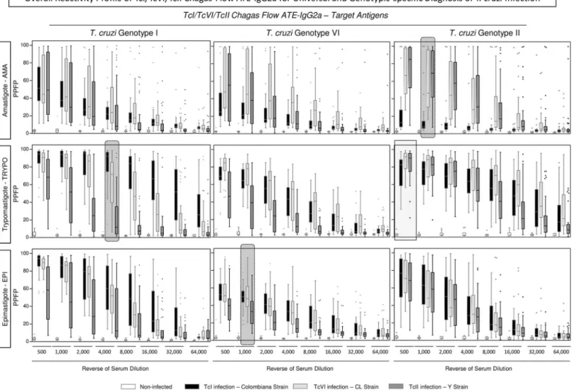
The overall profiles of TcI/TcVI/TcII Chagas-Flow ATE-IgG2a reactivity observed forT.cruzi
infected mice (TcI/Colombiana strain, TcVI/CL strain and TcII/Y strain) and non-infected controls are presented in theFig 2. The reactivity of individual samples were assessed for dis-tinct target-antigen (amastigote-AMA, trypomastigote-TRYPO and epimastigote-EPI) from
T.cruzigenotype I—Colombiana strain (Fig 2—left panels), genotype VI—CL strain (Fig 2— middle panels) and genotype II—Y strain (Fig 2—right panels) along the titration curves (serum dilutions ranging from 1:500 to 1:64,000).
Comparative analysis allowed the selection of pair of attributes (“target antigen/serum dilu-tion”) with the most promising perspective to be used for universal and genotypic-specific diagnosis ofT.cruziinfection.
The pair of attributes “anti-TcII TRYPO reactivity at 1:500” presented the highest signifi-cant difference between non-infected mice and allT.cruzi-infected hosts (TcI/Colombiana, TcVI/CL and TcII/Y strains), and therefore was further evaluated for universal diagnosis pur-pose (Fig 2—light gray continuous rectangle).
The pairs of attributes with putative applicability to genotype-specific diagnosis ofT.cruzi
infection comprise: (“anti-TcII AMA reactivity at 1:1,000”; “anti-TcI TRYPO reactivity at 1:4,000” and “anti-TcVI EPI reactivity at 1:1,000”). The pair of attributes “anti-TcII AMA reactivity at 1:1,000” presented the highest ability to distinguish the lower reactivity of hosts infected with TcI/Colombiana strain from the higher reactivity observed for hosts infected with TcVI/CL or TcII/Y strains (Fig 2—right dark gray dotted frame). The pair of attributes “anti-TcI TRYPO reactivity at 1:4,000” presented the highest ability to discriminate lower reac-tivity of hosts infected with TcII/Y strain from the intermediate reacreac-tivity observed for hosts infected with TcVI/CL strain and the higher reactivity observed for hosts infected with TcI/ Colombiana strain (Fig 2—left dark gray dotted frame). The pair of attributes “anti-TcVI EPI reactivity at 1:1,000” presented the most relevant potential to distinguish the lower reactivity of hosts infected with TcII/Y strain from the higher reactivity observed for hosts infected with TcI/Colombiana or TcVI/CLT.cruzistrains (Fig 2—middle dark gray dotted frame). Together, these pairs of attributes were selected for further performance assessment applicable to the genotype-specific diagnosis ofT.cruziinfection.
Performance of TcI/TcVI/TcII Chagas-Flow ATE-IgG2a for universal
diagnosis of
T
.
cruzi
infection
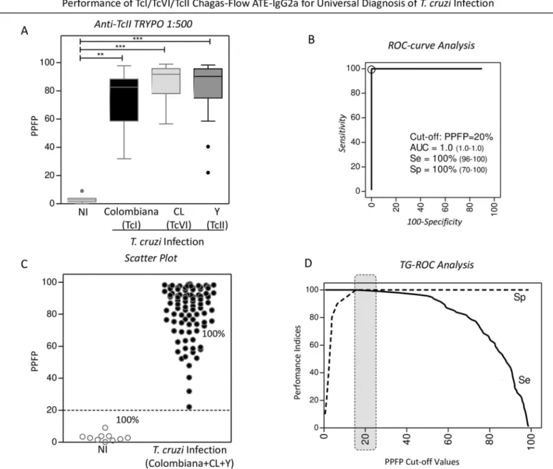
The performance of the pre-selected pair of attributes “anti-TcII TRYPO reactivity at 1:500” applied to the universal diagnosis ofT.cruziinfection is present in theFig 3.
ROC curve analysis indicated the PPFP value of 20% as the cut-off edge with excellent per-formance indices (area under the curve-AUC = 1.0 along with Sensitivity-Se and Specificity-Sp of 100%) (Fig 3B). Scatter plot distribution of individual values illustrates the ability of this set of attributes to completely segregate the serum samples of the NI andT.cruzi-infected hosts (Fig 3C). Additional analysis by TG-ROC confirmed the selected PPFP value of 20% as the best cut-off for universal diagnosis ofT.cruziinfection using the selected set of attributes (Fig 3D).
Fig 2. Overall reactivity profile of TcI/TcVI/TcII Chagas-Flow ATE-IgG2a for universal and genotypic-specific diagnosis ofT.cruziinfection.The Chagas-Flow ATE-IgG2a reactivity was determined for sera samples fromT.cruzi-infected mice, including TcI/TcVI/TcII genotype-representative strains, including: TcI/Colombiana strain (black dotted frame, n = 29), TcVI/CL strain (light gray dotted frame, n = 29) and TcII/Y strain (dark gray dotted frame, n = 35) as well as non-infected mice (white dotted frame, n = 10). Genotype-specific IgG2a reactivity to each target-antigen (amastigote-AMA, trypomastigote-TRYPO and epimastigote-EPI) fromT.cruzigenotype I (left panels), genotype VI (middle panels) and genotype II (right panels) was assessed at eight serum dilutions (1:500 to 1:64,000). The results are expressed as the percentage of positive fluorescent parasites (PPFP), using the box plot format, stretching from min to max values with outliers represented by gray-shaded dots and the box defining the 25thand 75thpercentile and the median value (line across the box).
Comparative analyses were performed by the Kruskal-Wallis followed by Dunn’s post test for multi-group comparisons. Significant differences were considered at p<0.05. The light gray continuous rectangle selects the pair of attributes (“target antigen/serum dilution”) with the most consistent ability to discriminate non-infected mice from allT.cruzi-infected hosts (Colombiana, CL and Y strains). Therefore, these features (anti-TcII TRYPO reactivity at 1:500) were selected for universal diagnosis ofT.cruziinfection. The dark gray dotted frame select the pair of attributes “target antigen/serum dilution” with the most promising perspective to distinguish the reactivity of sera samples amongst host infected with Colombiana, CL or YT.cruzistrains. Therefore, these features (anti-TcII AMA reactivity at 1:1,000; anti-TcI TRYPO reactivity at 1:4,000 and anti-TcVI EPI reactivity at 1:1,000) were selected for genotype-specific diagnosis ofT.cruziinfection.
Fig 3. Performance of TcI/TcVI/TcII Chagas-Flow ATE-IgG2a for universal diagnosis ofT.cruziinfection.(A) The anti-TcII TRYPO reactivity at 1:500, pre-selected as attributes pairs for universalT.cruziinfection diagnosis, were compared by Kruskal-Wallis followed by Dunn’s post test for multi-group comparisons and significant differences at*p<0.05,**p<0,001 and***p<0,0001, highlighted by connecting lines. Data are expressed as median PPFP values for non-infected mice (white bar) andT.cruzi-infected hosts (TcI/Colombiana strain = black bar, TcVI/CL strain = light gray bar and TcII/Y strain = dark gray bar). The similarity amongst the anti-TcII TRYPO IgG2a reactivity at 1:500 observed for the threeT.cruziinfected groups (Colombiana + CL + Y strains) allows the establishment of a single group referred to asT.cruziinfected hosts (n = 93) and the performance of the TcI/TcVI/TcII Chagas-Flow ATE-IgG2a in the universal diagnosis ofT.cruziinfection carried out as compared to a group of non-infected mice (NI, n = 10). (B) ROC-curve analysis was applied to define the appropriated cut-off to discriminate the PPFP values from NI andT.cruzi-infected host (Colombiana + CL + Y strains). Additional performance indices were also calculated and provided in the figure, including the area under the curve (AUC), defined as global accuracy, the sensitivity (Se) and the specificity (Sp). (C) Representative scatter plot illustrates the ability of the selected set of attributes (“target-antigen/serum dilution/cut-off”) to discriminate the reactivity of the sera from non-infected (NI) andT.cruzi-infected hosts (Colombiana+CL+Y). The dotted line represented the cut-off of PPFP = 20% defined by the ROC-curve analysis. (D) TG-ROC analysis was also performed to confirm the cut-off selection at higher “Se” and “Sp”, highlighted by dark gray dotted frame.
Overall reactivity of TcI/TcVI/TcII Chagas-Flow ATE-IgG2a applied for
genotype-specific diagnosis of
T
.
cruzi
infection
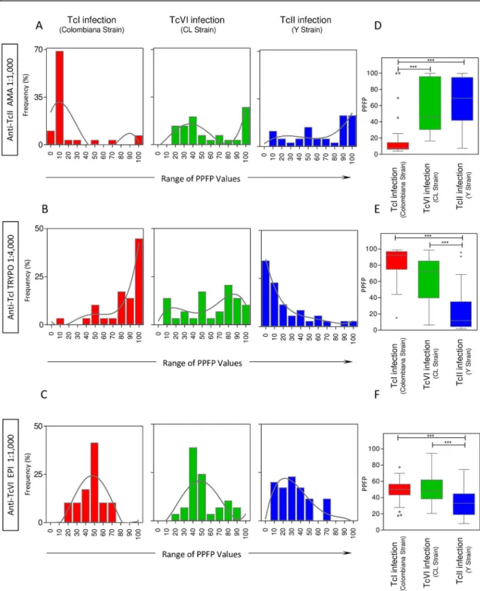
The overall reactivity profile of the pre-selected pairs of attributes (“anti-TcII AMA reactivity at 1:1,000”; “anti-TcI TRYPO reactivity at 1:4,000” and “anti-TcVI EPI reactivity at 1:1,000”) are shown in theFig 4.
Data mining was carried out by histogram graph and trendlines drawn by non-linear regression analysis. The results showed that the “anti-TcII AMA reactivity at 1:1,000” of serum samples from hosts infected with TcI/Colombiana strain displayed a nearly unimodal distribu-tion in the region of PPFP values = 10%, contrasting with the bimodal distribudistribu-tion of serum samples from hosts infected with TcVI/CL and TcII/Y strains that shows a shift towards higher PPFP values (Fig 4A).
The analysis of “anti-TcI TRYPO reactivity at 1:4,000” revealed a clear polarization of serum samples from hosts infected with TcI/Colombiana strain with a unimodal distribution in the region of PPFP values around 100%, contrasting with a unimodal distribution in the region of PPFP values around 0% observed for serum samples from hosts infected with TcII/Y strain. Again, a typical bimodal distribution was noticed for serum samples from hosts infected with TcVI/CL strain (Fig 4B).
The histogram distribution of “anti-TcVI EPI reactivity at 1:1,000” revealed a clear Gauss-ian unimodal distribution of serum samples from hosts infected with TcI/ColombGauss-iana and TcVI/CL strains within the region of PPFP values around 50%. On the other hand, the unimo-dal distribution observed for serum samples from hosts infected with TcII/Y strain showed a clear shift towards PPFP values<50% (Fig 4C).
Comparative analysis of median reactivity pattern of the selected pairs of attributes con-firmed the trend observed by histogram and non-linear regression analysis, pointing out the ability of “anti-TcII AMA reactivity at 1:1,000” to segregate hosts infected with TcII/Y strain (and TcVI/CL strain) apart from those infected with TcI/Colombiana strain (Fig 4D). On the other hand, the “anti-TcI TRYPO reactivity at 1:4,000” was able to segregate hosts infected with TcI/Colombiana strain (and TcVI/CL strain) apart from those infected with TcII/Y strain (Fig 4E). Moreover, the “anti-TcVI EPI reactivity at 1:1,000” was capable to discriminate the hosts infected with TcVI/CL strain (and TcI/Colombiana strain) apart from those infected with TcII/Y strain (Fig 4F).
Establishment of cut-off edges and performance of TcI/TcVI/TcII
Chagas-Flow ATE-IgG2a for genotype-specific diagnosis of
T
.
cruzi
infection
Aiming at making the TcI/TcVI/TcII Chagas-Flow ATE-IgG2a applicable to the genotypic-specific diagnosis ofT.cruziinfection, the overlaid trendlines for the overall reactivity (S1A Fig) along with the global median PPFP value of each pair of pre-selected attributes (S1B Fig) were employed to establish potential cut-off edges to categorize individual samples as they present negative (<cut-off) or positive (>cut-off) reactivity.
Using this approach, specific cut-off edges were defined for each pre-selected pairs of attri-butes (“anti-TcII AMA reactivity at 1:1,000”; “anti-TcI TRYPO reactivity at 1:4,000” and “anti-TcVI EPI reactivity at 1:1,000”), comprising PPFP = 40%, PPFP = 50% and PPFP = 45%, respectively (S1B Fig).
55% of TcVI/CL strain) apart from 14% of those infected with TcI/Colombiana strain (S1C Fig). Moreover, the set of attributes “anti-TcI TRYPO reactivity at 1:4,000, cut-off = 50%” showed positive results in 83% of hosts infected with TcI/Colombiana strain (and 59% of TcVI/CL strain) contrasting with 14% of those infected with TcII/Y strain (S1C Fig). Further-more, the set of attributes “anti-TcVI EPI reactivity at 1:1,000, cut-off = 45%” showed positive results in 72% of hosts infected with TcI/Colombiana strain (and 55% of TcVI/CL strain) dis-tinct from 27% of those infected with TcII/Y strain (S1C Fig).
Scatter plot distribution further illustrated the pre-selected sets of attributes segregated the reactivity of hosts infected with distinctT.cruzigenotypes, emphasizing the performance of “anti-TcII AMA reactivity at 1:1,000, cut-off = 40%” to discriminate the majority of the hosts infected with TcI/Colombiana strain and the ability of “anti-TcI TRYPO reactivity at 1:4,000, cut-off = 50%” and “anti-TcVI EPI reactivity at 1:1,000, cut-off = 45%” to discriminate the majority of the hosts infected with TcII/Y strain. In general, considerable proportion of hosts infected with the hybrid TcVI/CL strain presented positive results using the pre-selected set of attributes (S1D Fig).
Performance of combined TcI/TcVI/TcII Chagas-Flow ATE-IgG2a for
genotype-specific diagnosis of
T
.
cruzi
infection in two population/
prototype scenarios
The performance of combined TcI/TcVI/TcII Chagas-Flow ATE-IgG2a was evaluated in two population/prototypes (TcI/ColombianavsTcVI/CLvsTcII/Y strains and TcI/Colombianavs
TcII/Y strains) selected to exemplify the distribution of humanT.cruziinfection in distinct geographical regions around the world. Data analysis was carried out using the sets of pre-selected attributes (“anti-TcII AMA reactivity at 1:1,000, cutoff = 40%”; “anti-TcI TRYPO reac-tivity at 1:4,000, cut-off = 50%” and “anti-TcVI EPI reacreac-tivity at 1:1,000, cut-off = 45%”), as presented in theFig 5.
Three-dimensional plots were built to obtain a panoramic snapshot provided by the com-bined reactivity of the three sets of pre-selected attributes (Fig 5A and 5D). Data analysis was carried out in two population/prototypes (TcI/ColombianavsTcVI/CLvsTcII/Y strains,Fig 5A) and (TcI/ColombianavsTcII/Y strains,Fig 5D).
The results obtained for the first population/prototype (TcI/ColombianavsTcVI/CLvs
TcII/Y strains) demonstrated clearly that sera samples from hosts infected with TcI/Colombi-ana strain was confined in a region of high “anti-TcI TRYPO reactivity at 1:4,000” (left lateral axis) and low “anti-TcII AMA reactivity at 1:1,000” (vertical axis). In contrast, sera from hosts infected with TcII/Y strain presented a shift towards lower “anti-TcI TRYPO reactivity at 1:4,000” (left lateral axis) and higher “anti-TcII AMA reactivity at 1:1,000” (vertical axis). A slight translocation of samples from hosts infected with TcII/Y strain towards lower “anti-TcVI EPI reactivity at 1:1,000” (right lateral axis) was also observed. A notable evidence was that the sera samples from hosts infected with TcVI/CL strain displayed a typical interweaved distribution pattern (Fig 5A).
The dichotomic reactivity pattern of the three sets of pre-selected attributes was more evi-dent when data analysis was performed in the second population/prototype which included only hosts infected with TcI/ColombianavsTcII/Y strains (Fig 5D).
including: (D) anti-TcII AMA reactivity at 1:1,000, (E) the anti-TcI TRYPO reactivity at 1:4,000 and (F) the anti-TcVI EPI reactivity at 1:1,000. The results are expressed as median PPFP values in box plot format with the outliers underscored by gray-shaded dots. Data analyses were carried out by Kruskal-Wallis followed by Dunn’s post test for multi-group comparisons The significant differences were indicated by asterisk at*p<0.05,**p<0,001 and***p<0,0001 and highlighted by connecting lines.
Fig 5. Performance of combined TcI/TcVI/TcII Chagas-Flow ATE-IgG2a for genotype-specific diagnosis ofT.cruzi
Decision tree analyses were built for the two population/prototypes (Fig 5B and 5E). The algorithm proposed for the first population/prototype indicated the “anti-TcI TRYPO reactiv-ity at 1:4,000, cut-off = 50%” as the root attribute, followed by “anti-TcII AMA reactivreactiv-ity at 1:1,000, off = 40%” as the first branch and “anti-TcVI EPI reactivity at 1:1,000, cut-off = 45%” as the second branch to classify sera samples from hosts infected with TcI/Colombi-anavsTcVI/CLvsTcII/Y strains with a moderate global accuracy (68.8%, LOOCV = 58.0%) (Fig 5B). Data obtained for the second population/prototype indicated that the same decision tree algorithm presented high global accuracy (93.8%, LOOCV = 87.5%) (Fig 5E).
Bar charts were constructed to illustrate the categorical classification proposed by the deci-sion trees, demonstrating the number of animals that ranked within each branch amongst the
T.cruzi-infected hosts for the first population/prototype (TcI/ColombianavsTcVI/CLvsTcII/ Y strains,Fig 5C) and the second population/prototype (TcI/ColombianavsTcII/Y strains,Fig 5F). Data demonstrated that the algorithm applied to the first population/prototype was not able to clusterize the serum samples from hosts infected with the TcVI/CL strain that display a spread ranking within branches (Fig 5C). On the other hand, the algorithm applied to the second population prototype yielded lower classification error with only four samples mis-placed within branches (one sample from TcI/Colombiana strain and three from TcII/Y strain) (Fig 5F).
Discriminant analysis of combined TcI/TcVI/TcII Chagas-Flow ATE-IgG2a for genotype-specific diagnosis ofT.cruziinfection groups performed for the two population/prototypes are provided in theS2 Fig. Data analysis demonstrate that for the first population/prototype, the combined TcI/TcVI/TcII Chagas-Flow ATE-IgG2a categorize 82.8% of serum samples from hosts infected with TcI/Colombiana strain apart from 82.9% of those infected with TcII/Y strain. However, only 38% of serum samples from hosts infected with TcVI/CL strain were clusterized in a particular branch (S2A Fig). If we consider the scenario represented by the sec-ond population/prototype, the data showed that 96.6% of serum samples from samples TcI/ Colombiana strain were correctly classified apart from 91.4% of those infected with TcII/Y strain (S2B Fig).
Criteria to define universal and genotype-specific diagnosis of
T
.
cruzi
infection by TcI/TcVI/TcII Chagas-Flow ATE-IgG2a in two population/
prototype scenarios
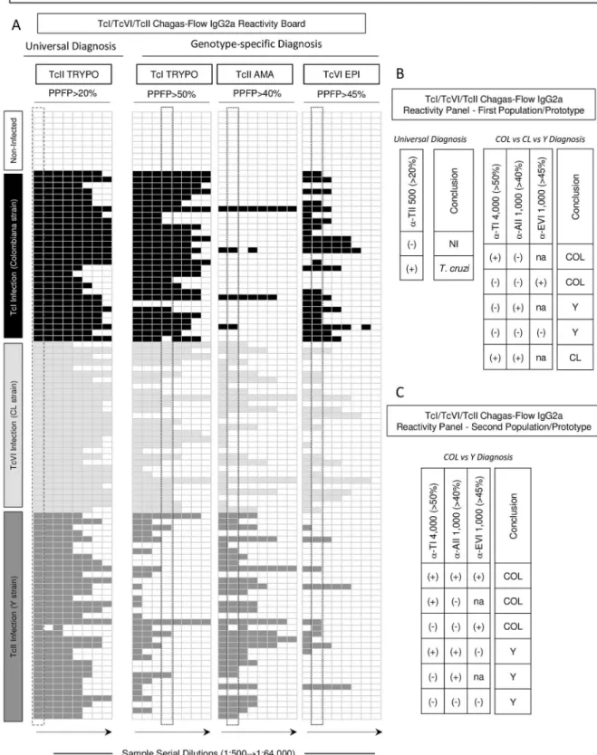
Reactivity boards were constructed using the pre-selected set attributes, including “anti-TcII TRYPO reactivity at 1:500, cut-off = 20%” for universal diagnosis purpose and “anti-TcI TRYPO reactivity at 1:4,000, off = 50%”, “anti-TcII AMA reactivity at 1:1,000, cut-off = 40%” and “anti-TcVI EPI reactivity at 1:1,000, cut-cut-off = 45%” for genotype-specific diag-nosis (Fig 6). The reactivity at selected serum dilutions (Fig 6—dashed frames) were used to further create a receiver operating reactivity panel, applicable for universal diagnosis (NI vsT.cruzi-infected hosts,Fig 6B) and for genotype-specific diagnosis applied to the first are expressed as Log of PPFP values for anti-TcI TRYPO at 1:4,000 (left lateral axis), anti-TcII AMA at 1:1,000 (vertical axis) and anti-TcVI EPI at 1:1,000 (right lateral axis). Decision trees were constructed using the set of attributes (“target-antigen/ serum dilution/cut-off”) to create algorithms (root and branch attributes) to classifyT.cruziinfected mice in a population/ prototype including (B) TcI/Colombiana (red rectangle)vsTcVI/CL (green rectangle)vsTcII/Y (blue rectangle) or including (E) TcI/Colombiana (red rectangle)vsTcII/Y(blue rectangle). Global accuracy and leave-one-out-cross-validation-LOOCV are provided in the Figure. Bar charts representing the performance of the decision trees demonstrate the number of animals that ranked within each branch amongst theT.cruzi-infected hosts for (C) (TcI/Colombiana = red barvsTcVI/CL = green bar vsTcII/Y = blue bar) and (F) (TcI/Colombiana = red barvsTcII/Y = blue bar).
population/prototype (TcI/ColombianavsTcVI/CLvsTcII/Y strains,Fig 6B) and the second population/prototype (TcI/ColombianavsTcII/Y strains,Fig 6C).
When using the TcI/TcVI/TcII Chagas-Flow ATE-IgG2a applied for the universal diagnosis purpose, the attributeα-TII 500 (>20%) presenting a positive score (+) define the presence of
T.cruziinfection, while a negative score (-) ruled out the presence ofT.cruziinfection. The reactivity panel for TcI/TcVI/TcII Chagas-Flow ATE-IgG2a applied for the genotype-specific diagnosis regardless the population/prototype scenario indicated scores sequences of α-TI (trypomastigote TcI) 4,000(>50%)/α-AII (amastigote TcII) 1,000(>40%)/α-EVI
(epi-mastigote TcVI) 1,000(>45%) to define theT.cruziinfection with distinct genotypes defined
as: (+/-/not applicable (na) or -/-/+) for TcI/Colombiana and (-/+/na or -/-/-) for TcII/Y strain infection. The extensions of the score (+/+) do not allow the proper identification theT.cruzi
infection genotype, since in can belongs to hosts infected with TcI/Colombiana (+/+/+), TcVI/ CL (+/+/na) or even TcII/Y strain (+/+/-) (Fig 6B and 6C).
Together, the proposed receiver operating reactivity panels for TcI/TcVI/TcII Chagas-Flow ATE-IgG2a provided a feasible tool to classify the serum samples as they belong to the true respective groups, supporting the potential of this method for universal and genotype-specific diagnosis ofT.cruziinfection.
Discussion
The broad genetic variability ofT.cruzihas been related to biological characteristics (infectiv-ity, parasitemia, tissue tropism, mortality during the acute phase of infection [13,26–30] and susceptibility/resistance to drugs [5,8,31–34] in murine model and infectivity, replication and differentiation in vector o) [35], epidemiological characteristics [4,36,37] and clinical manifes-tations [38–40] of Chagas disease. Therefore, the knowledge of parasite genetics may offer insights about the biology of the parasite, patient’s treatment outcome, clinical aspects of human disease, as well as how to establish epidemiological surveillance and control of Chagas disease [41,42]. So, the genetic diversity ofT.cruziinfection may also influence the sensitivity of the techniques used for Chagas disease diagnosis [43–45].
The currently available methods for genotype-specific diagnosis ofT.cruziinfection, most based on molecular biology approaches present distinct levels of complexity and in general dis-play high specificity but moderate sensitivity [4,10–12,46–52]. Moreover, a combination of several genetic markers is necessary to detect and distinguish theT.cruzigenotypes [10–12]. Furthermore, the majority of these methods can not directly be performed in biological and clinical samples, requiring previous parasite isolation by hemoculture/xenoculture followed by
in vitrogrowth and maintenance that may lead to clonal selection [14,53–55]. Besides, it is known that the parasitemia in patients and reservoirs ofT.cruziare variable and that the suc-cess of parasite isolation is dependent on the host parasitemia. The full extent of lineage distri-bution in nature using genetic markers it is not known due to the low levels of circulating parasitemia and possible lineage-specific tissue sequestration [34,56–58]. In addition, it has PPFP>40% and TcVI EPI, PPFP>45%) based on the differential positive reactivity of sera samples (TcI/Colombiana strain = black rectangle, TcVI/CL strain = light gray rectangle and TcII/Y strain = dark gray rectangle) from negative reactivity (white rectangle) observed forT.cruziinfected hosts and non-infected mice. The pre-selected sera dilutions defined by decision tree analysis are underscored by dotted rectangles and include TcII TRYPO PPFP>20% at 1:500 for universal diagnosis and the set of attributes (TcI-TRYPO/PPFP>50%/4,000 followed by TcII-AMA/PPFP>40%/1,000 and TcVI-EPI/PPFP>45%/1,000) for genotype-specific diagnosis criteria. Reactivity panels were constructed to define the diagnosis conclusion when applying TcI/ TcVI/TcII Chagas-Flow ATE-IgG2a for (B) universal diagnosis ofT.cruziinfection and genotype-specific diagnosis in a population/prototype including TcI/Colombiana strain (COL), TcVI/CL strain (CL) or TcII/Y (Y) strain or (C) including TcI/ Colombiana strain (COL) or TcII/Y strain (Y).
been proposed that parasite isolates from blood may not necessarily represent the full set of strains current in the individual, hence some strains ofT.cruzican be confined to tissues [11,13,16,15]. In general, PCR (polymerase chain reaction) based genotyping has limitations that hamper the analysis of large numbers of samples. Therefore, the development of methods for diagnosis and serotyping of Chagas disease are urgently required.
Attempting to address this matter, Mendeset al. (2013) [18] have described a set of B-cell epitopes able to discriminate TcI and TcII infections, demonstrating the potential of these tar-gets for Chagas disease serotyping. Later on, the putative TcI epitope reported by Mendeset al. (2013) [18] was found to be conserved across allT.cruzilineages by studies developed by Bhattacharyyaet al. (2014) [19]. Moreover, samples from animals infected with TcVI pre-sented cross-reactivity with a range ofT.cruzi-derived peptides, suggesting the need of improved antigen search and the development of a robust panel of strain-specific epitopes to achieve a method applicable in large epidemiological studies [18].
Aiming at developing innovative serological approaches for universal and improved geno-typic-specific diagnosis ofT.cruziexperimental infection, our goal was optimize the Chagas-Flow ATE methodology, proposed originally by Alessioet al. (2014) [21]. The present approach is based on parallel batches of distinctT.cruzigenotypes as target antigens, employ-ing parasites strains of three more important genotypes associated with human infection and Chagas disease (TcI, TcVI and TcII) [4,38,59] although others genotypes exist such as TcIII [60–62], TcIV recently described as associated to oral transmission [63,64] and TcV associated to the classical clinical forms of Chagas disease (cardiac and digestive) [39].
Previous studies have demonstrated that patients from distinct geographic areas, infected with different genotypes ofT.cruziseem to display differential serological pattern upon sero-diagnosis of Chagas disease, when employing distinct methodological approaches and differ-entT.cruzitarget antigens [4,43,44,65]. Veraniet al. (2009) [44] have demonstrated that the performance of two serological tests, using serum samples from distinct geographical regions (Bolivia and Peru) displayed distinct sensitivity, ranging from 26.6%-87.5%, corroborating the hypothesis that intrinsic features of regional parasite strains may influence the serological tests. Studies using six recombinant antigens ofT.cruzitested in samples from Argentina, Bra-zil, Chile, Colombia, El Salvador, Guatemala, Honduras and Venezuela also reported discrep-ancy in the serological reactivity ranging from 79% to 100% [43].
In this study, we have intended to evaluate the performance of combined TcI/TcVI/TcII Chagas-Flow ATE-IgG2a for universal diagnosis ofT.cruziinfection, simulating two popula-tion prototypes that may represent the geographic distribupopula-tion ofT.cruziinfection in the Latin America. The first population prototype, represent regions were TcI, TcVI and TcII genotypes are co-endemic. In the second population setting, we intended to evaluate the per-formance of combined TcI/TcVI/TcII Chagas-Flow ATE-IgG2a to discriminate the infections with TcI/ColombianavsTcII/Y strains. Our findings demonstrated that regardless the popula-tion prototype, the TcI/TcVI/TcII Chagas-Flow ATE-IgG2a presented an outstanding perfor-mance for universal diagnosis ofT.cruziinfection using the set of attributes “anti-TcII TRYPO reactivity at 1:500, cut-off = 20%” (Fig 3). In fact, although the sensitivity of TcI/TcVI/ TcII Chagas-Flow ATE-IgG2a varies according to the target antigen employed, the TcII TRYPO antigen was able to detect seroreactivity in all mice infected with distinctT.cruzi
genotypes (Figs2and3). Corroborating with our study, Bhattacharyyaet al. (2014) [19] also observed that all sera from patients with chronic Chagas disease recognized theT.cruziTcII lysate antigen preparation.
genotypic-specific antigenic features may be involved in the induction of lytic antibodies [67–69]. More-over, Di Noiaet al. (2002) [70] have reported that twoT.cruziantigens, named small surface antigen of trypomastigotes (TSSAI and TSSAII) presented the ability of genotypic-specific rec-ognition ofT.cruziinfection. These studies were expanded by Bhattacharyyaet al. (2010, 2014 and 2015) [17,19,20] that reported that TSSA pepII/V/VI isoforms were able to distinguish samples of hosts infected with distinctT.cruzigenotypes. Based on these findings, the authors proposed that TSSA isoforms are feasible serological markers to identify aT.cruzilineage in human and experimental infection. However, the TSSA pepI did not yield significant reactiv-ity, suggesting that novel targets for TcI is still required.
The innovative TcI/TcVI/TcII Chagas-Flow ATE-IgG2a methodology presented a high-quality performance to segregate infections with TcI/Colombiana, TcVI/CL or TcII/Y strain. The performance of combined TcI/TcVI/TcII Chagas-Flow ATE-IgG2a for genotype-specific diagnosis ofT.cruziinfection differs depending on the population prototypes used to repre-sent distinct geographic regions ofT.cruziinfection in the Latin America. In the first proto-type (TcI/TcVI/TcII), our data demonstrated that the proposed method showed a moderate global accuracy (68.8%, LOOCV = 58.0%) to discriminate the infections with TcI/Colombiana
vsTcVI/CLvsTcII/Y strains. On the other hand, the combined TcI/TcVI/TcII Chagas-Flow ATE-IgG2 was capable to discriminate the infections with TcI/ColombianavsTcII/Y strains in the second population prototype (TcI/TcII) with high global accuracy (93.8%, LOOCV = 87.5%) (Fig 5). Overall, hosts infected with TcI/Colombiana and TcII/Y strains dis-played opposite patterns of reactivity with “anti-TcI TRYPO” and “anti-TcII AMA” and hosts infected with TcVI/CL strain showed a typical interweaved distribution pattern (Figs4and
S1). This phenomenon may reflect the phylogenetic origin of DTUs [4] where TcI and TcII are ancestral DTUs presenting polar characteristics [5,27,28,30,71–73], whereas the TcVI has a hybrid origin, showing intermediate characteristics of both polar genotypes [73,74].
In conclusion, based on the receiver operating characteristic, the TcI/TcVI/TcII Chagas-Flow ATE-IgG2a seems to be a feasible tool to classify the serum samples as they belong to the true respective groups infected with distinctT.cruzigenotypes (Fig 6), suggesting its applica-bility for both, universal and genotype-specific diagnosis ofT.cruziinfection in clinical labora-tories. The proposed methodology includes essential advantages such as high sensitivity and specificity, ease to perform, using a wide range of antigenic preparation into a single flow cyto-metric platform [21,75–79]. Future derivation of TcI/TcVI/TcII Chagas-Flow ATE-IgG2a as the development of a suitable ELISA or multiplex beads assay would contribute to practical applications in routine clinical laboratories, since the original version of this fluorescence-based methodology is more reliable for applications in reference laboratories. Additional tests are under investigation to establish accuracy of TcI/TcVI/TcII Chagas-Flow ATE-IgG2a to identify mixed infections with distinctT.cruzigenotypes. An extension of this study may be applicable to other genetic groups not included in this work (TcIII, TcIV and TcV). Further studies including serum samples from patients with genotypic-specific diagnosis Chagas dis-ease performed by molecular methods are currently under investigation as a proof-of-concept to propose a prototype for clinical purposes, epidemiological studies and post-therapeutic monitoring applications.
Supporting information
S1 Fig. Cut-off edges and performance of TcI/TcVI/TcII Chagas-Flow ATE-IgG2a for genotype-specific diagnosis ofT.cruziinfection.(A) The trendlines of anti-TcII AMA at
1:1,000, anti-TcI TRYPO at 1:4,000 and anti-TcVI EPI at 1:1,000 reactivity observed forT.
dashed line and TcII/Y strain = dark gray dashed line) were overlaid aiming to differentiate the reactivity pattern. The results were expressed as the proportion of samples displaying a given PPFP values amongstT.cruzi-infected hosts. (B) The whole set of reactivity data were used to calculate the global median PPFP values applied as the cut-off edge to segregate the individual samples as they present negative (white square) or positive (black square) reactivity at the selected target-antigen/serum dilution. The results were expressed as the range of PPFP values in scatter plots for individual serum samples (C) Diagrams were used to compile the reactivity patterns and calculate the proportion of negative and positive results for each selected set of attributes (“target-antigen/serum dilution/cut-off”). (D) Representative scatter plots were also used to illustrate the ability of each set of attributes (“target-antigen/serum dilu-tion/cut-off”) to discriminate the reactivity of serum samples amongst theT.cruzi-infected mice (TcI-infection/Colombiana, n = 29; TcVI/CL, n = 29 and TcII/Y, n = 35). The results were expressed as the range of PPFP values in scatter plots for individual serum samples. The dotted line represents the cut-off for each target-antigen/serum dilution. Clusters of distinct reactivity patterns are highlighted by light-gray doted frames whereas indiscriminate distribu-tion pattern underscore by white-background frame.
(TIF)
S2 Fig. Discriminant analysis of combined TcI/TcVI/TcII Chagas-Flow ATE-IgG2a for genotype-specific diagnosis ofT.cruziinfection.(A) Discriminant analyses of combined
TcI/TcVI/TcII Chagas-Flow ATE-IgG2a were performed for genotype-specific diagnosis ofT.
cruziinfection in a population/prototype including TcI/Colombiana strain, TcVI/CL strain or TcII/Y strain. (B) Discriminant analyses of combined TcI/TcVI/TcII Chagas-Flow ATE-IgG2a were performed for genotype-specific diagnosis ofT.cruziinfection in a population/prototype including TcI/Colombiana strain or TcII/Y strain. The global accuracy and leave-one-out-cross-validation-LOOCV provided in the Figure.
(TIF)
Author Contributions
Conceptualization:GDA ATC OAMF MdL.
Data curation:GDA FFdA DFC PASJ DCL.
Formal analysis:GDA MdSG LRdA MAPX OAMF.
Funding acquisition:ATC OAMF MdL.
Investigation:GDA FFdA DFC PASJ DCL OAMF.
Methodology:GDA DFC PASJ DCL.
Project administration:ATC OAMF MdL.
Resources:GDA FFdA ATC OAMF MdL.
Software:MdSG LRdA MAPX.
Supervision:OAMF MdL.
Validation:GDA DFC DCL OAMF MdL.
Visualization:GDA FFdA ATC OAMF MdL.
Writing – review & editing:GDA FFdA PASJ ATC OAMF MdL.
References
1. Chagas C. Nova Tripanozomiase Humana. Estudo sobre a morfologia e o ciclo evolutivo do Schizotry-panum cruzi. n. gen., n. sp. agente etiolo´gico de nova entidade mo´rbida do homem. Memo´rias do Insti-tuto Oswaldo Cruz. 1909; 1: 159–218.
2. WHO- World Health Organization- Chagas disease (American trypanosomiasis) Fact sheet N˚ 340; 2015.
3. Dias JC. Epidemiology of Chagas disease. In Wendel S, Brener Z, Camargo MS, Rassi A (eds), Chagas disease (American Trypanosomiasis): its Impact on Transfusion and Clinical Medicine. ISBT Brazil, São Paulo. 1992; 49–80.
4. Zingales B, Miles MA, Campbell DA, Tibayrenc M, Macedo AM, Teixeira MG, Schijman AG, Llewellyn MS, Lages-Silva E, Machado CR, Andrade SG, Sturm NR. The revisedTrypanosoma cruzisubspecific nomenclature: Rationale, epidemiological relevance and research applications. Infect. Gene Evolut. 2012; 12: 240–253.
5. Toledo MJ, Bahia MT, Carneiro CM, Martins-Filho OA, Tibayrenc M, Barnabe C, Tafuri WL, Lana M. Chemotherapy with benznidazole and itraconazole for mice infected with differentTrypanosoma cruzi clonal genotypes. Antimicrob Agents Chemother. 2003; 47: 223–230.https://doi.org/10.1128/AAC.47. 1.223-230.2003PMID:12499195
6. Toledo MJ, Bahia MT, Veloso VM, Carneiro CM, Machado-Coelho GL, Alves CF, Martins HR, Cruz RE, Tafuri WL, Lana M. Effects of specific treatment on parasitological and histopathological parameters in mice infected with differentTrypanosoma cruziclonal genotypes. J Antimicrob Chemother. 2004; 53(6): 1045–53.https://doi.org/10.1093/jac/dkh224PMID:15102747
7. Martins HR, Figueiredo LM, Valamiel JC, Carneiro CM, Machado-Coelho GL, Bahia MT, Macedo AM, Lana M. Persistence of PCR-positive tissue in benznidazole-treated mice with negative blood parasito-logical and seroparasito-logical tests in dual infections withTrypanosoma cruzistocks from different genotypes. J. Antimicrob Chemother. 2008; 61(6): 1319–27.https://doi.org/10.1093/jac/dkn092PMID:18343804 8. Oliveira-Silva JC, Machado-de-Assis GF, Oliveira MT, Paiva NC, Arau´jo MS, Carneiro CM,
Martins-Filho OA, Martins HR, Lana M. Experimental benznidazole treatment ofTrypanosoma cruziII strains isolated from children of Jequitinhonha Valley, Minas Gerais, Brazil, with Chagas disease. Mem Inst Oswaldo Cruz. 2015; 110: 86–94.https://doi.org/10.1590/0074-02760140260PMID:25742267 9. Zingales B, Andrade SG, Briones MR, Campbell DA, Chiari E, Fernandes O, Guhl F, Lages-Silva E,
Macedo AM, Machado CR, Miles MA, Romanha AJ, Sturm NR, Tibayrenc M, Schijman AG. A new con-sensus forTrypanosoma cruziintraspecific nomenclature: second revision meeting recommends TcI to TcVI. Mem Inst Oswaldo Cruz. 2009; 104(7): 1051–1054. PMID:20027478
10. Lewis MD, Jonathan MA, Matthew YE, Herna´n J, Carrasco MS, Llewellyn AN, Michael AM. Genotyping ofTrypanosoma cruzi: Systematic Selection of Assays Allowing Rapid and Accurate Discrimination of All Known Lineages. Am J Trop Med Hyg. 2009; 81(6): 1041–1049.https://doi.org/10.4269/ajtmh.2009. 09-0305PMID:19996435
11. D’a´villa DA, Macedo AD, Valadares HM, Gontijo ED, De Castro AM, Machado CR, Chiari E, Galvão LM. Probing Population Dynamics ofTrypanosoma cruziduring Progression of the Chronic Phase in Chagasic Patients. J Clin Microbiol. 2009; 1718–1725.https://doi.org/10.1128/JCM.01658-08PMID: 19357212
12. Oliveira MT, Machado-de-Assis GF, Oliveira-Silva JC, Machado EM, Da Silva GN, Veloso VM, Macedo AM, Martins HR, Lana M.Trypanosoma cruziDiscret Typing Units (TcII and TcVI) in samples of patients from two municipalities of the Jequitinhonha Valley, MG, Brazil, using two molecular typing strategies. Parasit Vectors. 2015; 8: 568.https://doi.org/10.1186/s13071-015-1161-2PMID:26520576 13. Vago AR, Andrade LO, Leite AA, D’avilla RD, Macedo AM, Adad SJ, Tostes SJ, Moreira MC, Filho GB, Pena SD. Genetic characterization ofTrypanosoma cruzidirectly from tissues of patients with chronic Chagas disease: differential distribution of genetic types into diverse organs. Am J Pathol. 2000; 156 (5): 1805–9. PMID:10793092
14. Macedo AM, Pena SD. Genetic variability ofTrypanosoma cruzi: implications for the pathogenesis of Chagas disease. Parasitol Today. 1998; 14: 119–123. PMID:17040719
15. Macedo AM, Machado CR, Oliveira RP, Pena SD.Trypanosoma cruzi: genetic structure of populations and relevance of genetic variability to the pathogenesis of Chagas disease. Mem Inst Oswaldo Cruz. 2004; 99: 1–12.
cruzidiscrete typing units in end-stage chronic Chagas heart disease and reactivation after heart trans-plantation. Clin Infect Dis. 2010; 51(5): 485–95.https://doi.org/10.1086/655680PMID:20645859 17. Bhattacharyya T, Brooks J, Yeo M, Carrasco HJ, Lewis MD, Llewellyn MS, Miles MA. Analysis of
molec-ular diversity of theTrypanosoma cruzitrypomastigote small surface antigen reveals novel epitopes, evidence of positive selection and potential implications for lineage-specific serology. Int J Parasitol. 2010; 40(8): 921–8.https://doi.org/10.1016/j.ijpara.2010.01.002PMID:20097201
18. Mendes TA, Reis-Cunha JL, de Almeida LR, Rodrigues LG, Lemos LD, dos Santos AR, da Caˆmara AC, Galvão LM, Bern C, Gilman RH, Fujiwara RT, Gazzinelli RT, Bartholomeu DC. Identification of strain-specific B-cell epitopes inTrypanosoma cruziusing genome-scale epitope prediction and high-throughput immunoscreening with peptide arrays. Plos Negl Trop Dis. 2013; 7(10): e2524.https://doi. org/10.1371/journal.pntd.0002524PMID:24205430
19. Bhattacharyya T, Falconar AK, Luquetti AO, Costales JA, Grijalva MJ, Lewis MD, Messenger LA, Tran TT, Ramirez JD, Guhl F, Carrasco HJ, Diosque P, Garcia L, Litvinov SV, Miles MA. Development of peptide-based lineage-specific serology for chronic Chagas disease: geographical and clinical distribu-tion of epitope recognidistribu-tion. Plos Negl Trop Dis. 2014; 8(5): e2892.https://doi.org/10.1371/journal.pntd. 0002892PMID:24852444
20. Bhattacharyya T, Mills EA, Jansen AM, Miles MA. Prospects forT.cruzilineage-specific serological sur-veillance of wild mammals. Acta Trop. 2015; 151: 182–6.https://doi.org/10.1016/j.actatropica.2015.06. 017PMID:26116784
21. Alessio GD, Coˆrtes DF, Machado-de-Assis GF, Sales-Ju´nior PA, Ferro EA, Antonelli LR, Teixeira-Car-valho A, Martins-Filho OA, Lana M. Inovations in diagnosis and post-therapeutic monitoring of Chagas disease: Simultaneous flow cytometric detection of IgG1 antibodies anti-live amastigote, anti-live trypo-mastigote, and anti-fixed epimastigote forms ofTrypanosoma cruzi. Journal of Immunological Methods. 2014; 413: 32–44.https://doi.org/10.1016/j.jim.2014.07.005PMID:25064148
22. Federeci EE, Albemann WH, Neva FA. Chronic and progressive myocarditis and myositis in C3H mice infected withTrypanosoma cruzi. Am J Trop Med Hyg. 1964; 13: 272–80. PMID:14125879
23. Brener Z, Chiari E. Morphological variations observed in different strains ofTrypanosoma cruzi. Rev Inst Med Trop São Paulo. 1963; 5: 220–4. PMID:14110094
24. Pereira da Silva LH, Nussenzweig. Sobre uma cepa deTrypanosoma cruzialtamente virulenta para o camundongo branco. Folia Clin Biol. 1953; 20: 191–208.
25. Camargo EP. Growth and differentiation inTrypanosoma cruzi. I. Origin of metacyclic trypanosomes in liquid media. Rev Inst Med Trop São Paulo. 1964; 6: 93–100. PMID:14177814
26. Andrade SG. Caracterizac¸ão de cepas deTrypanosoma cruziisoladas no Recoˆncavo Baiano. Rev Pat Trop. 1974; 65–121.
27. Andrade SG, Magalhães JB. Biodemes and zimodemes ofTrypanosoma cruzistrains: correlations with clinical data and experimental pathology. Rev Soc Bras Med Trop. 1997; 27–35. PMID:8993106 28. Laurent JP, Barnabe C, Quesney V, Noel S, Tibayrenc M. Impact of clonal evolution on the biological
diversity ofTrypanosoma cruzi. Parasitology. 1997; 114: 213–218. PMID:9075341
29. Toledo MJ, Lana M, Carneiro CM, Bahia MT, Machado-Coelho GL, Veloso VM, Barnabe´ C, Tibayrenc M, Tafuri WL. Impact ofTrypanosoma cruziclonal evolution on its biological properties in mice. Exp Parasitol. 2002; 100(3): 161–7. PMID:12173401
30. Andrade SG, Campos RF, Steindel M, Guerreiro ML, Magalhães JB, Almeida MC, Reis JN, Santos VC, Valadares HM, Reis MG, Macedo AM. Biological, biochemical and molecular features ofTrypanosoma cruzistrains isolated from patients infected through oral transmission during a 2005 outbreak in the state of Santa Catarina, Brazil: its correspondence with the newT.cruziTaxonomy Consensus (2009). Mem Inst Oswaldo Cruz. 2011; 106(8): 948–56. PMID:22241116
31. Andrade SG. Morphological and behavioural characterization ofTrypanosoma cruzistrains. Rev Soc Bras Med Trop. 1985; 39–4624.
32. Filardi LS, Brener Z. Susceptibily and natural resistance osTrypanosoma cruzistrains to drugs used clinically in Chagas disease. Trans R Soc Trop Med Hyg. 1987; 81(5): 755–759. PMID:3130683 33. Andrade SG, Rassi A, Magalhães JB, Ferriolli FF, Luquetti AO. Specific chemotherapy of Chagas
dis-ease: a comparison between the response in patients and experimental animals inoculated with the same strains. Trans R Soc Trop Med Hyg. 1992; 86: 624–626. PMID:1287919
34. Teston AP, Monteiro WM, Reis D, Bossolani GD, Gomes ML, de Arau´jo SM, Bahia MT, Barbosa MG, Toledo MJ.In vivosusceptibility to benznidazole ofTrypanosoma cruzistrains from the western Brazilian Amazon. Trop Med Int Health. 2013; 18(1): 85–95.https://doi.org/10.1111/tmi.12014PMID:23130989 35. Lana M, da Silveira Pinto A, Barnabe´ C, Quesney V, Noe¨l S, Tibayrenc M.Trypanosoma cruzi:
36. Tibayrenc M, Ayala FJ. The population genetics of Trypanosoma cruzi revisited in the light of the pre-dominant clonal evolution model. Acta Trop. 2015; 151: 156–65.https://doi.org/10.1016/j.actatropica. 2015.05.006PMID:26188332
37. Miles MA, Llewellyn MS, Lewis MD, Yeo M, Baleela R, Fitzpatrick S, Gaunt MW, Mauricio IL. The molecular epidemiology and phylogeography ofTrypanosoma cruziand parallel research on Leish-mania: looking back and to the future. Parasitology. 2009; 136(12): 1509–28.https://doi.org/10.1017/ S0031182009990977PMID:19691868
38. Lages-Silva E, Ramı´rez LE, Pedrosa AL, Crema E, Da Cunha GL, Junho PS, Macedo AM, Chiari E. Variability of kinetoplast DNA gene signatures ofTrypanosoma cruziII strains from patients with differ-ent clinical forms of Chagas’ disease in Brazil. J Clin Microbiol. 2006; 44: 2167–71.https://doi.org/10. 1128/JCM.02124-05PMID:16757616
39. Virreira M, Serrano G, Maldonado L, Svoboda M.Trypanosoma cruzi: typing of genotype (sub)lineages in megacolon samples from bolivian patients. Acta Trop. 2006; 100(3): 252–5.https://doi.org/10.1016/j. actatropica.2006.11.005PMID:17157796
40. Valadares HM, Pimenta JR, De Freitas JM, Duffy T, Bartholomeu DC, Oliveira RP, Chiari E, Moreira MC, Filho GB, Schijman AG, Franco GR, Machado CR, Pena SD, Macedo AM. Genetic profiling of Try-panosoma cruzidirectly in infected tissues using nested PCR of polymorphic microsatellites. Int J Para-sitol. 2007; 38(7): 839–50.https://doi.org/10.1016/j.ijpara.2007.10.017PMID:18154957
41. Coura JR, Junqueira AC. Surveillance, health promotion and control of Chagas disease in the Amazon Region-Medical attention in the Brazilian Amazon Region: a proposal. Mem Inst Oswaldo Cruz. 2015; 110(7): 825–30.https://doi.org/10.1590/0074-02760150153PMID:26560976
42. Martinez-Perez A, Poveda C, Ramı´rez JD, Norman F, Girone´s N, Guhl F, Monge-Maillo B, Fresno M, Lo´pez-Ve´lez R. Prevalence ofTrypanosoma cruziDiscrete Typing Units in a cohort of Latin American migrants in Spain. Acta Trop. 2016; 157: 145–50.https://doi.org/10.1016/j.actatropica.2016.01.032 PMID:26851167
43. Umezawa ES, Bastos SF, Camargo ME, Yamauchi LM, Santos MR, Gonzalez A, Zingales B, Levin MJ, Souza O, Rangel-Aldao R, Silveira JF. Evaluation of recombinant antigens for serodiagnosis of Chagas disease in South and Central America. Journal of Clinical Microbiology. 1999; 37: 1554–1560. PMID: 10203520
44. Verani JR, Seitz A, Gilman RH, Lafuente C, Galdos-Cardenas G, Kawai V, De Lafuente E, Ferrufino L, Bowman NM, Pinedo-Cancino V, Levy MZ, Todd CW, Kirchhoff LV, Cabrera L, Verastegui M, Bern C. Geographic variation in the sensitivity of recombinant antigen-based rapid tests chronicTrypanosoma cruziinfection. Am J Trop Med Hyg. 2009; 80(3): 410–415. PMID:19270291
45. Reis-Cunha JL, Mendes TA, De Almeida LR, Ribeiro DR, Machado-De-Avila RA, Oliveira MT, Lemos DS, Caˆmara AC, Olo´rtegui CC, Lana M, Da Cunha GLM, Fujiwara RT, Bartholomeu DC. Genome-wide screening and identification of newTrypanosoma cruziantigens with potential application of chronic Chagas disease diagnosis. Plos One. 2014; 9(9).
46. Souto RP, Zingales B. Sensitive detection and strain classification ofTrypanosoma cruziby amplifica-tion of a ribossomal RNA sequence. Mol Biochem Parasitol. 1993; 62: 45–52. PMID:8114825 47. Clark CG, Pung OJ. Host specificity of ribosomal DNA variation in sylvaticTrypanosoma cruzifrom
North America. Mol Biochem Parasitol. 1994; 66: 175–179. PMID:7984184
48. Souto RP, Fernandes O, Macedo AM, Campbel DA, Zingales B. DNA markers define two major phylo-genetic lineages ofTrypanosoma cruzi. Mol Biochem Parasitol. 1996; 83: 141–152. PMID:9027747 49. Brisse S, Barnabe C, Tibayrenc M. Identification of sixTrypanosoma cruziphylogenetic lineages by
ran-dom amplified polymorphic DNA and multilocus enzyme electrophoresis. Int J Parasitol. 2000; 30: 35– 40. PMID:10675742
50. Consentino RO, Aguero F. A Simple Strain Typing Assay forTrypanosoma cruzi: Discrimination of Major Evolutionary Lineages from a Single Amplification Product. PLoS Negl Trop Dis. 2012; 6(7): 1777. 51. Higuera SL, Guhl F, Ramı´rez JD. Identification ofTrypanosoma cruziTyping Units (DTUs) through the
implementation of a High-Resolution Melting (HRM) genotyping assay. Parasites & Vector. 2013; 6: 112–117.
52. Cura CI, Duffy T, Lucero RH, Bisio M, Pe´neau J, Jimenez-Coello M, et al. Multiplex real-time PCR assay using TaqMan probes for the identification ofTrypanosoma cruziDTUs in biological and clinical samples. Plos Negl Trop Dis. 2015; 9(5): 1371–88.
53. Brener Z, Canc¸ado JR, Galvão LM, Luz ZP, Filardi LS, Pereira ME, Santos LM, Canc¸ado CB. An exper-imental and clinical assay with ketoconazole in the treatment of Chagas disease. Memo´rias do Instituto Oswaldo Cruz. 1993; 88: 149–153. PMID:8246750
55. Marcet PL, Duffy T, Cardinal MV, Burgos JM, Lauricella MA, Levin MJ, et al. PCR-based screening and lineage identification ofTrypanosoma cruzidirectly from fecal samples of triatomine bugs from north-western Argentina. Parasitology. 2006; 132(1): 57–65.
56. Abolis NG, Arau´jo SM, Toledo MJ, Fernandez MA, Gomes ML.Trypanosoma cruziI-III in southern Bra-zil causing individual and mixed infections in humans, sylvatic reservoirs and triatomines. Acta Trop. 2011; 120(3): 167–72.https://doi.org/10.1016/j.actatropica.2011.08.001PMID:21855523
57. Oliveira-Silva JC, Machado-de-Assis GF, Oliveira MT, Valadares HM, Valle IF, Paiva NC, Martins HR, Lana M. Molecular and biology characterization ofTrypanosoma cruzistrains isolated from children from Jequitinhonha Valley of Minas Gerais, Brazil. Rev Soc Bras Med Trop. 2013; 46(4): 433–40. https://doi.org/10.1590/0037-8682-0077-2013PMID:23982097
58. Sales-Campos H, Kappel HB, Andrade CP, Lima TP, de Castilho A, Giraldo LE, Lages-Silva E. Trypa-nosoma cruziDTU TcII presents higher blood parasitism than DTU TcI in an experimental model of mixed infection. Acta Parasitol. 2015; 60(3): 435–41.https://doi.org/10.1515/ap-2015-0060PMID: 26204180
59. Ramı´rez JD, Guhl F, Rendo´n LM, Rosas F, Marin-Neto JA, Morillo CA. Chagas cardiomyopathy mani-festations andTrypanosoma cruzigenotypes circulating in chronic Chagasic patients. PLoS Negl Trop Dis. 2010; 4(11): e899.https://doi.org/10.1371/journal.pntd.0000899PMID:21152056
60. Cardinal MV, Lauricella MA, Ceballos LA, Lanati L, Marcet PL, Levin MJ, et al. Molecular epidemiology of domestic and sylvaticTrypanosoma cruziinfection in rural northwestern Argentina. Int J Parasitol. 2008; 38: 1533–1543.https://doi.org/10.1016/j.ijpara.2008.04.010PMID:18585717
61. Marcili A, Lima L, Valente VC, Valente SA, Batista JS, Junqueira AC, et al. Comparative phylogeogra-phy ofTrypanosoma cruziTcIIc: new hosts, association with terrestrial ecotopes, and spatial clustering. Infect Genet Evol. 2009; 9: 1265–1274.https://doi.org/10.1016/j.meegid.2009.07.003PMID:19632356 62. Caˆmara AC, Varela-Freire AA, Valadares HM, Macedo A, D’A´ vila DA, Machado CR, et al. Genetic anal-yses ofTrypanosoma cruziisolates from naturally infected triatomines and humans in northeastern Bra-zil. Acta Trop. 2010; 115: 205–211.https://doi.org/10.1016/j.actatropica.2010.03.003PMID:20303924 63. Coura JR, Junqueira AC, Fernandes O, Valente SA, Miles MA. Emerging Chagas disease in
Amazo-nian Brazil. Trends Parasitol. 2002; 18: 171–176. PMID:11998705
64. Valente SA, da Costa V.V, das Neves P.A, de Jesus B.M, dos Santos MP, Miranda CO, et al. Analysis of an acute Chagas disease outbreak in the Brazilian Amazon: human cases, triatomines, reservoir mammals and parasites.Trans R Soc Trop. Med Hyg. 2009; 103: 291–297.https://doi.org/10.1016/j. trstmh.2008.10.047PMID:19118852
65. Umezawa ES, Luquetti A, Levitus G, Ponce C, Ponce E, Henriquez D, Revollo S, Espinoza B, Souza O, Khan B, Silveira JF. Serodiagnosis of chronic and acute Chagas disease withTrypanosoma cruzi recombinant proteins: Results of a collaborative study in six Latin American Countries. Journal of Clini-cal Microbiology. 2004; 42: 449–452.https://doi.org/10.1128/JCM.42.1.449-452.2004PMID:14715803 66. Dos Santos DM, Talvani A, Guedes PM, Machado-Coelho GL, Lana M, Bahia MT.Trypanosoma cruzi:
Genetic diversity influences the profile of immunoglobulins during experimental infection. Exp Parasitol. 2009; 121(1): 8–14.https://doi.org/10.1016/j.exppara.2008.09.012PMID:18848935
67. Krettli AU, Weisz-Carrington P, Nussenzweig RS. Membrane-bound antibodies to bloodstream Trypa-nosoma cruziin mice: strain differences in susceptibility to complement-mediated lysis. Clin Exp Immu-nol. 1979; 37(3): 416–23. PMID:116784
68. Wallace A, Sanchez G, Venegas J, Solari A. Lack of cross-reactivity of lytic antibodies with bloodstream forms ofTrypanosoma cruzizymodemes generated in a mouse experimental model. Exp Parasitol. 1995; 80(2): 176–85.https://doi.org/10.1006/expr.1995.1022PMID:7895829
69. Zulantay I, Venegas J, Apt W, Solari A, Sanchez G. Lytic antibodies inTrypanosoma cruzi-infected per-sons with low parasitemia. Am J Trop Med Hyg. 1998; 58(6): 775–9. PMID:9660462
70. Di Noia JM, Buscaglia CA, De Marchi CR, Almeida IC, Frasch AC. ATrypanosoma cruzismall surface molecule provides the first immunological evidence that Chagas0disease is due to a single parasite line-age. J Exp Med. 2002; 195(4): 401–413.https://doi.org/10.1084/jem.20011433PMID:11854354 71. Revollo S, Oury B, Laurent JP, Barnabe C, Quesney V, Carriere V, Noel S, Tibayrenc M.Trypanosoma
cruzi: impact of clonal evolution of the parasite on its biological and medical properties. Exp.Parasitol. 1998; 89: 30–39.https://doi.org/10.1006/expr.1998.4216PMID:9603486
72. Guedes PM, Veloso VM, Tafuri WL, Galvão LM, Carneiro CM, Lana M, Chiari E, Ataide SK, Bahia. The dog as model for chemotherapy of the Chagas0s disease. Acta Trop. 2002; 84(1): 9–17. PMID: 12387906