During infection of mice with a virulent Y strain of Trypanosoma cruzi, macrophages appear as the predominant site of parasite multiplication. This macrophagotropism is a prominent feature of Type 1 strains, which include the Y strain and the Peruvian strain as prototypes1.
By sequentially and histologically studying the evolution of a virulent Y strain infection in young mice, two striking features were observed: a) the progressive increase of macrophage parasitism in the spleen followed by a massive destruction of both macrophages and parasites at the height of infection, which coincided with a sudden drop in parasitemia and which o f t e n p r e c e d e d a n i m a l d e a t h ; b ) w h e n mounting splenic parasitism was abrogated by chemotherapy, infected animals survived for prolonged periods of time, splenic parasitism completely disappearing, while chronic infection followed its course. Thus, the dramatic and massive parasite destruction represents a transition from susceptibility to immunity. Although several well-conducted sequential histological studies of the spleen in acute T. cruzi infection with the Y strain have been
made, most of the characteristics of this transitional phase here considered have been overlooked2 5 9 10.
The present work attempts to investigate by light and electron microscopy the splenic changes leading or preceding the transition from susceptibility to resistance during a virulent T. cruziinfection in vivo.
MATERIAL AND METHODS
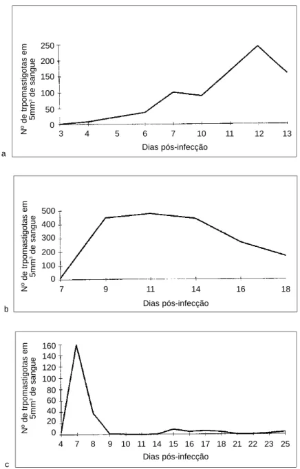
White swiss mice, 10/12g, of both sexes were intraperitoneally injected with 1,000 to 20,000 blood forms of T. cruzi in eight series of experiments, each involving about 50 animals. Parasitemia was evaluated daily for the first five groups and, for the other three groups, every other day, starting on day 5th. Mortality was very high. Even with small inoculum (5,000 trypomastigotes) mortality was total by the 10-11th day, eventually reaching the 15th/18thday in those receiving the smallest inoculum (1,000 trypomastigotes). Animals found dead were discarded. Following inoculation the animals were sacrificed on the 5th, 7th, 10th, 12th, 14th, 15th and 18thday.
To avoid high mortality, a group of 95 mice infected with 2,000 to 5,000 trypomastigotes was treated on the 7th day of infection with 50mg/kbw of benznidazole daily, during 5 days. They were sacrificed at different periods of time, up to 30 days after infection. Parasitemia was considerably reduced after treatment and
KINETICS OF
TRYPANOSOMA CRUZI
DESTRUCTION
IN THE MOUSE SPLEEN
Zulmira M.S. Cordeiro, Ana Cristina Gonzalez Dahia and Zilton A. Andrade
Massive destruction of parasitized splenic macrophages was histologically observed at the height of a virulent infection caused by Trypanosoma cruzi (Y strain) in the mouse. This was coincident with a sudden drop in parasitemic curve. Most of the animals died at this point, probably due to the liberation of toxic products, such as TNF, following the massive destruction of parasitized cells. However, parasitized-cell destruction indicated the transition from susceptibility to resistance. Although it has been extensively studied in vitro, this study contributes with the morphological counterpart observed in vivo by optical and electron microscopy. When infected animals were specifically treated during early infection transition to chronic phase was immediately observed without splenic parasitism. Animals that apparently recovered from massive cell-destruction in the spleen showed evidences of a rapid restoration of splenic architecture.
Key-words: Trypanosoma cruzi.Macrophages. Spleen.
Endereço para correspondência: Dr. Zilton A. Andrade.
Laboratório de Patologia Experimental, Centro de Pesquisas Gonçalo Moniz/FIOCRUZ. R.Valdemar Falcão 121, 40295-001 Salvador, B, Brasil.
remained low thereafter (Figure 1) and the animals presented normal development with no signs of disease.
Sacrificed animals were submitted to complete autopsies. The spleen was weighted. Fragments of the spleen, liver, heart and
Figure 1 - Parasitemic curves: a) animals inoculated with 20,000 trypomastigotes; b) shows a somewhat less severe course seen in animals inoculated with 4,000 trypomastigotes; c) it demonstrates the effect of treatment, which was administered from the 7th up to the 12th day of infection.
a
b
c
3 4 5 6 7 10 11 12 13
Nº de trpomastigotas em
5mm
3de sangue
Nº de trpomastigotas em
5mm
3de sangue
Nº de trpomastigotas em
5mm
3de sangue 250 200 150 100 50 0
Dias pós-infecção
500 400 300 200 100 0
7 9 11 14 16 18
Dias pós-infecção
160 140 120 100 80 60 40 20 0
intestines were fixed in neutral formalin (Millonig fixative). and later embedded in paraffin. The sections obtained were stained with hematoxilyn and eosin. Small pieces of splenic tissues were immediately fixed in 4% gluteraldehyde in 0.2M sodium cacodylate buffer, pH 7.4, during one hour, post-fixed for one hour in 1% pH 7.4 osmium tetroxide in 0.3M cacodylate buffer. These fragments were embedded in 812 polybed resin. The ultra-thin sections were contrasted with lead citrate and 7% uranyl acetate, and examined under an M-9 Zeiss electron microscope at 50Kv.
Parasitemia was performed in a drop of blood obtained from the mouse tail, covered with a cover slip and immediately examined with 40x objective x 10x ocular. One-hundred fields were counted and results were expressed as averages (Figure 1).
RESULTS
Parasitemic curves are shown in Figure 1. At light microscopy parasitism of splenic macrophages was detected on day 5thin isolated cells present specially within the marginal zone of the lymph follicles. Occasionally, macrophages in the red pulp also appeared parasitized at this time. Parasitism became more evident from the 7th day on. Amastigotes
in cells of the white pulp appeared later (15th day). Progressive accumulation of parasitized cells in the spleen was followed at first by relatively mild changes in splenic histology. Only after the 7th day of infection, three main changes became progressively more constant: hyperplasia of non-parasitized macrophages, depletion of lymphoid cells in the red pulp and activation of germinal centers of the white pulp. Depletion of lymphocytes was probably due to apoptosis, since many of them exhibited cytoplasmic condensation, nuclear clumping, picnosis and cariorrhexis. Non-parasitized macrophages, presented clear and enlarged cytoplasm, tending toward an epithelioid appearance. The center of the lymphoid follicles and the marginal perifollicular zones were expanded and the latter gradually fused with the red pulp. Dark dots, probably nuclear fragments, were frequently found within phagocytes in the center of the white pulp. After the 7th day of infection a few, isolated, parasitized macrophages disintegrated and the liberated amastigotes were seen within the shrunken and vacuolated macrophage cytoplasm or free in the interstitial tissue. This was r e g u l a r l y a c c o m p a n i e d b y a n i n f l u x o f polymorphonuclear neutrophils, which sometimes were seen with phagocytized amastigotes (Figure 2).
Macrophage parasitism, lymphoid-cell depletion and macrophage hyperplasia became accentuated in the following days. However, the most striking change was observed around the 12th-15th days when intracellular parasitism was at its highest point. Then, all parasitized cells appeared swollen, fragmented, containing d i s i n t e g r a t i n g p a r a s i t e s ( F i g u r e 3 ) . N o polymorphonuclear leukocytes were seen at this time, when a single parasitized macrophage could ever be detected, even after a thorough search of the histological sections. As indicated before these changes were coincident with a sharp decline in parasitemic curve and this event was regularly followed by the death of the animal.
Macrophages in the liver (Kupffer cells) also contained amastigotes and revealed the same changes observed in splenic macrophages (Figure 4). However, sections of the myocardium showed well preserved parasites within myocytes at the same time parasitized cells in t h e s p l e e n a n d l i v e r w e r e u n d e rg o i n g destruction (Figure 4).
Some animals were sacrificed next day after parasitemic drop and so the degradative changes could be analyzed by electron microscopy. Then, every intra and extra-cellular parasites showed evidences of degradation (Figure 5). Amastigotes were found within the cytoplasm of disintegrating macrophages or free in the interstitial tissue. In both cases they exhibited
Figure 3 - Aspect of the red splenic pulp during the phase of massive cellular destruction. Parasitized macrophages appear swollen, pale and vacuolated. Internalized parasites exhibit irregularities in nuclear size and staining (arrows) Hematoxylin & Eosin, 400X.
nuclear and cytoplasmic degenerative alterations. They usually presented an irregular outline and shrunken, dark, vacuolated cytoplasm and nuclear picnosis or cariorrhexis (Figure 6). The kinetoplast/mitochondrial complexes were markedly swollen and/or vacuolated. All parasitized macrophages were partially or totally destroyed, but the non-parasitized ones appeared normal. Frequently aggregated and partially degranulated platelets were observed within splenic capillaries (Figure 7), which showed excessive signs of pinocytosis and irregular vacuolization. Sometimes cells with
the characteristics of myofibroblasts (fusiform cells with dark submembranar contractile apparatus) exhibited focal proliferation, forming clusters in the middle or in close proximity to disintegrated macrophages. (Figure 8).
depletion was not a striking feature as in the previous cases. Accumulation of plasmocytoid and other basophilic lymphoid cells appeared at several scattered foci within the red pulp. Focal accumulation of macrophages, sometimes with a micro-granulomatous structure, was also observed. The marginal zone and the lymph follicles became more apparent,
suggesting a returning to the normal splenic structure. However, a few clear, empty areas were present within the red pulp, suggesting the dropping out of splenic cells.
The spleens taken from treated mice were essentially within normal limits and no parasites were seen within macrophages or elsewhere in the spleen after 5 days of
Figure 4 - Parasitized macrophages inside the hepatic sinusoids show disintegration in a similar way and at the same time as the splenic macrophages during the phase of massive cell destruction (arrow). Hematoxylin & Eosin, 160X.
Figure 5 - Section of the myocardium showing a large well preserved amastigote collection inside a myocyte. There is no reaction within the myocardial area seen in this picture. Hematoxylin & Eosin, 160X.
benznidazole treatment. Parasites were detected in other organs within muscle cells, but not in macrophages.
DISCUSSION
Sequential observation of splenic changes during a virulent T. cruzi infection in the mouse revealed that parasitism of splenic macrophages progressed up to a point of massive involvement and then, suddenly, there was a diffuse destruction of parasitized cells and parasites. Significance of this finding involves immunologic and pathogenetic
a s p e c t s o f g r e a t i m p o r t a n c e . I m m u n e destruction of infected cells probably means the expression of foreign antigen(s) in the macrophage external membranes. Such antigens are then a target to sensitized cytotoxic lymphocytes, specific antibodies, and/or antibody-mediated cell citotoxicity. Why the destruction occurred suddenly is a matter p r o b a b l y r e l a t e d t o t h e r e a c h i n g o f a n equivalence or critic point amongst immunologic factors15.
This would mean that the host became strongly immune, but paradoxically this transition is often fatal, probably due to the liberation of toxic products by the disintegrating cells. One of such product could well be TNF (tumor necrosis factors), since it is synthesized in great amount by macrophages parasitized by T. cruzi13and has been hold responsible for generalized toxic symptoms and even shock10. E v e n t u a l d i s i n t e g r a t i o n o f p a r a s i t i z e d macrophages occurring before the episode of massive cell destruction may depend on a different pathogenesis, since the host reaction was then limited to a focal infiltration by polymorphonuclear leukocytes. Similar findings observed in the connective tissue of mice have been correlated with local antigen-antibody reaction and hypocomplementemia12.
Macrophages are probably the first cells to be infected by T. cruzi. Trypomastigotes entering or being phagocytized by naive macrophages readly escape from the phagocytic vacuole and get into the cell cytosol, where they successively multiply as amastigotes. However, if macrophages are stimulated by sensitized T-cell lymphocyte soluble products, they become able to rapidly destroy internalized trypomastigotes6 7. These early events are crucial and may determined the outcome of the infection14. Suppressive chemotherapy accelerates this transitional phase and thus allows for a direct passage to the chronic stage, as has been demonstrated here and by others3. Probably, the elimination of parasites from the spleen would expose adequate antigens to the immune system and/or would interferes with the generation of suppressive splenic cells or other suppressive factors4.
Findings observed in two animals that apparently survived the critical period of massive parasitized-cell destruction are suggestive that reconstitution of splenic structure may be quite r a p i d a n d c o m p l e t e . T h a t c a p a c i t y f o r regeneration shown by the spleen is in
Figure 7 - Partially degranulated platelets (arrows) appeared frequently within the splenic capillaries, as shown in this electron micrograph. 7,000X,
keeping with observations made in other lymphoid tissues8.
RESUMO
Um estudo histologico sequenciado mostrou que o parasitismo dos macrófagos esplênicos por uma cepa virulenta (cepa Y) do Trypanosoma cruzi tem um curso progressivo, mas chega até um ponto em que todos as células parasitadas são subitamente destruídas. Tal achado coincidiu com uma quéda brusca da curva parasitêmica e com a morte da maioria dos animais, provavelmente devido à liberação de produtos tóxicos (como o TNF) pelas células desintegradas. O achado foi interpretado como o auge da transição entre uma fase de susceptibilidade e outra de resistência. Embora esta transição tenha sido bem estudada in vitro, este estudo contribui com os dados do substrato morfológico observados in vivo, através da microscopia ótica e eletrônica. O tratamento específico e supressivo feito na fase inicial da infecção acarreta uma transição imediata para a fase crônica e aí o parasitismo esplênico desaparece completamente. Os animais que aparentemente se recuperaram expontaneamente após a fase de destruição maciça dos macrófagos parasitados exibiram evidências de que a reconstituição da estrutura esplênica pode se fazer rapidamente.
Palavras-chaves: Trypanosoma cruzi. Macrófagos. Baço.
REFERENCES
1. Andrade SG. Morphological and behavioural
characterization of Trypanosoma cruzi strains.
Revista da Sociedade Brasileira de Medicina Tropical 18: 39-46, 1985.
2. Andrade ZA, Paiva A. Reações imunocelulares na tripanosomíase cruzi experimental. Revista Médica da Bahia 18: 27-33, 1962
3. Brener Z. Contribuição ao estudo da terapêutica experimental da doença de Chagas. Tese de Docência Livre, Facudade de Odontologia e Farmácia da Universidade de Minas Gerais, Belo Horizonte, MG., 1961.
4. Hoft DT, Lynch RG, Kirchoff LV. Kinetic analysis of antigen-specific immune responses in resistant and susceptible mice during infection with
Trypanosoma cruzi. Journal of Immunology 151: 7038-7047, 1993.
5. Maria TA. A resposta do baço na doença de C h a g a s m u r i n a ex p e r i m e n t a l : e s t u d o a o microscópio eletrônico. Tese de Doutorado, Universidade Federal de Minas Gerais, Belo Horizonte, MG 1987.
6. Nogueira N, Cohn Z. Trypanosoma cruzi:
mechanism of entry and intracellular fate in mammalian cells. Journal of Experimental Medicine 143: 1402-1420, 1976.
7. Nogueira N, Cohn Z.Trypanosoma cruzi: in vitro
induction of macrophage microbicidal activity. Journal of Experimental Medicine 148: 288-300, 1978.
8. Oliveira Filho J, Andrade ZA. Stromal collapse in acute atrophy of lymphoid organs. Experimental and Toxicologic Pathology 46: 81-85, 1994 9. Pizzi T, Knierin F. Modificaciones del baso en
relación con la tasa de anticuerpos circulantes en ratones experimentalmente infectados con
Trypanosoma cruzi. Bolletin Chileno de Parasitologia 10: 42-52, 1955.
10. Playfair JH, Taverne J. Anti-parasite effects of
tumor necrosis factor in vivo and in vitro. Ciba
Foundation Symposium 131: 192-205, 1987. 11. Rego SFM. Estudo das lesões provocadas pelo
Trypanosoma cruzi, Chagas 1909, no baço e
fígado de camundongo branco (Mus musculus)
com diversos graus de resistência. Tese de Doutorado, Faculdade de Medicina de Ribeirão Preto, Ribeirão Preto, SP, 1957.
12. Silva JC, Pirmez C, Morgado MG, Galvão Castro B. Immunopathological aspects of experimental
Trypanosoma cruzi infection: correlation of immune complexes and other serological festures with muscle lesions during infection. Parasite Immunology 7: 457-466, 1985.
13. Tarleton RL. Tumor necrosis factor (cachectin) production during experimental Chagas disease. Clinical and Experimental Immunology 73: 186-190, 1988.
14. Tarleton RL, Scott DW. Initial induction of immunity followed by suppression of responses
to parasite antigens during Trypanosoma cruzi
infection of mice. Parasite Immunology 9: 579-589, 1987.