256 Radiol Bras. 2011 Jul/Ago;44(4):256–262
Anatomical variations of paranasal sinuses at multislice
computed tomography: what to look for
*
Variações anatômicas das cavidades paranasais à tomografia computadorizada multislice: o que procurar?
Christiana Maia Nobre Rocha de Miranda1, Carol Pontes de Miranda Maranhão2, Fabiana Maia Nobre Rocha Arraes3, Igor Gomes Padilha4, Lucas de Pádua Gomes de Farias4, Mayara Stephanie de Araujo Jatobá5, Anna Carolina Mendonça de Andrade5, Bruno Gomes Padilha5
Multislice computed tomography is currently the imaging modality of choice for evaluating paranasal sinuses and adjacent structures. Such a method has been increasingly utilized in the assessment of anatomical variations, allowing their accurate identification with high anatomical details. Some anatomical variations may predispose to sinusal diseases, constituting areas of high risk for injuries and complications during surgical procedures. Therefore, the recognition of such variations is critical in the preoperative evaluation for endoscopic surgery.
Keywords: Multislice computed tomography; Paranasal sinuses; Anatomical variations.
A tomografia computadorizada multislice é, atualmente, a modalidade de imagem de escolha para a avaliação dos seios paranasais e das estruturas adjacentes. Ela tem sido cada vez mais utilizada para a avaliação das variações anatômicas, identificando-as de forma precisa e com elevados detalhes anatômicos. Algumas variações anatômicas podem predispor a sinusopatias e constituir regiões de alto risco para lesões e complicações durante atos operatórios. Portanto, o reconhecimento dessas variações é de fundamental importância no pré-operatório de cirurgia endoscópica. Unitermos: Tomografia computadorizada multislice; Seios paranasais; Variações anatômicas.
Abstract
Resumo
* Study developed at Clínica de Medicina Nuclear e Radiolo-gia de Maceió (Medradius), Maceió, AL, Brazil.
1. PhD, Coordinator for the Sector of Computed Tomography at Clínica de Medicina Nuclear e Radiologia de Maceió (Medra-dius), Professor of Radiology and Imaging Diagnosis, Universi-dade Federal de Alagoas (UFAL), Maceió, AL, Brazil.
2. Titular Member of Colégio Brasileiro de Radiologia e Diag-nóstico por Imagem (CBR), MD, Radiologist, Sector of Comput-ed Tomography, Clínica de MComput-edicina Nuclear e Radiologia de Maceió (Medradius), Maceió, AL, Brazil.
3. PhD, MD, Otorhinolaryngologist, Clínica Sinus, Maceió, AL, Brazil.
4. Graduate Students of Medicine, Student Monitor of the Discipline of Radiology and Imaging Diagnosis, Universidade Federal de Alagoas (UFAL), Maceió, AL, Brazil.
5. Graduate Students of Medicine, Universidade Federal de Alagoas (UFAL), Maceió, AL, Brazil.
Mailing Address: Dra. Christiana Maia Nobre Rocha de Mi-randa. Rua Hugo Corrêa Paes, 104, Farol. Maceió, AL, Brazil, 57050-730. E-mail: [email protected]
Received February 13, 2011. Accepted after revision June 3, 2011.
Miranda CMNR, Maranhão CPM, Arraes FMNR, Padilha IG, Farias LPG, Jatobá MSA, Andrade ACM, Padilha BG. Anatomical variations of paranasal sinuses at multislice computed tomography: what to look for. Radiol Bras. 2011 Jul/Ago;44(4):256–262.
the ethmoid bone and septal cartilage) re-sult in morphological variations such as septal deviation, chondrovomeral junction deformity and nasal bone spur. Septal de-viation is a sift of the midline associated with deformities or asymmetry of the ad-jacent turbinates (Figure 1A) or of the na-sal wall structure, with variable presenta-tions in the population(6,7).
Nasal septal spur (Figure 1B) is a gen-erally asymptomatic bone deformity that may cause restriction of the nasal air flow, and that may be associated with septal de-viation. Depending on the degree of ob-struction and symptoms severity, surgical correction may be required(7).
VARIATIONS OF THE MIDDLE TURBINATES
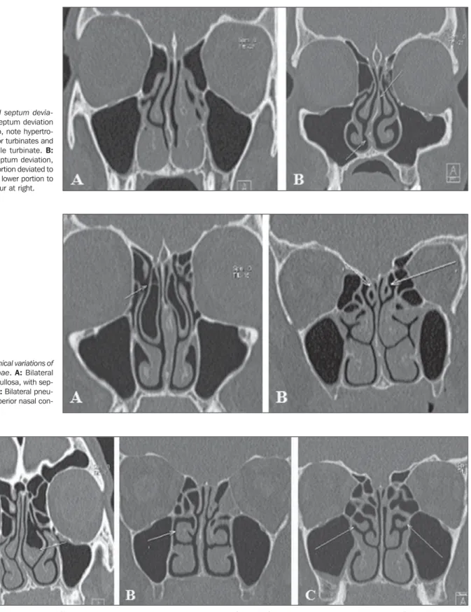
Concha bullosa (Figure 2A) is a varia-tion originated from pneumatizavaria-tion of the bone plate by extension of ethmoid sinus cells. Such variation may be either uni- or bilateral (Figure 2B). Varied degrees of pneumatization of the concha may be ob-served, possibly causing middle meatus or fected(1,2). Anatomical variations, in
asso-ciation with their inherent conditions, are added to such risks so the knowledge on these structures is critical for endoscopic surgeons as well as for radiologists in-volved in the preoperative evaluation, in order to avoid therapeutic failure and iatro-genic complications(2–5).
The acquisition of an excellent defini-tion of the sinusal anatomy for a preopera-tive endoscopic evaluation can be done by means of computed tomography that is the gold standard in the study of such struc-tures, for providing accurate information on soft tissues, bone structures and air, thus characterizing a highly sensitive imaging method(5,6).
The present pictorial essay is aimed at demonstrating the main anatomical varia-tions that may be detected in paranasal si-nuses by means of 40-row multislice com-puted tomography.
NASAL SEPTUM
Alterations of the nasal septum (delim-ited by the vomer, perpendicular plate for
INTRODUCTION
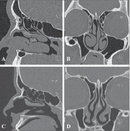
af-Figure 3. Anatomical variations of the nasal conchae. A: Middle concha bullosa at left, with inflammatory process inside. Hypoplasia of right middle nasal concha in association with septal deviation. B: Paradoxical middle turbinate at right. C: Bilateral, accessory middle nasal concha.
infundibulum obstruction, besides being related to deviation of the nasal septum to the contralateral side(8). Other variation that
is frequently associated with septal
devia-tion and spur is unilateral hypoplasia (Fig-ure 3A). In cases of bilateral hypoplasia, it is associated with low fovea ethmoidalis(7).
Paradoxical turbinates occur as the
convex-ity of the middle turbinate is directed to-wards the medial wall of the maxillary si-nus. Depending on the degree of curvature of the paradoxical turbinate (Figure 3B),
Figure 1. Nasal septum devia-tion. A: Nasal septum deviation to the right. Also, note hypertro-phy of the inferior turbinates and of the left middle turbinate. B: Double nasal septum deviation, with the upper portion deviated to the left, and the lower portion to the right with spur at right.
Table 1 Types of ostiomeatal complex abnormalities(7)
. Type
1 2 3 4 5 6
Uncinate process orientation Vertical
Vertical Vertical Horizontal Horizontal Horizontal
Ethmoid bulla appearance Enlarged or prolapsed
Normal Absent or hypoplastic Enlarged or prolapsed
Normal Absent or hypoplastic
compression of the infundibulum and sinusal obstruction may be observed(8).
UNCINATE PROCESS VARIATIONS
The uncinate process is a superior ex-tension of the lateral nasal wall that is ana-tomically relevant for draining the frontal recess. Variations such as hypertrophy, de-viation and pneumatization (Figure 4) may affect the drainage, generating abnormali-ties in the ostiomeatal complex (Table 1) and predisposing to obstruction(6,7,9).
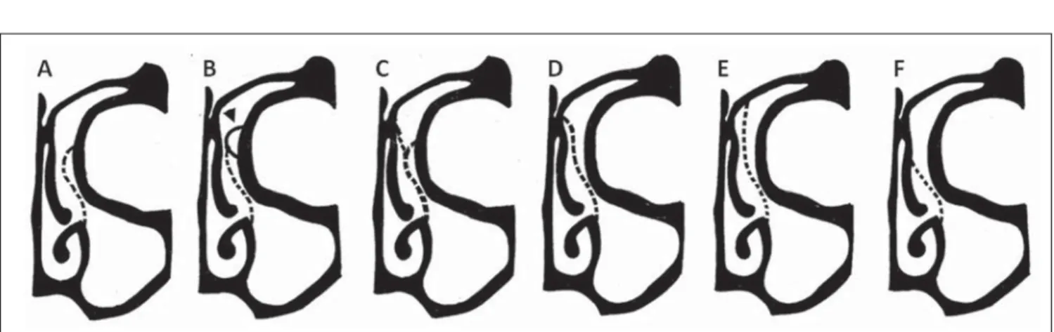
Traditionally, the uncinate process is identified from its lower segment through the architecture of the ostiomeatal unit. Variations in the superior insertion of the uncinate process are classified according to the criteria developed by Landsberg & Friedman(10) (Figure 5).
PARANASAL SINUSES PNEUMATIZATION EXTENT
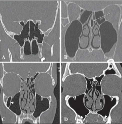
The sphenoid sinuses arise from pre-sphenoid bone centers, with variable pneu-matization extent (Figure 6A). In most of cases, pneumatization presents recesses related to the greater sphenoid wing, al-though lateral extensions may also be ob-served in the smaller sphenoid wing, infe-rolateral and septal recesses(7) (Figure 6B). Figure 4. Anatomical variations of the uncinate process. A: Right, horizontal uncinate process as-sociated with voluminous ethmoid bulla (type 4). B: Hypertrophy of left uncinate process and inser-tion into the vertical lamella of left middle nasal concha. C: Pneumatization of left uncinate process. D: Vertical left uncinate process associated with voluminous ethmoid bulla (type 1).
Figure 7. Keros classification. A: Type 1 (lateral cribriform lamella of 1–3 mm, the cribriform plate and the ethmoid cell roof are practically parallel to each other). B: Type 2 (lateral lamella of 4-7 mm, cribriform plate is much below the nasal cavity as compared with the ethmoid roof). C: Type 3 (lateral lamella of cribriform plate of 8–16 mm, ethmoid cell roof is located much above the plate).
Frontal sinus extension (Figure 6C) is a rare condition characterized by increased sinusal aeration beyond the normal margin of the frontal bone that originates from anterior extension of the anterior ethmoid
air cells. Extensions related to the lamina of the frontal bone, crista galli, besides in-ferior, symmetric extension of the frontal sinus towards the anterior ethmoid air cells may also be found predominantly in male
individuals in the age range between 20 and 40 years. Cases in children have not been reported(11).
As regards the maxillary sinus, four re-cesses have been described, as follows: the palatine recess that extends inferomedially to the hard palate towards the midline; the alveolar recess, closely related to the mo-lar and premomo-lar teeth roots (Figure 6D); the infraorbital recess, projecting anteriorly along the roof of the maxillary sinus; and the zygomatic recess that extends over the malar bone at variable distances(7).
VARIATIONS OF THE CRIBRIFORM PLATE
The cribriform plate may present at variable levels and, in such cases it is clas-sified according to the criteria developed by Keros (Figure 7), that is based on the height of the olfactory fossa in relation to the roof of the ethmoid sinus as compared with the length of the lateral lamella of cribriform plate. The higher the Keros grade, the greater the chance of injury of the cribriform plate and olfactory fossa, with consequential risk for iatrogenic cere-brospinal fluid fistula and olfactory im-pairment(12).
ETHMOID CELLS VARIATIONS
Infraorbital ethmoid cells (Figure 8), or Haller cells, are ethmoid air cells located anteriorly to the ethmoid bulla, along the orbital floor, adjacent to the natural ostium of the maxillary sinus, which may cause
Figure 6. Extension of pneumatization of paranasal sinuses. A: Extension of pneumatization of sphenoid sinus towards the lateral recess of the sphenoid bone (bilateral) and towards the anterior clinoid pro-cesses. B: Nasal septum pneumatization characterizing septal recess. C: Pneumatization of crista galli (arrow). Note the presence of septation in the right maxillary sinus (arrowhead). D: Extension of pneu-matization of the maxillary sinus towards the palatine recess (arrows) and alveolar recess (arrowheads).
Figure 8. Haller cell. Bilateral infraorbital ethmoid cells, larger at right.
Figure 10. Onodi cell. A,B: Bilateral Onodi cells (arrows). C: Note the sphenoid sinus ostium without connection with the Onodi cell (arrow). Figure 9. Agger nasi cell. Voluminous agger nasi cell at left.
mucociliary drainage obstruction, predis-posing to the development of sinusitis(5,7).
Agger nasi cells (Figure 9), which are the most anterior ethmoid cells, are located anteriorly to the upper margin of the naso-lacrimal duct and anteriorly to the plane of
Figure 11. Bulla frontalis. Bulla frontalis (arrow).
the maxillary sinus infundibulum. Studies demonstrate that their major dimensions are correlated with frontal sinus diseases and lacrimation(6,8,9).
Onodi cells (Figure 10) are ethmoid cells that have migrated to the anterior region of
the sphenoid sinus, with anterosuperior location, and intimately related to the op-tic nerve, causing opop-tic neuropathy in case of certain conditions that affect such cells(13).
Bulla frontalis (Figure 11) are character-ized by anterior ethmoid cells which invade
the frontal bone, bulging its floor, but with no connection with this sinus. They are more easily demonstrated at sagittal com-puted tomography, where they appear as ethmoid air cells located above the eth-moid bulla and as an extension towards the frontal sinus. Depending on their size and pneumatization extent, such cells may af-fect the frontal sinus drainage, represent-ing a real diagnostic challenge in addition to the difficulty of the surgical manage-ment of inflammatory diseases involving such a sinus.
in-fluencing its architecture that is determined by the patency of walls and boundaries of adjacent structures(9), such as frontal cells
which are classified into four types(14), as
follows:
a) type 1: detected in 37% of frontal re-cesses; defined as a single ethmoid cell located anteriorly to the frontal recess and above the agger nasi cell;
b) type 2: detected in 19% of cases; two or more ethmoid cells located anteriorly to the frontal recess, above the agger nasi cell (Figures 12A and 12B);
c) type 3: represents 6–8% of cases; a single, voluminous cell detected above the agger nasi cell, with extension to the fron-tal sinus;
d) type 4: rarely found, it represents 2– 4% of cases, corresponding to an ethmoid cell within the frontal sinus, with no con-nection with the agger nasi cell.
Figure 12. Anatomical variations in the frontal recess. A,B: Type II frontal cell at left. C,D: Normal frontal recess at right, anteriorly delimitated by agger nasi cell, laterally by the lamina papyracea, and medially by the anterior and superior portions of the middle concha.
MAXILLARY SINUSES SEPTA
Maxillary sinus septa (Figure 6C) are thin walls of cortical bone present within the maxillary sinus, with variable number, thickness and length. Such septa may di-vide the sinus into two or more cavities arising from the inferior and lateral walls of the sinus. Septa originating from teeth may be classified according to their devel-opment at the different phases of the den-tal eruption(15,16).
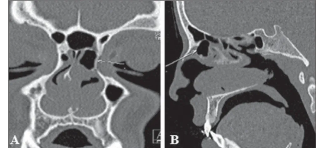
ACCESSORY MAXILLARY OSTIA
Accessory maxillary ostia (Figure 13) are generally solitary, but occasionally may be multiple. Such variation may be con-genital or secondary to sinusal diseases. Possible mechanisms involved in the de-velopment of such variation include: main
ostium obstruction, maxillary sinusitis or anatomical/pathological factors in the middle meatus, resulting in rupture of membranous areas(17).
CONCLUSION
Computed tomography plays a funda-mental role in the diagnosis of sinusal ana-tomical variations as well as of sinusal dis-eases, for a better guidance in the decision making about clinical therapeutic and sur-gical approaches, acting as an essential tool for a better performance of less invasive operative techniques.
REFERENCES
1. Ohnishi T, Tachibana T, Kaneko Y, et al. High-risk areas in endoscopic sinus surgery and preven-tion of complicapreven-tions. Laryngoscope. 1993;103: 181–5.
2. Souza SA, Souza MMA, Idagawa M, et al. Aná-lise por tomografia computadorizada do teto et-moidal: importante área de risco em cirurgia en-doscópica nasal. Radiol Bras. 2008;41:143–7. 3. Kantarci M, Karasen RM, Alper F, et al.
Remark-able anatomic variations in paranasal sinus region and their clinical importance. Eur J Radiol. 2004;50:296–302.
4. Schnipper D, Spiegel JH. Management of intrac-ranial complications of sinus surgery. Otolaryngol Clin North Am. 2004;37:453–72.
5. Kinsui MM, Guilherme A, Yamashita HK. Varia-ções anatômicas e sinusopatias: estudo por tomo-grafia computadorizada. Rev Bras Otorrinolarin-gol. 2002;68:645–52.
6. Riello APFL, Boasquevisque EM. Variações ana-tômicas do complexo ostiomeatal: achados tomo-gráficos em 200 pacientes. Radiol Bras. 2008;41: 149–54.
7. Earwaker J. Anatomic variants in sinonasal CT. Radiographics. 1993;13:381–415.
8. Araújo Neto SA, Martins PSL, Souza AS, et al. O papel das variantes anatômicas do complexo ostiomeatal na rinossinusite crônica. Radiol Bras. 2006;39:227–32.
9. Huang BY, Lloyd KM, DelGaudio JM, et al. Failed endoscopic sinus surgery: spectrum of CT findings in the frontal recess. Radiographics. 2009;29:177–95.
10. Turgut S, Ercan I, Sayin I, et al. The relationship between frontal sinusitis and localization of the frontal sinus outflow tract: a computer-assisted anatomical and clinical study. Arch Otolaryngol Head Neck Surg. 2005;131:518–22.
11. Hajiioannou J, Owens D, Whittet HB. Evaluation of anatomical variation of the crista galli using computed tomography. Clin Anat. 2010;23:370– 3.
12. Keros P. Über die praktische Bedeutung der Niveauunter-schiede der Lamina cribrosa des Ethmoids. Z Laryngol Rhinol Otol. 1962;41:809– 13.
13. Klink T, Pahnke J, Hoppe F, et al. Acute visual loss by an Onodi cell. Br J Ophthalmol. 2000;84:801– 2.
14. Lee WT, Kuhn FA, Citardi MJ. 3D computed to-mographic analysis of frontal recess anatomy in
patients without frontal sinusitis. Otolaryngol Head Neck Surg. 2004;131:164–73.
15. González-Santana H, Peñarrocha-Diago M, Gua-rinos-Carbó J, et al. A study of the septa in the maxillary sinuses and the subantral alveolar pro-cesses in 30 patients. J Oral Implantol. 2007; 33:340–3.