321 Radiol Bras. 2011 Set/Out;44(5):321–326
Computed tomography of intra- and extramural ethmoid
cells: iconographic essay
*
Tomografia computadorizada das células etmoidais intra e extramurais: ensaio iconográfico
Fabrício Guimarães Gonçalves1, Cássio Lemos Jovem2, Leonardo de Oliveira Moura3
The development of the paranasal sinuses is an intricate process that begins in the intrauterine life and terminates in early adulthood. Among the paranasal sinuses, the ethmoid cells or labyrinth are probably the most complex structures, being associated with the highest number of normal variants. Variations in the pattern of pneumatization of the ethmoid cells can be divided into intra- and extramural cells. Intramural cells are those which develop within the ethmoid labyrinth. Extramural cells are those that develop isolatedly. Computed tomography is the most useful tool in the evaluation of inflammatory processes of the paranasal sinuses. Computed tomography also plays a relevant role in the preoperative planning as well as in the postoperative follow-up, since it demonstrates exact anatomical details of normal structures with accuracy in the detection of variants. In the present pictorial essay, the authors describe the most common anatomical variants of the ethmoid labyrinth and their relationship with adjacent structures. Endoscopic sinonasal surgery has become increasingly less invasive, requiring more detailed anatomical imaging of this region.
Keywords: Computed tomography; Ethmoid sinus.
O desenvolvimento dos seios paranasais é um processo intricado que se inicia na vida intrauterina e termina na idade adulta. Dos seios da face, as células etmoidais são provavelmente as estruturas mais complexas e as que estão as-sociadas com o maior número de variantes da normalidade. Variações no padrão de pneumatização das células et-moidais podem ser divididas em intra ou extramurais. Intramurais são aquelas que ao se desenvolverem mantêm contato com o labirinto etmoidal, e extramurais as que se desenvolvem isoladamente. A tomografia computadorizada é a fer-ramenta mais útil na avaliação de processos inflamatórios dos seios paranasais. De igual modo, ela é importante para o planejamento pré-operatório e controle pós-operatório, pois possibilita grande detalhe anatômico das estruturas normais e detecção precisa de suas variantes. Neste ensaio iconográfico os autores descrevem as principais variantes da normalidade do labirinto etmoidal e suas relações com estruturas adjacentes. Cirurgias endoscópicas para o trata-mento de afecções dos seios paranasais têm-se tornado cada vez menos invasivas, o que certamente aumentará a demanda por relatórios de imagem cada vez mais ricos em detalhes desta região.
Unitermos: Tomografia computadorizada; Seio etmoidal.
Abstract
Resumo
* Study developed at McGill University Health Center (MUHC), Montreal General Hospital, Montreal, Quebec, Canada.
1. MD, Radiologist, Member of Colégio Brasileiro de Radiolo-gia e Diagnóstico por Imagem (CBR), Clinical Fellow in Neuro-radiology, McGill University Health Center (MUHC), Montreal General Hospital, Montreal, Quebec, Canada.
2. MD, Radiologist, Fellow in Neuroradiology, Med Imagem – Hospital Beneficência Portuguesa e São Paulo, São Paulo, SP, Brazil.
3. MD, Radiologist, Member of Colégio Brasileiro de Radiologia e Diagnóstico por Imagem (CBR), Cetrim – Centro de Treinamento em Imagenologia, Ecoclínica Multi Diagnose, João Pessoa, PB, Brazil.
Mailing Address: Dr. Fabrício Guimarães Gonçalves. Depart-ment of Diagnostic Radiology, Montreal General Hospital. 1650 Cedar Avenue, Room D5 137, Montreal, Quebec, Canada H3G 1A4.
Received March 11, 2011. Accepted after revision August 24, 2011.
Gonçalves FG, Jovem CL, Moura LO. Computed tomography of intra- and extramural ethmoid cells: iconographic essay. Radiol Bras. 2011 Set/Out;44(5):321–326.
Present at birth, ethmoid cells present a variable and heterogeneous development until the early adulthood(11). At different
stages of the ethmoid labyrinth develop-ment, there are two main groups of normal variants: the intramural and extramural eth-moid cells. Extramural etheth-moid cells are structures that pneumatize and develop protruding externally to the ethmoid laby-rinth. This group is comprised of agger nasi cells, frontal cells, supraorbital ethmoid cells and Haller and Onodi cells. On their turn, intramural ethmoid cells are structures that pneumatize and remain intimately re-lated to the ethmoid labyrinth, character-ized by the frontal bulla cells, suprabullar cells, and ethmoid bulla(12). The
recogni-tion of different anatomical variants is of of uncomplicated paranasal sinuses
inflam-matory processes(1,2). Additionally, CT is
extremely useful in the preoperative plan-ning and in postoperative control in cases of endonasal interventions for providing important details on the normal anatomy and its variants(3,4). Most recently,
multipla-nar and three-dimensional reconstruction has been utilized as a part of the routine in the study of paranasal sinuses, for providing higher quality diagnostic images and data than conventional CT, particularly in the identification of some anatomic variants(4–6).
Anatomical variants arising from ethmoidal cells development process are the most com-mon, frequently associated with inflam-matory processes and responsible for most of paranasal sinuses revision surgeries(7–10).
INTRODUCTION
utmost importance for the rhinologist. Be-cause of their proximity with the main drainage pathways of the paranasal sinuses, some cells may reduce the mucociliary clearance thus predisposing to inflamma-tory processes and causing endonasal en-doscopic revision surgery.
In the present pictorial essay, extramu-ral and intramuextramu-ral cells are described in detail, with illustrative images from each one. Additionally, the authors emphasize some relevant aspects of the ostiomeatal unit, frontal recess and sphenoethmoidal recess, structures of great importance in the mucociliary drainage of paranasal sinuses. All the images in the present essay were se-lected from paranasal sinuses computed tomography studies of patients from the otorhinolaryngology clinic at Hospital Universitário de Brasília, by the authors F.G.G. and C.L.J., with respectively eight-and three-year experience in radiology. All the paranasal sinuses studies were per-formed with the volumetric acquisition technique in a 4-channel multidetector CT apparatus. The images were reviewed and reconstructed in multiple planes on General Electric Advantage 4.2 workstations.
Ostiomeatal unit and frontal and sphenoethmoidal recesses
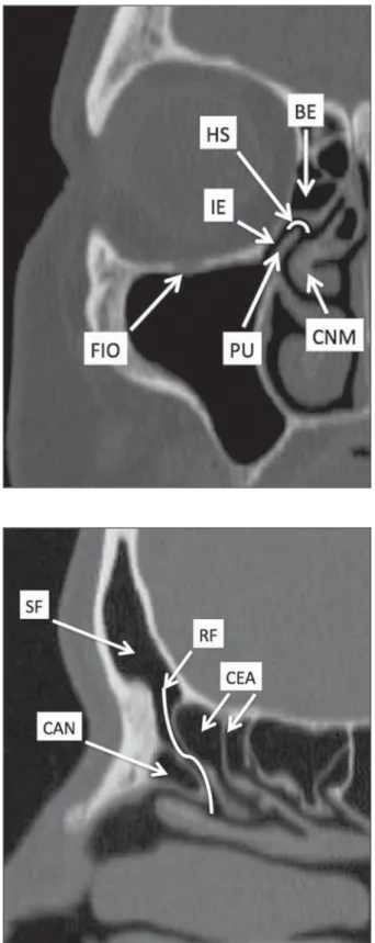
The ostiomeatal unit is the common drainage pathway of the anterior paranasal sinuses, acting as a unit that controls and modulates the mucociliary drainage of the frontal sinuses, anterior ethmoid cells and maxillary sinus. This is composed of the following structures: uncinate process, eth-moid bulla, middle turbinate, and the spaces between these structures (infun-dibulum, middle meatus and semilunar hia-tus) (Figure 1)(13,14).
The frontal recess is the drainage path-way of the frontal sinuses. Each frontal recess is surrounded by the following struc-tures: the agger nasi cells, ethmoid bulla, middle turbinate, basal lamella and anterior ethmoidal cells(1) (Figure 2). The
spheno-ethmoidal recess is the drainage pathway of the sphenoid sinus, a rather small struc-ture next to the midline, posteriorly to the upper turbinate, between the anterior sphe-noid sinus wall and the posterior wall of the ethmoid cells(13) (Figure 3).
Agger nasi cell
Agger nasi cell, first described by H. Meyer(15), is the most anterior ethmoid cell,
present in up to 98% of cases. Its anterior and posterior walls constitute part of the
frontal recess walls, endoscopically corre-sponding to a bulging in the nasal wall anteriorly to the middle turbinate(16). The
malities related to the agger nasi cells are the most frequent causes for endonasal endoscopic revision surgery(17).
Frontal cells
Frontal cells or Kuhn’s cells(12), are
eth-moid cells intimately related to agger nasi cells. According to their pneumatization pattern, frontal cells may be divided into four different types (types I thru IV). By far
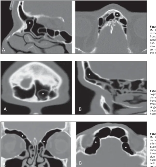
the most common ones, type I frontal cells are single cells, located above the agger nasi cell and inferiorly to the frontal sinus floor (Figure 5). Type II frontal cells cor-respond to two or more anterior ethmoid cells which pneumatize above the agger nasi cell, sometimes extending towards the interior of the frontal sinus (Figure 6). Type III frontal cells are single anterior ethmoid cells, which, because of their large volume,
pneumatize above the agger nasi cell supe-riorly extending into the frontal sinus (Fig-ure 7). Less frequent, type IV frontal cells are isolated cells located within the fron-tal sinus, above the agger nasi cell (Figure 8)(13). Park et al. have found a frontal cell
in 32% of the 105 studied patients, and the prevalence of types I thru IV frontal cells was, respectively, 24.2%, 4.2%, 3.1% and 0%(18).
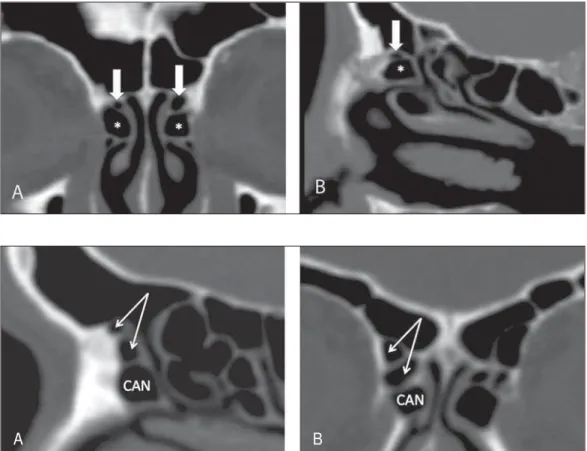
Figure 3. Axial image at the level of the sphenoethmoidal recesses (arrows). The sphenoeth-moidal recess is best visualized in the axial plane and drains the sphenoid sinuses and some posterior ethmoid cells.
Figure 4. Coronal image at the level of the agger nasi cells (highlighted – asterisk). Agger nasi cells are considered to be the most anterior ethmoid cells.
Figure 5. Coronal (A) and sag-ittal (B) images demonstrating type I frontal cells (arrows) and their intimate relationship with agger nasi cells (asterisks). Type I frontal cells are single and do not extend into the frontal sinus.
Figure 6. Sagittal (A) and coro-nal (B) images demonstrating type II frontal cells (arrows). Type II frontal cells present a ladder-like appearance of two or more cells, and are located above the agger nasi cells (CAN).
Supraorbital ethmoid cell
Supraorbital ethmoid cell is the ethmoid cell that extends superolaterally between the middle orbit wall and the ethmoid roof (Figure 9)(13). Supraorbital ethmoid cells
may simulate multiple frontal sinuses, type III frontal cells, suprabullar cells, frontal bulla cells or interfrontal sinus septal cells on coronal CT images. According to Zhang et al., its incidence may reach 5.4%(19).
Haller’s cells
Haller cells, also known as orbitomaxil-lary cells, were first described by Albert Von Haller in 1743(20). They are extramural
eth-moidal cells that pneumatize inferiorly to the orbital floor, extending from the ethmoid labyrinth, below the ethmoid bulla, towards the interior of the maxillary sinus (Figure 10). Because of its location, next to the ostiomeatal unit, and depending on its number and size, the presence of such cells may cause obstruction of mucociliary drain-age and be related to sinusopathy(13).
Ac-cording to Stackpole and Edelstein, Haller cells are present in 34% of the patients with sinusopathy. Additionally, such authors have demonstrated that the higher the num-ber of Haller’s cells, the greater the chances of maxillary sinus inflammation(21).
Onodi cells
Also known as sphenoethmoidal cells, Onodi cells were first described by the Hungarian laryngologist Adolf Onodi, in 1904(22). Onodi cells are considered as the
most posterior ethmoid cells. Recent data on the prevalence of Onodi cells at CT studies, particularly in cases where more modern techniques are utilized, are scarce. According to anatomy studies on cadavers, Onodi cells are very common, with a preva-lence ranging between 39% and 60%(23,24).
The presence of a horizontal septum divid-ing the sphenoid sinuses in “two floors” suggests the presence of an Onodi cell
(Fig-Figure 9. Coronal coronal (A) and axial (B) images demonstrating supraorbital ethmoid cells (asterisks). Such cells pneumatize above the orbits floor, some-times causing the impres-sion of septate frontal si-nuses. Supraorbital ethmoid cells are best identified on coronal and axial views. Figure 8. Coronal (A) and sagittal oblique (B) images demonstrating a type IV frontal cell (asterisk). Type IV frontal cells are generally single and are isolatedly lo-cated within the frontal si-nuses.
13)(7,25). According to Park et al., its
preva-lence is around 8%(18).
Ethmoid bulla
Classically, ethmoid bulla is described as the largest and most constant of the an-terior ethmoid cells. It is an intramural eth-moid cell in intimate relationship with the ostiomeatal unit, embracing the lamina papyracea and which drains into the middle meatus through a pneumatized retrobullar tract(26) (Figure 1). Because of its
consis-tency, the ethmoid bulla is an important repair for the rhinologist. In addition, to-gether with the uncinate process, it defines the hiatus semilunaris, the exit pathway to the ethmoidal infundibulum, located on the lateral wall of the nasal cavity(13–15). Wright
& Bolger(26), after detailed macroscopic
analyses of 14 nasal cavities in 8 speci-mens, have observed that all the ethmoid bullas presented incomplete posterior walls independently from their pneumatization degree and from the presence of internal septations. According to those authors, it is debatable whether the ethmoidal bullas actually represent a true cell or should be considered a lamella. In spite of being known as ethmoid bulla since 1893 (Zuckerkandl(27)), according to the authors,
“from an anatomical perspective, bulla is not, however, the best description for such a structure”(26).
CONCLUSION
The development of the ethmoid laby-rinth is a heterogeneous and variable pro-cess. From such an intricate process, two specific cell groups originate: the extramu-ral ethmoid cells and the intramuextramu-ral eth-moid cells. Computed tomography with
Figure 12. Sagittal oblique (A) and axial (B) images demonstrating a frontal bulla cell (asterisk) and its relationship with the skull base and the frontal sinus. The anterior edge of a frontal bulla cell does not ex-tend into the frontal sinuses. Figure 11. Coronal image demon-strating an Onodi cell (asterisk) immediately below the optical nerve channel. The “two-floor” as-pect of sphenoidal sinus in the coronal plane is useful in the iden-tification of this type of cell. Figure 10. Haller’s cells (asterisks) are ethmoid cells that pneumatize inferiorly to the orbits towards the interior of the maxillary sinuses. In special situations such cells may reduce the ostiomeatal unit and predispose to obstruction of the maxillary infundibulum.
ure 11). Onodi cells are intimately related to the optical nerves and internal carotid arteries, hence their clinical relevance in the event of sinusopathy(13).
Frontal bullar cell
Frontal bullar cell is the ethmoidal cell located above the ethmoid bulla that pneumatizes along the skull base towards the frontal sinus, sometimes causing con-vexity in its floor (Figure 12)(7,23).
Accord-ing to Park et al., its prevalence may reach 10%(18).
Suprabullar cell
multiplanar reformation provides greater anatomical detail and higher spatial reso-lution for the study of the paranasal sinuses anatomy. Recognition of the anatomical variants in paranasal sinuses studies may be useful for the assisting physician in the management of patients with sinuso-pathies. The improvements in minimally invasive endoscopic surgeries of the paranasal sinuses will probably increase the demand for reports with more anatomical details of the paranasal sinuses and their variants.
REFERENCES
1. Kountakis SE, Senior BA, Draf W. The frontal sinus. Berlin: Springer-Verlag; 2005. 2. Aygun N, Zinreich SJ. Imaging for functional
en-doscopic sinus surgery. Otolaryngol Clin North Am. 2006;39:403–16, vii.
3. Batra PS. Radiologic imaging in rhinosinusitis. Cleve Clin J Med. 2004;71:886–8.
4. Beale TJ, Madani G, Morley SJ. Imaging of the paranasal sinuses and nasal cavity: normal anatomy and clinically relevant anatomical vari-ants. Semin Ultrasound CT MR. 2009;30:2–16. 5. Isaacs SJ, Goyal P. Comparison between three-dimensional and triplanar computed tomography imaging of the frontal recess. Am J Rhinol Al-lergy. 2009;23:502–5.
6. Kew J, Rees GL, Close D, et al. Multiplanar re-constructed computed tomography images im-proves depiction and understanding of the
anatomy of the frontal sinus and recess. Am J Rhinol. 2002;16:119–23.
7. Bradley DT, Kountakis SE. The role of agger nasi air cells in patients requiring revision endoscopic frontal sinus surgery. Otolaryngol Head Neck Surg. 2004;131:525–7.
8. Ramadan HH. Revision endoscopic sinus surgery in children: surgical causes of failure. Laryngo-scope. 2009;119:1214–7.
9. Osguthorpe JD. Surgical causes of failure in en-doscopic sinus surgery. Laryngoscope. 2000;110: 177.
10. Ramadan HH. Surgical causes of failure in endo-scopic sinus surgery. Laryngoscope. 1999;109: 27–9.
11. Scuderi AJ, Harnsberger HR, Boyer RS. Pneuma-tization of the paranasal sinuses: normal features of importance to the accurate interpretation of CT scans and MR images. AJR Am J Roentgenol. 1993;160:1101–4.
12. Bent JP, Cuilty-Siller C, Kuhn FA. The frontal cell as a cause of frontal sinus obstruction. Am J Rhinol. 1994;8:185–91.
13. Coates MH, Whyte AM, Earwaker JW. Frontal recess air cells: spectrum of CT appearances. Australas Radiol. 2003;47:4–10.
14. Laine FJ, Smoker WR. The ostiomeatal unit and endoscopic surgery: anatomy, variations, and imaging findings in inflammatory diseases. AJR Am J Roentgenol. 1992;159:849–57. 15. Meyer H. Lehrbuch der Anatomie. Leipzig; 1861. 16. Earwaker J. Anatomic variants in sinonasal CT.
Radiographics. 1993;13:381–415.
17. Eskiizmir GA. The role of agger nasi air cells in revision endoscopic sinus surgery. Otolaryngol Head Neck Surg. 2005;133:464; author reply 465.
18. Park SS, Yoon BN, Cho KS, et al. Pneumatiza-tion pattern of the frontal recess: relaPneumatiza-tionship of the anterior-to-posterior length of frontal isthmus and/or frontal recess with the volume of agger nasi cell. Clin Exp Otorhinolaryngol. 2010;3:76–83. 19. Zhang L, Han D, Ge W, et al. Computed tomo-graphic and endoscopic analysis of supraorbital ethmoid cells. Otolaryngol Head Neck Surg. 2007;137:562–8.
20. Haller A. Praelectiones academicae in proprias institutiones rei medicae/Hermanni Boerhaave. Vol. IV. Vandenhoeck, Gottingae; 1743. p. 43. 21. Stackpole AS, Edelstein DR. The anatomic
rel-evance of the Haller cell in sinusitis. Am J Rhinol. 1997;11:219–23.
22. Onodi A. Die Sehstoerungen und Erblidung nasalen Ursprunges, bedingt durch Erkrankungen der hinteren Nebenhoehlen. Z Augenheilkd. 1904;12:23–46.
23. Driben JS, Bolger WE, Robles HA, et al. The re-liability of computerized tomographic detection of the Onodi (sphenoethmoid) cell. Am J Rhinol. 1998;12:105–11.
24. Thanaviratananich S, Chaisiwamongkol K, Kraitkrakul S, et al. The prevalence of an Onodi cell in adult Thai cadavers. Ear Nose Throat J. 2003;82:200–4.
25. Landsberg R, Friedman, M. A computer-assisted anatomical study of the nasofrontal region. Laryn-goscope. 2001;111:2125–30.
26. Wright ED, Bolger WE. The bulla ethmoidalis: lamella or a true cell? J Otolaryngol. 2001;30: 162–6.
27. Zuckerkandl E. Normale und pathologische Ana-tomie der Nasenhöhle und ihrer pneumatischen Anhänge. Vol 1. Wien: Wilhelm Braumüller; 1893.