ABSTRACT
http://dx.doi.org/10.1590/1678-775720140371
Interrelationship between implant and orthognathic
surgery for the rehabilitation of edentulous cleft
palate patients: a case report
José Fernando Scarelli LOPES1, João Henrique Nogueira PINTO1, Monica Moraes Waldemarin LOPES1, Reinaldo MAZOTTINI2, Simone SOARES3
1- Prosthodontics Sector, Hospital for Rehabilitation of Craniofacial Anomalies - University of São Paulo (HRAC/USP), Bauru, SP, Brazil. 2- Surgery Sector, Hospital for Rehabilitation of Craniofacial Anomalies - University of São Paulo (HRAC/USP), Bauru, SP, Brazil.
3- Department of Prosthodontics, Bauru School of Dentistry, University of São Paulo; Prosthodontics Sector, Hospital for Rehabilitation of Craniofacial Anomalies - University of São Paulo (HRAC/USP), Bauru, SP, Brazil.
Corresponding address: Simone Soares - Setor de Prótese Dentária - Hospital de Reabilitação de Anomalias Craniofaciais - Universidade de São Paulo (HRAC/USP) - Rua Sílvio Marchioni, 3-20 - Vila Nova Universitária - Bauru - SP - Brazil - 17012-900 - e-mail: [email protected] / [email protected]
Submitted: September 17, 2014 - Modiication: November 4, 2014 - Accepted: November 5, 2014
A
43-year-old woman with a unilateral cleft lip and palate, presenting a totally edentulous maxilla and mandible with marked maxillomandibular discrepancy, attended the Prosthodontics section of the Hospital for Rehabilitation of Craniofacial Anomalies, Universityof São Paulo for treatment. She could not close her mouth and was dissatisied with
her complete dentures. Treatment planning comprised placement of six implants in the maxilla, four in the mandible followed by prostheses installation and orthognathic surgery. The mandibular full arch prosthesis guided the occlusion for orthognathic positioning of the maxilla. The maxillary complete prosthesis was designed to assist the orthognathic surgery with a provisional prosthesis (no metal framework), allowing reverse treatment planning. Maxillary and mandibular realignment was performed. Three months later, a relapse in the position of the maxilla was observed, which was offset with a new maxillary prosthesis. This isa complex interdisciplinary treatment and two-year follow-up is presented and discussed. It should be considered that this type of treatment could also be applied in non-cleft patients.
Keywords: Orthognathic surgery. Cleft palate. Implant-supported dental prothesis.
INTRODUCTION
Treatment of adult patients with cleft lip and palate who do not receive proper treatment at the appropriate age is challenging for prosthodontists. The situation becomes worse in adulthood if teeth are lost and patients become edentulous, making prosthetic reconstruction even more challenging5.
The lack of information and limited surgical and dental expertise often preclude the proper facial reconstruction and dental rehabilitation for patients with clefts presenting maxillomandibular discrepancies and malocclusion in adolescence. The appropriate timing and planning of surgical
reconstruction signiicantly reduce functional and
esthetic disorders in patients with cleft lip and palate. Ideally, the treatment of patients with clefts should begin at an early age.
Many patients with clefts are partially or totally edentulous, and this condition is nearly always associated with a deficient maxillomandibular skeletal relationship. Thus, orthognathic surgery for
these patients should fulill the essential needs for
prosthesis placement and also re-establish balance in the facial architecture, which is fundamental to stability3.
Studies have demonstrated that the functionality of the stomatognathic system is significantly reduced in patients with complete dentures compared with that in individuals with natural teeth14. Endosseous implants increase the treatment eficacy of edentulous patients, who
may be rehabilitated by the placement of four to six implants, followed by fabrication of an
implant-supported ixed partial denture. Since the 1990s,
developmental maxillocraniofacial defects, including the rehabilitation of patients with clefts18.
In 1995, Posnick and Tompson15 reviewed
the complications and long-term results of a consecutive series of adolescents with clefts who underwent primary repair in childhood and later developed maxillary deformity and malocclusion requiring orthognathic surgery. The methods used to manage maxillary deformity, malocclusion,
residual oronasal istula, and bone defects in these
adolescents were safe and reliable and offered the patients an enhanced quality of life.
These aforementioned problems affect patients’ psychosocial well-being, function, and esthetics, to some extent impairing their ability to integrate into society. A follow-up study of young adults with
cleft lip and palate showed that up to 30% of them
experienced psychosocial maladjustment, with highest levels of dissatisfaction with appearance, hearing, speech, dentition, and social life. As for education, occupation, and marital status, the
psychosocial dificulties observed during childhood
persisted, with consequences in adolescence and adulthood, presenting problems in terms of education, self-confidence, assertiveness, and independence, which generate inhibition16.
Craniofacial morphology in patients with cleft palate shows a short and retrusive maxilla in relation to the cranial base6. Also, the mandible is
small and retrusive, with an obtuse gonial angle and a steep mandibular plane. The Le Fort I osteotomy is commonly used for correction of maxillary
deiciency in patients with and without clefts, but
differences in post-operative skeletal stability and soft-tissue changes exist. The tendency toward relapse and the extent of relapse tend to be higher in patients with clefts10. Several factors related to the cleft itself, such as surgery, method of ixation,
neuromuscular adaptation, and orthodontics, might contribute to the tendency toward relapse7.
Reverse planning is fundamental when implants are used for the rehabilitation of edentulous patients, especially when orthognathic surgery is required. Treatment planning in these cases should focus on the esthetic and functional outcomes expected. The present clinical case involved functional features, mainly, but esthetic concepts were evaluated, since the patient was totally edentulous and could not close her mouth completely.
The rehabilitation of patients with cleft lip and palate at the Hospital for Rehabilitation of Craniofacial Anomalies (HRAC-USP) in Bauru, Brazil, includes interventions by an interdisciplinary team at appropriate times to achieve complete rehabilitation. This paper reports the interdisciplinary orthognathic surgery/prosthesis treatment in a patient presenting marked maxillomandibular discrepancies. The treatment was planned in
collaboration by a maxillofacial surgeon and a prosthodontist. Scarce clinical cases are reported in the literature with remarkable maxillomandibular discrepancy in cleft patients.
Intensive scarring and tension of the soft tissue have been reported as potential causes of relapse in patients with clefts13.
As Bailey, et al.1 (2004) wrote about the
stability and predictability of orthognathic surgery, downward movement of the maxilla is troublesome: if the maxilla is moved both forward and downward, the vertical component is likely to relapse, although the horizontal component has a good chance of being retained.
The purpose of this article was to report the interdisciplinary orthognathic surgery/prosthesis treatment in a patient presenting significant maxillomandibular discrepancies and totally edentulous. A surgeon and a prosthodontist planned the treatment. Although this is a rare case, it should be taken into consideration the relevance of
interdisciplinary treatments because the beneicial aspects could be more eficient to the patient.
CASE REPORT
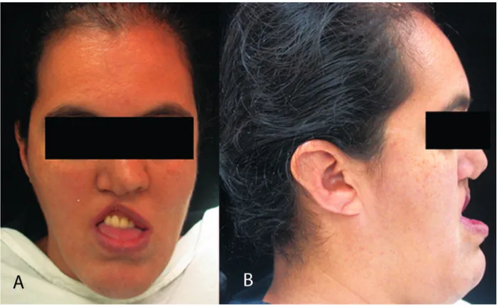
A 43-year-old female patient, presenting unilateral cleft lip and palate (UCLP), sought care at the Hospital for Rehabilitation of Craniofacial Anomalies (HRAC-USP). Her complaints were difficulties in functioning, poor esthetics, and poor prostheses (two complete dentures), since the maxillomandibular relationship was distorted (Figure 1A and B). Her psychological state was also a concern, and the patient’s expectations were high. Six implants were placed in the maxilla and four in the mandible (Figures 2 and 3). Maxillary and mandibular protocols were planned and mandibular full arch prosthesis was concluded before the orthognathic surgery, to guide the occlusion.
The first prosthesis was fabricated without consideration for the correct maxillomandibular relationship. Conversely, an adequate relationship between the prosthesis with soft tissue and lip support was achieved. This new prosthesis was fabricated in only acrylic resin, without metal framework, and its primary function was anchored to the orthognathic surgery.
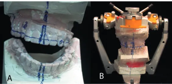
Since the maxilla is presented in two segments, the maxillary prosthesis was sectioned to facilitate the orthognathic surgery planning, and leveling the maxilla (Figure 4A). The newly achieved position was registered (Figure 4B) and duplicated, and two casts were obtained and mounted in a semi-adjustable articulator (Figure 5A) to aid in the planned surgery (Figure 5B).
this new position, and a new maxillomandibular relationship was established. Note that the implants inserted before the surgery were important in assisting the surgeon to obtain and maintain the suitable maxillomandibular position.
A f t e r 3 m o n t h s , a s e x p e c t e d , t h e maxillomandibular relationship relapsed (Figure 6A). A new maxillary complete prosthesis, with a favorable maxillomandibular relationship and metal framework, was carried out to correct the occlusion
Tooth/area Implant dimensions
4 3.75x10 mm
3 3.75x13 mm
9 3.75x8.5 mm
11 3.75x11.5 mm
12 3.75x10 mm
14 3.75x10 mm
Figure 2- Location and size of implants placed in the patient’s maxilla (Titanium Fix, A. S. Technology, São José dos Campos, São Paulo, Brazil)
Tooth/area Implant dimensions
23 3.75x15 mm
20 3.75x15 mm
26 3.75x15 mm
29 3.75x15 mm
Figure 3- Location and size of implants placed in the patient’s mandible (Conexão, Conexão Sistemas de Prótese, São Paulo, São Paulo, Brazil)
Figure 1- Initial condition presented by the patient when seeking care at the hospital (A) observe the discrepancy in the maxillomandibular relationship (B)
(Figure 6B).
The case was concluded after orthognathic surgery and the patient was satisfied with the cosmetic and functional outcome and was reintegrated into society. Two years later we can see the stability of the case report (Figures 7A, 7B).
DISCUSSION
An interdisciplinary team should treat patients with clefts from early ages. These individuals may present esthetic, functional, and psychological problems and occasionally, when they seek Figure 5- Casts mounted in a semi-adjustable articulator (A) and positioned correctly in the semi-adjustable articulator (B)
Figure 6- Relapse, 3 months after surgery (A), and case concluded, with a correct maxillomandibular relationship (B)
subsequent treatment, may present remarkable maxillomandibular discrepancies.
In this case report, a 43-year-old woman with unilateral cleft lip and palate could not close her
mouth and was dissatisied with her complete
dentures. Six upper and four lower implants were installed; however, her maxillomandibular relationship was incorrect.
Patients who undergo lip and palate repair in childhood often show restricted maxillary growth to a greater or lesser extent, which may cause significant changes in the maxillomandibular relationship. These patients often exhibit a retruded maxilla in relation to the anterior cranial base, compared with individuals without clefts2,9. Xu, et
al.19 (2014) reported that the maxilla in patients
with clefts is shorter and retroclined, which causes
a signiicant maxillomandibular discrepancy.
This problem must be addressed in the treatment planning for patients with clefts17. Many cases at
HRAC-USP require a combined approach between orthognathic surgery and prosthodontics, as reported in the present case.
Orthognathic surgery to correct facial disharmony is part of the normal follow-up for patients with complete cleft lip and palate. Many factors are taken into account in the planning of corrective surgery,
such as facial proile, intermaxillary discrepancies,
and the dentoalveolar relationship.
Le Fort I maxillary osteotomy and advancement in patients with cleft lip and palate are the procedures of choice, but have inherent limitations, such as vertical and horizontal relapse7,8.
Orthognathic surgery is recommended for patients with cleft lip and palate and having moderate to severe maxillary deficiency after
growth. Analysis of unoficial data indicates that around 40% of patients with complete cleft lip and
palate at HRAC-USP undergo orthognathic surgery, especially maxillary advancement surgery or combined maxillary advancement and mandibular retroposition. In such cases, the degree of maxillary advancement required is considered clinically
signiicant, usually around 4 mm; therefore, partial
relapses are frequent4.
In a clinical study, Kumari, et al.12 (2013)
evaluated nine patients, pre- and postsurgically, and concluded that Le Fort 1 advancement in surgically repaired cases of cleft lip and cleft palate has inherent potential for relapse. In our case, three months later, we observed relapse and fabricated a new prosthesis for the patient.
In cases with severe maxillomandibular discrepancy, reverse planning allows for analysis of the actual situation, to highlight the importance of orthognathic surgery11. Reverse planning is critical
before implant insertion, because implants placed without planning can lead to prosthesis failure.
In this case, reverse planning consisting initially of registration of the desired maxillomandibular relationship was used to address the patient’s situation. Reverse planning consists of the mounting of models in a semi-adjustable articulator and
waxing the case in its intended inal position. A
guide is fabricated based on this new relationship, then used during surgery to bring the maxilla and mandible into the new position.
Cases of maxillomandibular discrepancies are observed in patients with cleft lip and palate, yet severe cases, such as with limited mouth-opening, as in the present case, are rare, making this case report unique in terms of prosthetic planning and surgery. Other cases with smaller discrepancies may involve surgery, orthodontics, or even prosthodontics to a lesser extent, yet they should also be guided by careful planning, similar to the present report. The present treatment combined orthognathic surgery/prostheses and implants, with
esthetic, functional, and psychologically beneicial
results.
CONCLUSION
The interdisciplinary team is important in managing patients with unique maxillomandibular relationships. Only orthognathic surgery can solve these kinds of discrepancies, and reverse planning will aid both the surgeon and the prosthodontist.
CONFLICTS OF INTEREST
None.
REFERENCES
1- Bailey LJ, Cevidanes LH, Profit WR. Stability and predictability
of orthognathic surgery. Am J Orthod Dentofacial Orthop.
2004;126:273-7.
2- Bishara SE. Cephalometric evaluation of facial growth in operated and non-operated individuals with isolated clefts of the
palate. Cleft Palate J. 1973;10:239-46.
3- Erkan M, Karaçay Ş, Atilla OA, Uzel G, Ülkür E, Yıldırım E.
Orthognathic surgery of a patient with oligodontia: alternative technique for intermaxillary fixation. J Craniofac Surg.
2012;23(5):e407-10.
4– Freitas JA, Garib DG, Trindade-Suedam IK, Carvalho RM,
Oliveira TM, Lauris , et al. Rehabilitative treatment of cleft lip and palate: experience of the Hospital for Rehabilitation of Craniofacial Anomalies–USP (HRAC-USP) – part 3: oral and maxillofacial
surgery. J Appl Oral Sci. 2012;20:673-9.
5– Güven O, Gürbüz A, Baltali E, Yilmaz B, Hatipoğlu M. Surgical
and prosthetic rehabilitation of edentulous adult cleft palate
patients by dental implants. J Craniofac Surg. 2010;21:1538-41.
6- Heliövaara A, Leikola J, Hukki J. Craniofacial cephalometric morphology and later need for orthognathic surgery in 6-year-old children with bilateral cleft lip and palate. Cleft Palate Craniofac
J. 2013;50(2):e35-40.
7- Heliövaara A, Ranta R, Hukki J, Rintala A. Skeletal stability of Le Fort I osteotomy in patients with isolated cleft palate and bilateral
8- Heliövaara A, Ranta R, Hukki J, Rintala A. Skeletal stability of Le Fort I osteotomy in patients with unilateral cleft lip and palate.
Scand J Plast Reconstr Surg Hand Surg. 2001;35:43-9.
9- Hermann NV, Kreiborg S, Darvann TA, Jensen BL, Dahl E,
Bolund S. Early craniofacial morphology and growth in children with unoperated isolated cleft palate. Cleft Palate Craniofac J.
2002;39:604-22.
10- Hirano A, Suzuki H. Factors related to relapse after Le Fort I
maxillary advancement osteotomy in patients with cleft lip and
palate. Cleft Palate Craniofac J. 2001;38:1-10.
11- Jones RH. Orthognathic surgery and implants. Ann R Australas
Coll Dent Surg. 2002;16:105-8.
12– Kumari P, Roy SK, Roy ID, Kumar P, Datana S, Rahman S. Stability of cleft maxilla in Le Fort I maxillary advancement. Ann
Maxillofac Surg. 2013;3:139-43.
13- Meazzini MC, Basile V, Mazzoleni F, Bozzetti A, Brusati R.
Long-term follow-up of large maxillary advancements with distraction osteogenesis in growing and non-growing cleft lip and palate
patients. J Plast Reconstr Aesthet Surg. 2015;68:79-86.
14- Mishellany-Dutour A, Renaud J, Peyron MA, Rimek F, Woda
A. Is the goal of mastication reached in young dentates, aged
dentates and aged denture wearers? Br J Nutr. 2008;99:121-8.
15– Posnick JC, Tompson B. Cleft-orthognathic surgery: complications and long-term results. Plast Reconstr Surg.
1995;96(2):255-66.
16- Roberts RM, Mathias JL. Psychosocial functioning in adults with congenital craniofacial conditions. Cleft Palate Craniofac J.
2012;49:276-85.
17- Scheuer HA, Höltje WJ, Hasund A, Pfeifer G. Prognosis of
facial growth in patients with unilateral complete clefts of the lip,
alveolus and palate. J Craniomaxillofac Surg. 2001;29(4):198-204. 18- Wang F, Wu Y, Zou D, Wang G, Kaigler D. Clinical outcomes
of dental implant therapy in alveolar cleft patients: a systematic
review. Int J Oral Maxillofac Implants. 2014;29(5):1098-105. 19- Xu Y, Yang C, Schreuder WH, Shi J, Shi B, Zheng Q, Wang Y.
Cephalometric analysis of craniofacial morphology and growth in unrepaired isolated cleft palate patients. J Craniomaxillofac Surg.