178
Testa ML et al. Diffusion-weighted MRI: biomarker for treatment response
Radiol Bras. 2013 Mai/Jun;46(3):178–180
Diffusion-weighted magnetic resonance imaging: biomarker
for treatment response in oncology
*
Ressonância magnética com difusão: biomarcador de resposta terapêutica em oncologia
Maria Luiza Testa1, Rubens Chojniak2, Letícia Silva Sene3, Aline Santos Damascena4
The authors report a case where a quantitative assessment of the apparent diffusion coefficient (ADC) of liver metastasis in a patient undergoing chemotherapy has shown to be an effective early marker for predicting therapeutic response, anticipating changes in tumor size. A lesion with lower initial ADC value and early increase in such value in the course of the treatment tends to present a better therapeutic response.
Keywords: Magnetic resonance imaging; Diffusion; Liver metastases; Breast cancer; Chemotherapy; Therapy monitoring.
Relatamos um caso no qual a avaliação quantitativa do coeficiente de difusão aparente (ADC) de metástases hepáticas submetidas a quimioterapia se mostrou um bom preditor e marcador precoce de resposta terapêutica, antecipando alterações de tamanho. Lesão com valor inicial do ADC mais baixo e com aumento precoce deste valor no curso do tratamento tende a apresentar melhor resposta terapêutica tardia.
Unitermos: Ressonância magnética; Difusão; Metástases hepáticas; Câncer de mama; Quimioterapia; Monitoramento da terapia.
Abstract
Resumo
* Study developed in the Department of Imaging Diagnosis at Hospital A. C. Camargo, São Paulo, SP, Brazil.
1. Master of Sciences – Oncology, MD, Radiologist, Hospi-tal A. C. Camargo, São Paulo, SP, Brazil.
2. PhD, Director of Department of Imaging Diagnosis, Hos-pital A. C. Camargo, Professor, School of Medicine, Universidade Nove de Julho, São Paulo, SP, Brazil.
3. Fellow Master degree in Science – Oncology, MD, Radi-ologist, Hospital A. C. Camargo, São Paulo, SP, Brazil.
4. Statistics, Centro Internacional de Pesquisa (CIPE) – Hospital A. C. Camargo, São Paulo, SP, Brazil.
Mailing Address: Dra. Maria Luiza Testa. Rua Professor Antônio Prudente, 211, Liberdade. São Paulo, SP, Brazil, 01509-900. E-mail: mluizatesta@gmail.com.
Received August 23, 2012. Accepted after revision Febru-ary 22, 2013.
Testa ML, Chojniak R, Sene LS, Damascena AS. Diffusion-weighted magnetic resonance imaging: biomarker for treatment response in oncology. Radiol Bras. 2013 Mai/Jun;46(3):178–180.
0100-3984 © Colégio Brasileiro de Radiologia e Diagnóstico por Imagem CASE REPORT
The assessment of cancer and its me-tastases by means of functional imaging techniques may lead to a more reliable di-agnosis and early knowledge of the treat-ment efficacy. Diffusion-weighted mag-netic resonance imaging (MRI) represents a promising imaging tool for tissue char-acterization, prediction and evaluation of therapeutic response in oncology(12). In
cases of liver disease, diffusion-weighted imaging presents higher sensitivity in the detection of lesions as compared with T2-weighted sequences(13).
Diffusion-weighted MRI is a noninva-sive imaging technique which measures the water mobility within the tissues(1,14). The
quantitative diffusion analysis is performed by means of the apparent diffusion coeffi-cient (ADC), which is inversely propor-tional to the cellularity(14). High ADC
val-ues correspond to greater freedom of mo-tion of water molecules, for example, when some intact cell structures, such as mem-branes, are present. Processes such as cell apoptosis and proliferation influence the cell density and therefore the ADC value(12).
Diffusion is a cell marker. Malignant le-sions, such as liver metastases, frequently demonstrate low ADC values, except in cases of treated lesions or necrosis(15). Low INTRODUCTION
Breast cancer is the most common ma-lignant tumor in women(1). The Brazilian
radiological literature has recently been very preoccupied with the role played by imaging methods in the improvement of the diagnosis of breast cancer(2–11). Most
pa-tients with metastatic breast cancer will receive numerous antitumor therapies in an attempt to minimize symptoms and prolong life. The oncology objective is to individu-alize the patient care to improve the thera-peutic responses(1). Thus, the availability of
a tool for early monitoring of the therapeu-tic response is desirable, avoiding the treat-ment toxicity and unnecessary expenses.
ADC values (indicative of lesions with high cellularity) measured before the begin-ning of the treatment correspond to a sub-sequent good therapeutic response, indicat-ing a greater decrease in tumor volume af-ter chemotherapy(16). Non significant
changes in ADC were observed in lesions not responding to chemotherapy or in healthy liver parenchyma(16). Pretreatment
ADC values in tumors submitted to chemo-therapy seem to be useful to evaluate the therapeutic response of liver metastasis(17).
CASE REPORT
179
Testa ML et al. Diffusion-weighted MRI: biomarker for treatment response
Radiol Bras. 2013 Mai/Jun;46(3):178–180 baseline disease. All the lesions were as-sessed with diffusion-weighted MRI and ADC measurement. The two largest lesions presented ADC = 1.6 × 10⁻³ mm2/s each,
and, in the other lesion, the ADC value corresponded to 0.6 × 10⁻³ mm2/s (Figure
1). Chemotherapy (anti-HER2) was initi-ated in May 2011.
At the subsequent imaging evaluation on June 20, 2011, the lesions presented a significant increase in ADC, the two larg-est lesions with 2.2 and 2.3 × 10⁻³ mm2/s,
and the third lesion with ADC = 1.9 × 10⁻³
mm2/s (Figure 2).
DISCUSSION
Patients with history of breast cancer are susceptible to develop liver metastasis in the course of the disease. Chemotherapy plays a relevant role in management and eradication of such lesions. However, the therapeutic response can hardly be pre-dicted(1). With the use of
diffusion-weighted MRI, it can be observed that, in most tumors, an increase in the ADC value after the treatment initiation occurs in re-sponse to the therapeutics, reflecting the cell death induced by the treatment(12).
Koh et al. have assessed 20 patients with 40 liver lesions by means of diffusion-weighted MRI, before and after chemo-therapy. After chemotherapy, the respon-sive lesions presented a significant increase in ADC. Non significant changes were observed in the metastatic lesions not re-sponding to chemotherapy. Such authors have concluded that a significant increase in ADC values was observed in the respon-sive metastatic lesions(18).
In another study, Cui et al. have ana-lyzed 87 liver metastases in 23 patients – 38 lesions responded and 49 did not
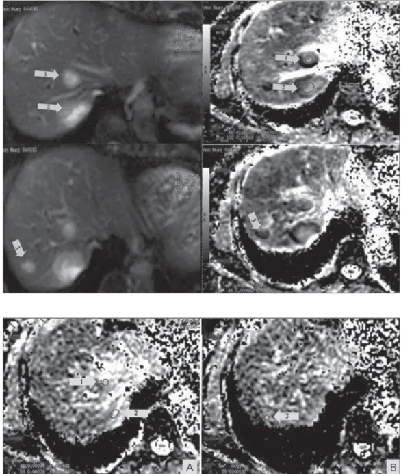
re-Figure 1. Magnetic resonance of the liver – image acquired on March 22, 2011. Liver metastases visualized on diffusion-weighted sequence (at left) and respective apparent diffusion coefficient val-ues on the ADC mapping (at right) previously to the chemotherapy initiation: lesion 1 = 1.6 × 10⁻³
mm2/s; lesion 2 = 1.6 × 10⁻³
mm2/s; lesion 3 = 0.6 × 10⁻³
mm2/s.
Figure 2. Magnetic resonance imaging of the liver – image ac-quired on June 20, 2011. Liver metastases with respective appar-ent diffusion coefficiappar-ent values on the ADC mapping, after about one month of chemotherapy: A: lesion 1 = 2.2 × 10⁻³ mm2/s; lesion 2 =
2.3 × 10⁻³ mm2/s. B: lesion 3 =
1.9 × 10⁻³ mm2/s. A significant
180
Testa ML et al. Diffusion-weighted MRI: biomarker for treatment response
Radiol Bras. 2013 Mai/Jun;46(3):178–180 spond to chemotherapy. An early increase
in ADC values was observed in the respon-sive lesions (p = 0.002). A statistically sig-nificant correlation was found between the decrease in the tumor size and the pretreat-ment ADC value (p = 0.006), besides early changes in the ADC (p < 0.001)(19).
In the presently described case, chemo-therapy was initiated as soon as it was evi-denced that the liver lesions were sugges-tive of involvement secondary to the base disease, and after follow-up with diffusion-weighted MRI. The lesions detected at the first follow-up study demonstrated early, significant increase in ADC, within about one month of treatment (seven-cycle che-motherapy).
Tumors are highly heterogeneous in terms of morphology, physiology (blood flow and vascular permeability) and genetic expression levels. Such variations in the tumor tissue may significantly regulate the therapeutic interventions efficacy. Because of the excellent imaging resolution and quantification provided by diffusion-weighted MRI, it can be utilized as guid-ance in the early phases of the treatment, with basis on the different regions where there is response or resistance to the therapy. For example, in an individual sub-mitted to chemotherapy for a primitive neu-roectodermal tumor, the mean diffusion values for the anterior and posterior regions of the tumor were measured in a deter-mined period. The anterior region demon-strated a significant increase in the diffu-sion values during the first three weeks of treatment, as compared with the posterior region of the tumor, indicating that the treatment was less effective in this region. In fact, 14 weeks later, the tumor had pro-gressed in the posterior region, revealing that such region was probably resistant to the chemotherapy. The use of diffusion-weighted in the follow-up of regional changes in tumors like the one described in the present case is potentially useful to guide the treatment. It is expected that such method can provide an early evidence of
the therapeutic efficacy, before the treat-ment is completed, offering an additional parameter to evaluate the response for con-tinuing or changing the treatment in case of ineffectiveness(20).
Because of molecular and cell changes induced by the treatment which precede macroscopic changes such as, for example, in tumor size, diffusion-weighted MRI can be utilized to detect early changes in the le-sion structure, thus allowing the use of such imaging marker as an indicator of early response in oncology. Additionally, the dif-fusion-weighted MRI technique allows a noninvasive evaluation of response on ana-tomical regions or histological subtypes, or after new molecular therapies which have not been accessible with conventional ra-diological methods (14).
CONCLUSION
Changes in ADC may occur in lesions submitted to chemotherapy, before changes in the lesion size can be perceived, thus allowing that therapeutic decisions can be made in advance. Diffusion-weighted MRI has shown to be a promising tool in the prediction of the tumor response and fol-low-up of oncological patients undergoing chemotherapy.
REFERENCES
1. Theilmann RJ, Borders R, Trouard TP, et al. Changes in water mobility measured by diffusion MRI predict response of metastatic breast cancer to chemotherapy. Neoplasia. 2004;6:831–7. 2. Miranda CMNR, Santos CJJ, Maranhão CPM, et
al. A tomografia computadorizada multislice é ferramenta importante para o estadiamento e se-guimento do câncer de mama? Radiol Bras. 2012;45:105–12.
3. Moreira BL, Lima ENP, Bitencourt AGV, et al. Metástase na mama originada de carcinoma ova-riano: relato de caso e revisão da literatura. Radiol Bras. 2012;45:123–5.
4. Azevedo AC, Canella EO, Djahjah MCR, et al. Conduta das funcionárias de um hospital na ade-são ao programa de prevenção do câncer de mama. Radiol Bras. 2012;45:215–8.
5. Barra FR, Barra RR, Barra Sobrinho A. Novos métodos funcionais na avaliação de lesões mamá-rias. Radiol Bras. 2012;45:340–4.
6. Urban LABD, Schaefer MB, Duarte DL, et al. Recomendações do Colégio Brasileiro de Radio-logia e Diagnóstico por Imagem, da Sociedade Brasileira de Mastologia e da Federação Brasileira das Associações de Ginecologia e Obstetrícia para rastreamento do câncer de mama por métodos de imagem. Radiol Bras. 2012;45:334–9. 7. Calas MJG, Alvarenga AV, Gutfilen B, et al.
Ava-liação de parâmetros morfométricos calculados a partir do contorno de lesões de mama em ultras-sonografias na distinção das categorias do sistema BI-RADS. Radiol Bras. 2011;44:289–96. 8. Marques EF, Medeiros MLL, Souza JA, et al.
Indicações de ressonância magnética das mamas em um centro de referência em oncologia. Radiol Bras. 2011;44:363–6.
9. Oliveira FGFT, Fonseca LMB, Koch HA. Respon-sabilidade civil do radiologista no diagnóstico do câncer de mama através do exame de mamogra-fia. Radiol Bras. 2011;44:183–7.
10. Vianna AD, Gasparetto TD, Torres GC, et al. Cancerização de lóbulos: correlação de achados mamográficos e histológicos. Radiol Bras. 2011; 44:275–8.
11. Vieira SC, Silva JS, Madeira EB, et al. Heman-gioma de mama simulando metástase no PET-CT. Radiol Bras. 2011;44:401–2.
12. Heijmen L, Ter Voert EE, Nagtegaal ID, et al. Dif-fusion-weighted MR imaging in liver metastases of colorectal cancer: reproducibility and biologi-cal validation. Eur Radiol. 2013;23:748–56. 13. Taouli B. Diffusion-weighted MR imaging for
liver lesion characterization: a critical look. Ra-diology. 2012;262:378–80.
14. Hamstra DA, Rehemtulla A, Ross BD. Diffusion magnetic resonance imaging: a biomarker for treatment response in oncology. J Clin Oncol. 2007;25:4104–9.
15. Taouli B, Koh DM. Diffusion-weighted MR im-aging of the liver. Radiology. 2010;254:47–66. 16. Charles-Edwards EM, deSouza NM.
Diffusion-weighted magnetic resonance imaging and its ap-plication to cancer. Cancer Imaging. 2006;6:135– 43.
17. Kele PG, van der Jagt EJ. Diffusion weighted im-aging in the liver. World J Gastroenterol. 2010;16: 1567–76.
18. Koh DM, Scurr E, Collins D, et al. Predicting re-sponse of colorectal hepatic metastasis: value of pretreatment apparent diffusion coefficients. AJR Am J Roentgenol. 2007;188:1001–8. 19. Cui Y, Zhang XP, Sun YS, et al. Apparent
diffu-sion coefficient: potential imaging biomarker for prediction and early detection of response to che-motherapy in hepatic metastases. Radiology. 2008;248:894–900.