Influence of the power level of an
ultra-sonic system on dental cavity
preparation
Influência do nível de potência de um sistema
de ultra-som em preparos cavitários
Abstract: The aim of this study was to evaluate the shape of dental cavities made with the CVDentus® system using different ultrasound power levels. One standard cavity was made on the buccal aspect of 15 bovine incisors with a CVDentus® cylindrical bur (82142). The sample was divided into three groups: G1 – ultrasound with power II; G2 – ultrasound with power III; and G3 – ultrasound with power IV. A standardizing device was used to obtain standardized preparations and ultrasound was applied during one minute in each dental preparation. The cavities were sectioned in the middle, allowing observation of the cavity’s proile with a magnifying glass, and width and depth measurement using the Leica Qwin program. The Kruskal-Wallis (p < 0.05) and Dunn statistical analyses demonstrated differences between the dental cavity shapes when powers III and IV were used. However, the cavities that were made with power III presented dimensions similar to those of the bur used for preparation. We concluded that the power recommended by the manufacturer (III) is the most adequate for use with the CVDentus® system.
Descriptors: Dental cavity preparation; Technology, dental; Ultrasonography; Diamond.
Resumo: O objetivo deste estudo foi avaliar o formato dos preparos cavitários realiza-dos com o sistema CVDentus® utilizando potências variadas do ultra-som. Uma cavidade padronizada foi realizada na face vestibular de 15 incisivos bovinos utilizando as pontas cilíndricas CVDentus® (82142). A amostra foi dividida em 3 grupos: G1 – ultra-som com potência II; G2 – ultra-som com potência III; e G3 – ultra-som com potência IV. Foi uti-lizada uma máquina padronizadora de preparos cavitários e o ultra-som foi aplicado du-rante 1 minuto em cada preparo. As cavidades foram seccionadas no centro, permitindo a visualização do peril cavitário em uma lupa estereoscópica, e esse foi medido em largu-ra e profundidade por meio do proglargu-rama Leica Qwin. O teste estatístico Kruskal-Wallis (p < 0,05) e o método de Dunn demonstraram diferenças entre os formatos das cavidades produzidas com as potências III e IV. Entretanto, as cavidades realizadas com a potência III apresentaram dimensões semelhantes às da ponta utilizada. Concluiu-se que a potên-cia indicada pelo fabricante (III) é a mais adequada para uso do sistema CVDentus®. Descritores: Preparo da cavidade dentária; Tecnologia odontológica; Ultrassonograia; Diamante.
Érika Botelho Josgrilberg(a)
Murilo de Sousa Guimarães(a)
Cyneu Aguiar Pansani(b)
Rita de Cássia Loiola Cordeiro(b)
(a) Graduate Students; (b)Associate Professors – Department of Pediatrics, School of Dentistry of Araraquara, São Paulo State University.
Corresponding author: Rita de Cássia Loiola Cordeiro Departamento de Clínica Infantil R. Humaitá, 1680
Araraquara - SP - Brazil CEP: 14801-903
E-mail: [email protected]
Introduction
The use of ultrasound in dentistry was irst in-troduced over ifty years ago for the preparation of cavities.2 The instrument was considered eficient
for cutting hard dental tissue due to its precise cut, the soft aspect of the cavities obtained and its good tolerance by patients.8 However, some limitations in
its use, such as a slow cut, the insuficient removal of tissue or weak materials, the need for constant maintenance and its high cost, have restricted the recommendations for its use and, consequently, it has had a low acceptance by professionals.10
During the years, this technology has been adapt-ed for modern Dentistry neadapt-eds, introducing water irrigation and adequate drill oscillation. Currently, it is used for endodontic, surgical, TMJ dysfunction, periodontic and other dental treatments, besides its application with the CVDentus® drill.
The CVDentus® system is a new Brazilian
tech-nology that is innovating the procedure of dental cavity preparation. The diamond bur is obtained by a chemical deposition of gas over a molybdenum stem, forming a unique diamond stone. This process results in an instrument that presents high durabili-ty, structural stability and biocompatibilidurabili-ty, without forming dentine residues or contaminating the tooth with metal.1,9,11,13 Its adaptation for ultrasound adds
to its advantages, providing improved visibility, low noise and often discarding the need for local anes-thesia, promoting more comfort and facilitating the treatment for patients and for the operator.3,13
These tips are available in different shapes, and each one demands an adequate ultrasound power to reach greater effectiveness of cut and durability. How-ever, there is no standardization with regard to the recommended power level for the various ultrasound systems available on the marketplace, thus requiring adaptations for their use. Usually, the power is in-creased to retain the effectiveness of cut. However, there is a need for professional training for the use of this technology since it operates in a different way than do conventional rotary devices, which seem to cut more quickly, causing a false impression that there is a need to increase the power of the ultrasound.
The aim of this in vitro study was to characterize the effect of the variation in ultrasound power level
on dental cavity morphology.
Material and Methods
This study was approved by the Ethics and Re-search Comittee, School of Dentistry of Araraquara, São Paulo State University (protocol n.71/03).
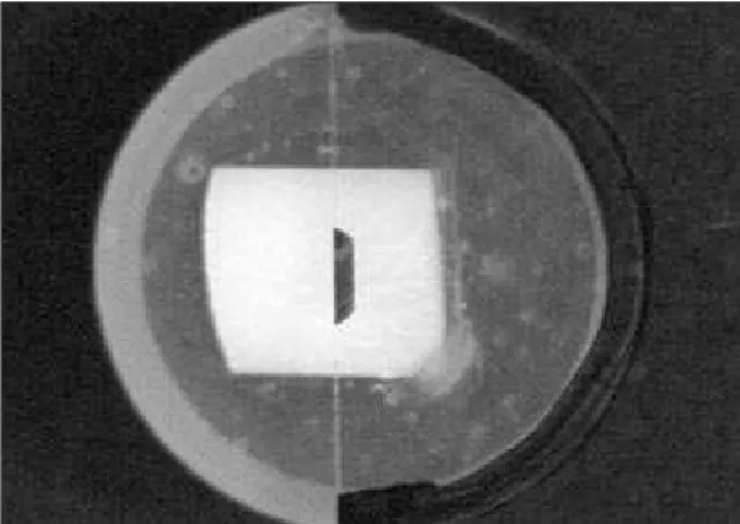
Fifteen bovine incisors without caries injuries, cracks or anomalies observed by a magnifying glass (10 X) were used in this study. The crowns were divid-ed and embdivid-edddivid-ed in a block-type PVC pipe illdivid-ed with resin (Ortoclass® - Clássico Ltda., Campo Limpo
Pau-lista, SP, Brazil). Standardization of the crown fragment position in the PVC pipe was made using a glass slide/ blade with a mark corresponding to the central region of the tooth, providing the ideal position for the crown in the PVC tube (Figure 1). The samples were then di-vided into three randomized groups, corresponding to each power level used with the ultrasound equipment (power II, power III and power IV – n = 10). Dental cavity preparations were performed with the CVDen-tus® system (Clorovale Diamantes - São José dos
Cam-pos, SP, Brazil) using a cylindrical drill (model 82142) connected to a Prof I ultrasound equipment (AS Ce-ramic - Dabi Atlante®, Ribeirão Preto, SP, Brazil) set at
power levels II, III or IV, equivalent to 50%, 75% and 100% of the device’s total power.
A standard device, produced exclusively for this type of study, was used for dental cavity prepara-tion;7 the cuts were made with 63 consecutive
move-ments of the bur on the dental surface with a speed
of 5.3 mm/s, corresponding to 1 minute of use of the CVDentus® tips (Figure 2A and Figure 2B).
The cavities were sectioned in the middle along the cervical-incisal plane and in their extremities to obtain two plane aspects of cavity proile for adapta-tion onto the Stereoscopic Magnifying glass (Leica, Carl Zeiss, Jena 16 X, Bensheim, Germany) to allow visualization of the cavity proiles, totaling 10 cavity proiles for each group (n = 10). The images were cap-tured using the Leica Qwin Program (Leica Q550IW, Bensheim, Germany), and two tracings were made corresponding to width and depth of the cavity prepa-ration. The program then gave out the measurements corresponding to width and depth. These measure-ments were performed twice for each sample, with an interval of one week between each other.
The cavity preparation images were visually an-alyzed by the examiner and classiied according to the position of the pulp wall, and to whether it was inished in dentine, enamel or both.
All values obtained were transferred to the Biostat program, where the Spearman Correlation test was ap-plied for examiner calibration veriication. Statistical analysis was performed by the Kolmogorov-Smirnov normality test and the Kruskal-Wallis test (5% sig-niicance), to assess possible differences between the groups. The Dunn method was used to assess the dif-ference between the ranks, when necessary.
Results
The results of the Spearman Correlation Test
(5% signiicance) demonstrated that there was no signiicant differences between the two measure-ments, allowing the use of the irst examination for the continuity of statistical analysis.
Differences in the cutting depths and widths, as demonstrated by the Kruskal-Wallis test, are pre-sented in Table 1.
The Dunn method demonstrated that statistical difference occurred in both the depth and width of the dental cavities when the power levels II vs. IV and III vs. IV were compared (Graph 1).
Figure 2 - Standard device for dental cavity preparation (A). CVDentus system on ultrasound device making the cavity on the specimen (B).
A B
B
Table 1 - Comparison between measurements of the cavi-ties made with power levels II, III or IV.
Dental cavity profile P
Width 0.003*
Depth 0*
*p < 0.05.
Graph 1 - Mean differences (micrometers) in width and depth (*p < 0.05).
2.3
18.3
16
0.3
14.9 15.2
0 3 6 9 12 15 18 21
II - III II - IV Power
III - IV Depth Width
Mediu
m
po
sts
P < 0.05 P < 0.05
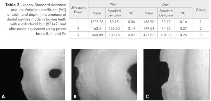
The cut measurements for each of the experimen-tal conditions are summarized in Table 2.
Visual cavity wall analysis demonstrated that all cavities had only reached the enamel (Figure 3).
Discussion
The association of ultrasound with the CVDen-tus® bur is not yet used on a daily clinical basis since
the use and capabilities of the technique are widely unknown to professionals and researchers. The sys-tem may be used for dental cavity preparation, but the use of adequate power is important to provide an eficient cut and greater durability of the instru-ment.
A different power level for each tip model is rec-ommended by the manufacturer, since each one pro-motes a different ampliication of the ultrasound up to its extremity. For example, the system adapted for ine conical tips requires a power level of 30% due to the intense vibration produced. Although spherical tips are thicker, they exert an impact only at one point, causing great force in the connecting rod, disabling any power greater than 50%. There-fore, when using the system, care is required with regard to the choice of tip design and power level in order to allow greater effectiveness of cut and safety for the procedure.
Indicated power levels for each tip format are in the manual’s instructions for the system. How-ever, some ultrasound devices present power levels that do not coincide with those indicated by the manufacturer.
The aim of this study was to assess the effect of ultrasound power variation on the morphology of the obtained cavity preparation, using a CVDentus®
system cylindrical bur. This model was chosen be-cause it is widely used in Pediatric Dentistry. Previ-ous studies have shown that this tip presents great stability when comparing the shape of dental cavi-ties in dentine and enamel.7
According to the manufacturer, the cylindri-cal tip must be used with 60% of the maximum ultrasound power. The equipment used in this re-search presented power levels of 25%, 50%, 75% and 100%. As our objective was to evaluate powers above and below the suggested average, we opted to use 50% (Group 1- power II), 75% (Group 2 - power III) and 100% (Group 3 - power IV) of the maximum power.
The methodology used for obtaining dental cavi-ties was performed previously by Lima et al.7 (2006),
and measurements were made using the Leica Qwin program. According to the Spearman Correlation test, no differences were observed between the irst
Ultrasound Power
Width Depth
Group
Mean Standard
deviation VC Mean
Standard
Deviation VC
II 1227.78 80.95 0.06 185.90 30.77 0.16 1
III 1163.41 163.20 0.14 199.64 74.65 0.37 2
IV 1400.88 109.48 0.07 417.85 106.33 0.25 3
Table 2 - Mean, Standard deviation and the Variation coefficient (VC) of width and depth (micrometers) of dental cavities made in bovine teeth with a cylindrical bur (82142) and ultrasound equipment using power levels II, III and IV.
Figure 3 - Dental cavities made using power levels II (A), III (B) and IV (C).
and second examinations, demonstrating the reli-ability of the methodology applied.
The Kruskal-Wallis test demonstrated signii-cant differences between the three groups (Table 1). The Dunn method (Graph 1) demonstrated that these differences occurred between the group where power IV was used when compared to powers III and II.
Unexpectedly, this study did not ind any statisti-cally signiicant differences between powers II and III. This result may have occurred due to an absence of variation during the transmission of power or, possibly, because a threshold may exist above which the system becomes more effective.
In addition, the group in which power III was used demonstrated a greater variation in data distri-bution. A supposed initial trepidation of the ultra-sound on the tooth surfaces or a changeable inclina-tion of specimen surfaces observed in the magnifying glass may be the reason for this variability.
When power III was used, the mean cavity width (Table 2) was more similar to the actual diameter of the bur (1.1 mm), thus our study agrees with the manufacturer’s recommendation for the use of this power level for the cylindrical tip. The association of this tip with ultrasound allows the professional to perform precise and consistent dental cavity prepa-rations, since it does not cut beyond its size and can decrease the risk of undesireble damage. To that ex-tent, we agree with Khambay, Walmsley4,5 (2000),
who compared ultrasound equipment with conven-tional rotary instruments and observed that the for-mer cut with more precision.
Wapington et al.14 (1995) compared different
power levels of ultrasound used with a standardized load and different formats of conventional tips and observed that, regardless of tip format, an increase in power results in an increase in amplitude of vi-bration and, consequently, an increase in the cut-ting ability of the instrument. In this study, we also observed that when power was increased to level IV (100%), the width and depth measurements of the dental cavities were greater when compared to those resulting from the other ultrasound powers
(Table 2). When power is increased beyond the ide-al, the CVDentus® system may remove chips of
sub-stratum, and make cavities that are larger or deeper than desired.
Although it was not the intention of this study to perform other analyses, cracks were observed in some of the samples. However, it is not possible to determine whether these were due to the preparation with the CVDentus® system or from the sectioning
of the cavities for attainment of proile cavities for observation in the magnifying glass.
Effectiveness of cut studies show that rotary in-struments cut faster than ultrasound.4,6,7,12 However,
Khambay, Walmsley4 (2000) suggest that ultrasound
cuts in a different manner and that the professional must be trained to use this instrument.
Power variation may cause alterations in cavity preparation size, in vibration and in the amount of noise, causing more discomfort to the patient and shorter tip durability, although, in this study no signs of frangibility or fracture of the bur were ob-served. Thus, it is important to obtain data regard-ing the adequate power levels for use with the CV-Dentus® system.
Ultrasound offers innumerous advantages for dental procedures. Moreover, the characteristics of ultrasound for use in Pediatric dentistry, such as comfort and the production of a noise that is differ-ent from that produced by rotary instrumdiffer-ents, may contribute to obtain a more favorable behavior of children.
Conclusion
In this study, the dental cavity preparations made with power level IV were statistically different from those made with power levels II and III, suggesting that the power recommended by the manufacturer (III) is the most adequate for use with the CVDen-tus® system.
Acknowledgements
References
1. Borges CFM, Magne P, Pfender E, Heberlein J. Dental dia-mond burs made with a new technology. J Prosthet Dent. 1999;82(1):73-9.
2. Catuna MC. Sonic energy. A possible dental application. Pre-liminary report of an ultrasonic cutting method. Ann Dent. 1956;12(100):256-60.
3. Conrado LA, Trava-Airoldi VJ, Corat E, Munin E, Rolim TS. The use of a CVD-Coated diamond bur coupled to an ultrasound handpiece in dental preparation [cited 2004 Oct 29]. Available from: http://www.cvd-diamante.com.br/ref-bibliograficas.htm.
4. Khambay BS, Walmsley AD. Investigations into the use of an ultrasonic chisel to cut bone. Part 1: forces applied by clini-cians. J Dent. 2000;28(1):31-7.
5. Khambay BS, Walmsley AD. Investigations into the use of an ultrasonic chisel to cut bone. Part 2: cutting ability. J Dent. 2000; 28(1):39-44.
6. Lima LM, Motisuki C, Santos-Pinto L. Análise da efetividade de corte da ponta de diamante CVD em ultra-som e ponta diamantada convencional em alta-rotação [resumo]. Braz Oral Res. 2004;18(Suppl):128.
7. Lima LM, Motisuki C, Santos-Pinto L, Santos-Pinto A, Corat EJ. Cutting characteristics of dental diamond burs made with CVD technology. Braz Oral Res. 2006;20(2):155-61. 8. Postle HH. Ultrasonic cavity preparation. J Prosthet Dent.
1958;8(1):153-60.
9. Silva AP, Menezes MM, Araújo RM. Influência da limpeza e esterilização sobre a capacidade de desgaste de pontas diaman-tadas. J Bras Clin Odontol Int. 2002;6(33):239-45.
10. Street EV. A critical evaluation of ultrasonics in dentistry. J Prosthet Dent. 1959;9(1):132-41.
11. Valera MC, Ribeiro JF, Trava-Airoldi VJ, Corat EJ, Pena AF, Leite NF. Pontas de diamantes - CVD. RGO. 1996;44(2):104-8. 12. Vieira ASB, Antunes LAA, Maia LC, Santos MPA, Primo LG.
Efetividade da abrasão ultra-sônica versus alta rotação: estudo in vitro [resumo]. Braz Oral Res. 2004;18(Suppl):172. 13. Vieira D, Vieira D. Pontas de diamante CVD: início do fim da
alta rotação? [Brazilian edition] J Am Dent Assoc. 2002;5:307-13.