CLINICAL SCIENCE
Regulation of hypoxia-inducible factor-1
a
(HIF-1
a
)
expression by interleukin-1
b
(IL-1
b
), insulin-like
growth factors I (IGF-I) and II (IGF-II) in human
osteoarthritic chondrocytes
Ange´lica Rossi Sartori-Cintra, Cristiane Sampaio de Mara, Danielle L. Argolo, Ibsen Bellini Coimbra State University of Campinas (UNICAMP), Department of Clinical Medicine, Division of Rheumatology, Laboratory of Molecular Biology of Cartilage, Campinas/SP, Brazil.
OBJECTIVE:Hypoxia-inducible factor 1 alpha regulates genes related to cellular survival under hypoxia. This factor is present in osteroarthritic chondrocytes, and cytokines, such as interleukin-1 beta, participate in the pathogenesis of osteoarthritis, thereby increasing the activities of proteolytic enzymes, such as matrix metalloproteinases, and accelerating cartilage destruction. We hypothesize that Hypoxia Inducible Factor-1 alpha (HIF-1a) can regulate cytokines (catabolic action) and/or growth factors (anabolic action) in osteoarthritis. The purpose of this study was to investigate the modulation of HIF-1a in human osteoarthritic chondrocytes by interleukin-1 beta (IL-1b) and insulin-like growth factors I (IGF-I) and II (IGF-II) and to determine the involvement of the phosphatidylinositol-3-kinase (PI-3K) pathway in this process.
METHODS:Human osteroarthritic chondrocytes were stimulated with IL-1b, IGF-I and IGF-II and LY294002, a specific inhibitor of PI-3K. Nuclear protein levels and gene expression were analyzed by western blot and quantitative reverse transcription-polymerase chain reaction analyses, respectively.
RESULTS:HIF-1aexpression was upregulated by IL-1bat the protein level but not at the gene level. IGF-I treatment resulted in increases in both the protein and mRNA levels of HIF-1a, whereas IGF-II had no effect on its expression. However, all of these stimuli exploited the PI-3K pathway.
CONCLUSION:IL-1bupregulated the levels of HIF-1aprotein post-transcriptionally, whereas IGF-I increased HIF-1aat the transcript level. In contrast, IGF-II did not affect the protein or gene expression levels of HIF-1a. Furthermore, all of the tested stimuli exploited the PI-3K pathway to some degree. Based on these findings, we are able to suggest that Hypoxia inducible Factor-1 exhibits protective activity in chondrocytes during osteoarthritis.
KEYWORDS: HIF-1a; Chondrocytes; Phosphatidylinositol-3-kinase (PI-3K), Cytokines; IL-1b; Growth factors; IGF-I, IGF-II, Osteoarthritis; Articular cartilage.
Sartori-Cintra AR, Mara CS, Argolo DL, Coimbra IB. Regulation of hypoxia-inducible factor-1a(HIF-1a) expression by interleukin-1b(IL-1b), insulin-like growth factors I (IGF-I) and II (IGF-II) in human osteoarthritic chondrocytes. Clinics. 2012;67(1):35-40.
Received for publication onAugust 5, 2011;First review completed onSeptember 14, 2011;Accepted for publication onSeptember 14, 2011
E-mail: [email protected]
Tel.: 55 19 3521-9587
INTRODUCTION
Articular cartilage is a highly specialized tissue present in all diarthrodial joints, and its breakdown is a crucial event in the etiopathogenesis of osteoarthritis (OA). OA is characterized by the degeneration of articular cartilage in association with subchondral bone erosions and sclerosis. Numerous inflammatory cytokines, such as interleukins 1 and 6 (IL-1 and IL-6) and tumor necrosis factor-a (TNFa),
participate in the pathogenesis of this disease, increasing the expression of proteolytic enzymes and metalloproteases (MMPs) and accelerating the destruction of cartilage (1-3). Chondrocytes are cartilage cells that exist in a hypoxic microenvironment because cartilage is an avascular tissue. Hypoxia-inducible factor 1 (HIF-1) is a transcription factor that activates the expression of target genes involved in essential pathways regulating cellular survival under con-ditions of hypoxia, such as angiogenesis and glycolysis (4). Under normal oxygen tension, the HIF-1asubunit is marked for ubiquitination and rapid proteosome-mediated degra-dation by the von Hippel-Lindau tumor suppressor (pVHL). During hypoxia, ubiquitination and degradation are inhib-ited, increasing the steady-state level of HIF-1a protein in the cytoplasm. HIF-1a subsequently translocates to the nucleus, where it dimerizes with HIF-1b, the constitutively
Copyrightß2012CLINICS– This is an Open Access article distributed under the terms of the Creative Commons Attribution Non-Commercial License (http:// creativecommons.org/licenses/by-nc/3.0/) which permits unrestricted non-commercial use, distribution, and reproduction in any medium, provided the original work is properly cited.
expressed HIF-1 subunit, forming the transcription complex HIF-1 (5,6). In addition to hypoxia, HIF-1aexpression can be induced by numerous other factors, including inflammatory cytokines, reactive oxygen species (ROS), nitric oxide, and hormone-like growth factors, such as Insulin-like growth factor (IGF) and TGF-b(Transforming Growth-factorb) (7). In previous studies, we identified expression of HIF-1a associated with human OA as well as in normal chondro-cytes under normal oxygen tension conditions and found that HIF-1a protein levels were increased by TNFa treat-ment (8). Because HIF-1ais related to cellular survival via the modulation of genes related to this function, we believe that when this transcription factor is present in osteoar-thritic chondrocytes, it could be related to cytokine modulation. Specifically, HIF-1a may modulate IL-1b, a major catabolic factor involved in OA that can induce potent changes in cartilage metabolism in OA joints, thereby inhibiting the synthesis of cartilage-specific collagen II and proteoglycans and increasing the production of numerous MMPs (2,3,9).
Similarly, we chose to investigate insulin-like growth factors I and II, which are reported to be involved in HIF-1a regulation in other cell lines (10,11). In chondrocytes, the anabolic role of these growth factors, especially IGF-I, is well recognized as stimulating cellular proliferation and extracellular-matrix synthesis (12,13). However, no studies have examined whether IGF-I and II regulate the expression of HIF-1ain these cells. Thus, the aim of the present study was twofold (1): to investigate the modulation of the expression of HIF-1a in human OA chondrocytes under normal oxygen conditions in response to treatment with IL-1b, IGF-I and IGF-II; and (2). to determine whether the phosphatidylinositol-3-kinase (PI-3K) pathway participates in this modulation, as is the case in other cell types.
MATERIALS AND METHODS
Isolation and culture of human OA chondrocytes
Human chondrocytes were obtained from patients with OA who underwent knee joint replacement surgery at the University Hospital of Campinas, Sa˜o Paulo, Brazil. This study was analyzed and approved by the local ethics committee. The patients signed informed consent docu-ments to allow the authors to use material from their replaced joints in the current study. Approximately 80% of the patients presented as class IV in the Kelgreen and Lawrence (14). radiographical scale. Chondrocytes were isolated from all remaining cartilage tissue as previously described (15). Briefly, the cartilage was minced and incubated in Hanks’ medium containing trypsin and bacterial collagenase (2 mg/ml each) for 1 h at 37˚C. The medium was subsequently discarded, and tissue frag-ments were incubated overnight at 37˚C in Dulbecco’s minimum essential medium (DMEM) containing 10% fetal bovine serum and 0.5 mg/ml bacterial collagenase. The released cells were filtered through a 70-mm nylon cell strainer, were collected by centrifugation at 2506g for
5 min and were washed with collagenase-free medium. Isolated chondrocytes were immediately frozen in freezing media (90% FBS, 10% DMSO) and stored for future experiments. For these experiments, cells were thawed and plated in suspension cultures in 6-well ultralow attachment plates (Corning, Acton, MA) at a density of 56106/ml. The cells were allowed to recover for 48 h in
DMEM containing 10% FBS, 2 mM glutamine, 1% vitamin supplements, 100 U/ml penicillin and 100mg/ml strepto-mycin. Amphotericin B and ascorbic acid were avoided because they can interfere with HIF-1aexpression (16). For experiments under normoxic conditions, cells were main-tained at 37˚C in 5% CO2and 95% air (21% O2). The cells were stimulated for 6 h with 10 ng/ml IL-1b Pierce Endogen (N. Meridian Road, Rockford, IIL). To study PI-3K, the specific inhibitor LY 294002 Sigma-Aldrich (Sigma-Aldrich, St. Louis, MO) was employed at a concentration of 10mM/ml. For treatment with IGF-I or IGF-II (at 10 ng/ ml each), cells were serum-depleted to 0.1% FBS 12 h before the indicated treatment, and nuclear protein extraction was performed 12 h after addition of the growth factor.
Preparation of nuclear extracts
Nuclear extracts were isolated from the chondrocytes according to the method of Dignam et al (17). using the CellLytic NuCLEAR extraction kit (Sigma-Aldrich, St. Louis, MO) at 4˚C to avoid nuclear protein denaturation according to the manufacturer’s instructions. All buffers contained a protease inhibitor cocktail with 2 mM 4-(2-aminoethyl) benze-nesulfonylfluoride, 1.4 pMtrans-epoxysuccinyl-L-leucylamido (4-guanidinobutane), 130 pM bestatin, 1mM leupeptin, 0.3 pM aprotinin (Sigma-Aldrich) and 2.6mM calpain inhibitor (Calbiochem, San Diego CA). The obtained protein concentra-tions were analyzed via the Bradford method at a wavelength of 595 nm using a spectrophotometer.
Western Blot Analysis
For western blot analysis, 30mg of nuclear chondrocyte extract in 2x SDS buffer and distilled water (final volume of 30ml) was denatured at 95˚C for 90 sec and then separated on an 8% SDS-polyacrylamide gel. Following electrophoresis, the proteins were transferred to Hybond-P membranes (Amersham Pharmacia Biotech) in buffer containing 20 mM Tris HCl, 150 mM glycine and 20% methanol at 40 V for 18 h at 4˚C. The membranes were blocked with Tris-Buffered Saline (TBS) wash solution containing 5% nonfat milk and 0.1% Tween 20 (TBSMT) for 2 h at pH 7.6. The membranes were incubated with the primary antibody, a mouse monoclonal anti-HIF-1 antibody (1/250 dilution, Trans-duction Laboratories, Lexington, KY), with shaking over-night at 4˚C followed by incubation with the secondary antibody, a rabbit anti-mouse IgG-horseradish peroxidase conjugate (1/2,000 dilution, Amersham Pharmacia Biotech). The membranes were washed three times (1615 min and
2610 min) between antibody incubations with TBMST, and
the blots were developed using an ECL detection kit (Amersham Pharmacia Biotech). The membranes were subjected to stripping and re-probed with a mouse mono-clonal anti-b-actin antibody to normalize the results. The bands were analyzed by densitometry and normalized using ImageMaster TotalLab v.2.0 software (Amersham).
RNA Isolation and Real-Time PCR (qRT-PCR) Analysis
analysis using real-time-PCR (Applied Biosystems 7500 Real Time PCR). Amplification of specific PCR products was detected using SYBR Green PCR Master Mix (Applied Biosystems). All primers employed in these experiments were designed using VectorH3.0 software and prepared by Invitrogen (Sa˜o Paulo, Brazil). The primers used were as follows: HIF-1a, forward 59CTGACCCTGCACTCAATCAA 39, reverse 59 CTTTGCTTCTGTGTCTTCAGCAGCA 39; b -actin, 59 GCTCGTCGTCGACAA CGGCTC 39, reverse 59 CAAACATGATCTGGGTCATCTTCTC 39.
The samples for qRT-PCR were prepared and analyzed in triplicate in a reaction volume of 10mL containing 5mL of SYBR Green PCR Master Mix, 3ml of cDNA from each sample as a template (between 10 and 18 ng of cDNA), 2ml of primer solution (forward and reverse) and DEPC water. The primer and cDNA concentrations were standardized. A melting curve analysis was performed to confirm the specificity of the amplification and the absence of primer dimers. Samples were heated for 10 min at 95˚C and amplified for 50 cycles of 15 sec at 95˚C and 60 sec at 60˚C. Blank controls were run in parallel to determine the amplification efficiency within each experiment. Quanti-fication was performed using a standard curve. Serial dilutions of cDNA in dH2O, which were used as a calibrator, were amplified to construct standard curves for target and control genes. The slopes of the standard curves ranged from -3.2 to -3.9. For each sample, the levels of the target and control genes were determined based on the appropriate standard curve. The target level was subsequently divided by the control gene level to obtain a normalized target value, which was calibrated using the standard RNA sample.
Statistical Analysis
The results of three separate experiments were expressed as means¡standard deviations. Data were analyzed using
nonparametric statistical analysis with the Kruskal-Wallis test, and in the significant cases, the Duncan test was applied to further discriminate group differences. For all analyses, a p-value of ,0.05 was considered to be significant.
RESULTS
IL-1b
Treatment with IL-1bdownregulated HIF-1agene expres-sion in OA chondrocytes, although the difference was not statistically significant. In contrast, when OA chondrocytes were incubated with both IL-1b and LY2 94002 (a PI-3K inhibitor), the observed inhibition was significantly more intense and was statistically different from the control (*p,0.05) (Fig. 1A). In contrast to the mRNA levels of HIF-1a, the expression of HIF-1a protein assayed in nuclear extracts was upregulated in OA chondrocytes stimulated with IL-1b (*p,0.05). When LY 294002 treatment was applied, we observed that there was a decrease in this effect of IL-1b, with HIF-1a protein levels exhibiting downregulation (*p,0.05) (Fig. 1B).
IGF-I
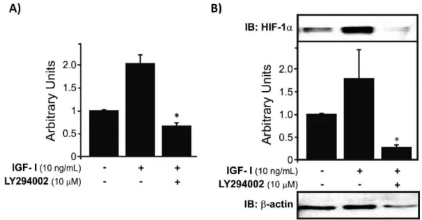
When the cells were treated with IGF-I, HIF-1a gene expression was upregulated, whereas the application of both IGF-I and LY294002 caused a significant reduction in HIF-1alevels compared to control, untreated cells (*p,0.05). These results suggest that IGF-I stimulation of HIF-1a expression is largely mediated by the PI-3K pathway (Fig. 2A). In addition to the upregulation of HIF-1a mRNA by IGF-I, treatment with this growth factor also significantly increased HIF-1aprotein levels. The combina-tion of IGF-I and LY294002 resulted in a complete abrogation of the stimulatory effect and provoked a notable
decrease in HIF-1anuclear protein levels compared to basal levels (*p,0.05) (Fig. 2B).
IGF-II
Surprisingly, we did not observe upregulation of HIF-1a mRNA expression when the cells were treated with IGF-II. However, the results further showed that the PI-3K path-way is involved in the basal regulation of HIF-1a gene expression. The involvement of the PI-3K pathway is
reinforced by the finding that in cells treated with both IGF-II and LY209002, the protein levels were significantly reduced and were statistically different (*p,0.05) from levels in the control, as well as from the levels in IGF-II-treated cells (Fig. 3A). Unlike the previously demonstrated upregulation of HIF-1a protein levels by IL-1b and IGF-I, IGF-II failed to increase the expression of this protein. In contrast, IGF-II downregulated HIF-1a protein levels. However, this result corroborates the findings from the Figure 2 -HIF-1amRNA expression and protein levels in human chondrocytes treated with IGF-I.A)IGF-I increases the mRNA expression of HIF-1a, and the combination of LY294002 and IGF-I produces downregulation of HIF-1amRNA to subbasal levels (*p,0.05).B)HIF-1a protein levels were increased by IGF-I treatment, and addition of LY294002 (a PI-3K pathway inhibitor) resulted in downregulation of HIF-1aprotein expression (*p,0.05) to reduced levels compared to controls. The results were normalized to the endogenous protein
b-actin.
RNA expression analysis of HIF-1afollowing treatment of the chondrocytes with IGF-II. Nevertheless, blocking the PI-3K pathway further decreased HIF-1ato sub-basal levels (*p,0.05) (Fig. 3B).
DISCUSSION
HIF-1a, a heterodimer transcription factor, plays a pivotal role in articular cartilage development and viability (18,19); however, its participation in the process of cartilage break-down remains unclear. In this study, we observed that IL-1b treatment downregulated HIF-1a mRNA expression in human OA chondrocytes, though the difference was not statistically significant. However, when the cells were treated with the combination of IL-1band LY 294002 (PI-3K pathway inhibitor), we observed stronger, statistically significant downregulation of HIF-1a mRNA expression. We found evidence of differential regulation of HIF-1a protein levels and mRNA expression. IL-1b increased HIF-1a levels through the PI-3K pathway, as IL-1b-specific upregulation was suppressed by blocking the PI-3K pathway. These findings reinforce previous observations (20-23). in other cell lines in which the regulation of HIF-1aby IL-1boccurs post-transcriptionally. In further agreement with our results, other investigators (24). who have performed real-time PCR analyses using different cartilage samples (degenerated and non-degenerated) have detected upregulation of HIF-1a, mostly in degenerated areas, suggesting that this factor could be related to a pathogenic mechanism involved in OA. As observed in the current investigation, other researchers have not found a significant effect of IL-1b treatment on mRNA levels. In contrast with our results, this effect has not previously been detected in cultured cells under normal oxygen tension. These discrepancies can be explained by the fact that other investigators have used chondrocyte mono-layer cultures, whereas we used suspension cultures. We previously demonstrated that HIF-1awas not present in OA chondrocytes in monolayer culture systems (8).
Nevertheless, when other investigators have analyzed the effect of IL-1b on chondrocytes cultured under hypoxic conditions, they have also observed this increase. Additionally, the same group (25). recently reported finding elevated HIF-1aprotein levels in human OA chondrocytes cultured under conditions of hypoxia and normoxia when they treated cells with IL-1bfor an extended period (24 h), suggesting a delayed response in OA chondrocytes, even in cells cultured in a monolayer. Therefore, our findings suggested that in chondrocytes, as in other cell lines, IL-1b acts as a positive regulator of HIF-1a protein levels at the post-transcriptional level. Our data also confirm that the regulation of HIF-1aby IL-1brelies, at least in part, on the PI-3K pathway, given that cells treated with inhibitors of this pathway showed no effects of this cytokine. This post-transcriptional regulation may suggest that HIF-1a has a protective role because elevated protein levels may increase the possibility of the binding of DNA to HIF-1 and the subsequent transcription of genes related to cell survival.
In osteoarthritis, IGF-I expression is associated with increasing synthesis of matrix molecules in early stages of the disease and with osteophyte formation later. The lack of an IGF-I pathway could be implicated in cartilage degen-eration (3,26). In this study, we demonstrated increases in HIF-1a mRNA expression and protein levels in IGF-I-treated OA chondrocytes. The PI-3K pathway was the
preferred mechanistic route for this upregulation because when this pathway was blocked, HIF-1a upregulation was not observed. Moreover, without PI-3K, HIF-1amRNA and protein levels decreased to sub-basal levels, suggesting that this pathway may be active even under baseline conditions. This upregulation has also been observed in other cell lines (27,28). Our findings suggested that IGF-I induces HIF-a expression, although additional studies will be essential to obtain a comprehensive understanding of the mechanisms involved in this induction. IGF-1 treatment increases the expression of HIF-1a at both the gene and protein levels, which reinforces its protective activity of HIF-1a because this growth factor is related to the maintenance of cartilage homeostasis.
Previous studies have shown that IGF-II promotes placental growth and function, and this process appears to be related to the regulation of HIF-1 and HIF-2a(29-31). To the best of our knowledge, this study is the first investigation that verifies the action of IGF-II on HIF-1a expression in human OA chondrocytes. Our results regarding HIF-1a protein levels are similar to previous observations made in murine throphoblast cells (29), but our findings diverge from observations in several other cell lines (27,30,31). We strongly suspect that these differences can be accounted for by length of treatment employed, given that our treatments were performed for 12 hours under low-serum conditions (0.1% FBS). In contrast, other investigators have used cells treated with-out serum deprivation with higher concentrations of IGF-II for a more extended period of time (30,31), and they did not use primary human OA chondrocytes. Our results may suggest that in human OA chondrocytes, IGF-II and IGF-I may play different roles; however, more studies are necessary to confirm this hypothesis. We also cannot discard the hypothesis that IGF-II may exert distinct effects in human OA chondrocytes across different stages of the disease because during our experiments, we treated cells from patients in different phases of OA. This possibility may bias our results. Interestingly, although we did not see any significant effect of IGF-II on HIF-1a mRNA or protein levels in human OA chondrocytes, blocking the PI-3K pathway produced a statistically significant down-regulation of HIF-1a mRNA and protein levels. This finding suggests that expression of HIF-1a under normal O2 levels in OA chondrocytes may also occur via this pathway (31).
In conclusion, we observed that IL-1b post-transcription-ally upregulated HIF-1a protein levels in human OA chondrocytes. We also found that IGF-I upregulated HIF-1a protein and mRNA levels, indicating that this action occurs at the gene level (no effect of IGF-II on HIF-1a regulation was observed). We have demonstrated that both IL-1, the main catabolic agent, and IGF-1, an anabolic agent, are able to stabilize HIF-1aunder normal oxygen conditions via the PI-3K pathway. These findings strongly suggest that Hypoxia inducible Factor-1 has a protective role in chondrocytes during osteoarthritis, and further studies are needed to clarify this relationship.
Funding: Grants were from FAPESP (Fundac¸a˜o de Amparo a` Pesquisa do Estado de Sa˜o Paulo).
ACKNOWLEDGMENTS
Msc Lucas Rossi Sartori and PhD Michael Niehues for excellent technical assistance and to FAPESP (Fundac¸a˜o de Amparo a` Pesquisa do Estado de Sa˜o Paulo) for financial support (Projects: 02/14132-1 and 05/00985-0).
AUTHOR CONTRIBUTIONS
Sartori-Cintra AR conceived and designed the study, performed the experiments, analyzed the data and wrote the paper. Coimbra IB conceived and designed the study and wrote the paper. Mara CS and Argolo DL performed the experiments.
REFERENCES
1. Martel-Pelletier J. Pathophysiology of osteoarthritis. Osteoarthritis Cartilage. 2004;12 Suppl A:S31-33, doi: 10.1016/j.joca.2003.10.002. 2. Malemud CJ. Cytokines as therapeutic targets for osteoarthritis.
BioDrugs. 2004;18:23-35, doi: 10.2165/00063030-200418010-00003. 3. van der Kraan PM, van den Berg WB. Anabolic and destructive
mediators in osteoarthritis. Curr Opin Clin Nutr Metab Care. 2000;3: 205-211, doi: 10.1097/00075197-200005000-00007.
4. Semenza GL. Expression of hypoxia-inducible factor 1: mechanisms and consequences. Biochem Pharmacol. 2000;59:47-53, doi: 10.1016/S0006-2952(99)00292-0.
5. Semenza GL. Hypoxia-inducible factor 1: control of oxygen homeostasis in health and disease. Pediatr Res. 2001;49:614-7, doi: 10.1203/00006450-200105000-00002.
6. Bardos JI, Ashcroft M. Negative and positive regulation of HIF-1: a complex network. Biochim Biophys Acta. 2005;1755:107-20.
7. Honorati MC, Cattini L, Facchini A. IL-17, IL-1beta and TNF-alpha stimulate VEGF production by dedifferentiated chondrocytes. Osteoarthritis Cartilage. 2004;12:683-91, doi: 10.1016/j.joca.2004.05.009. 8. Coimbra IB, Jimenez SA, Hawkins DF, Piera-Velazquez S, Stokes DG.
Hypoxia inducible factor-1 alpha expression in human normal and osteoarthritic chondrocytes. Osteoarthritis Cartilage. 2004;2:336-45, doi: 10.1016/j.joca.2003.12.005.
9. Chevalier X. Upregulation of enzymatic activity by interleukin-1 in osteoarthritis. Biomed Pharmacother. 1997;51:58-62, doi: 10.1016/S0753-3322(97)87727-X.
10. Treins C, Giorgetti-Peraldi S, Murdaca J, Semenza GL, Van Obberghen E. Insulin stimulates hypoxia-inducible factor 1 through a phosphatidyli-nositol 3-kinase/target of rapamycin-dependent signaling pathway. J Biol Chem. 2002;277:27975-81, doi: 10.1074/jbc.M204152200.
11. Feldser D, Agani F, Iyer NV, Pak B, Ferreira G, Semenza GL. Reciprocal positive regulation of hypoxia-inducible factor 1alpha and insulin-like growth factor 2. Cancer Res. 1999;59:3915-8.
12. Blumenfeld I, Livne E. The role of transforming growth factor (TGF)-beta, insulin-like growth factor (IGF)-1, and interleukin (IL)-1 in osteoarthritis and aging of joints. Exp Gerontol. 1999;34:821-829, doi: 10.1016/S0531-5565(99)00062-5.
13. Loeser RF, Shanker G. Autocrine stimulation by insulin-like growth factor 1 and insulin-like growth factor 2 mediates chondrocyte survival in vitro. Arthritis Rheum. 2000;43:1552-9, doi: 10.1002/1529-0131(200007) 43:7,1552::AID-ANR20.3.0.CO;2-W.
14. Kellgren J.H. and Lawrence J.S. Radiological assessment of osteo-arthrosis. Ann Rheum Dis. 1957;16(4):p.494-502
15. Reginato AM, Iozzo RV, Jimenez SA. Formation of nodular structures resembling mature articular cartilage in long-term primary cultures of human fetal epiphyseal chondrocytes on a hydrogel substrate. Arthritis Rheum. 1994;37:1338-49, doi: 10.1002/art.1780370912.
16. Jaakkola P, Mole DR, Tian YM, Wilson MI, Gielbert J, Gaskell SJ, et al. Targeting of HIF-alpha to the von Hippel-Lindau ubiquitylation complex
by O2-regulated prolyl hydroxylation. Science. 2001;292:468-72, doi: 10. 1126/science.1059796.
17. Dignam JD, Lebovitz RM, Roeder RG. Accurate transcription initiation by RNA polymerase II in a soluble extract from isolated mammalian nuclei. Nucleic Acids Res. 1983;11:1475-89, doi: 10.1093/nar/11.5.1475. 18. Schipani E, Ryan HE, Didrickson S, Kobayashi T, Knight M, Johnson RS.
Hypoxia in cartilage: HIF-1alpha is essential for chondrocyte growth arrest and survival. Genes Dev. 2001;15:2865-76.
19. Pfander D, Kobayashi T, Knight MC, Zelzer E, Chan DA, Olsen BR, et al. Deletion of Vhlh in chondrocytes reduces cell proliferation and increases matrix deposition during growth plate development. Development. 2004;131:2497-508, doi: 10.1242/dev.01138.
20. Hellwig-Burgel T, Rutkowski K, Metzen E, Fandrey J, Jelkmann W. Interleukin-1beta and tumor necrosis factor-alpha stimulate DNA binding of hypoxia-inducible factor-1. Blood. 1999;94:1561-7.
21. Qian D, Lin HY, Wang HM, Zhang X, Liu DL, Li QL, et al. Normoxic induction of the hypoxic-inducible factor-1 alpha by interleukin-1 beta involves the extracellular signal-regulated kinase 1/2 pathway in normal human cytotrophoblast cells. Biol Reprod. 2004;70:1822-7, doi: 10.1095/ biolreprod.103.025031.
22. Jiang BH, Jiang G, Zheng JZ, Lu Z, Hunter T, Vogt PK. Phosphatidylinositol 3-kinase signaling controls levels of hypoxia-inducible factor 1. Cell Growth Differ. 2001;12:363-9.
23. Haddad JJ. Recombinant human interleukin (IL)-1 beta-mediated regulation of hypoxia-inducible factor-1 alpha (HIF-1 alpha) stabiliza-tion, nuclear translocation and activation requires an antioxidant/ reactive oxygen species (ROS)-sensitive mechanism. Eur Cytokine Netw. 2002;13:250-60.
24. Yudoh K, Nakamura H, Masuko-Hongo K, Kato T, Nishioka K. Catabolic stress induces expression of hypoxia-inducible factor (HIF)-1 alpha in articular chondrocytes: involvement of HIF-1 alpha in the pathogenesis of osteoarthritis. Arthritis Res Ther. 2005;7:R904-914, doi: 10.1186/ar1765. 25. Murata M, Yudoh K, Nakamura H, Kato T, Inoue K, Chiba J, et al. Distinct signaling pathways are involved in hypoxia- and IL-1-induced VEGF expression in human articular chondrocytes. J Orthop Res. 2006;24:1544-54, doi: 10.1002/jor.20168.
26. Olney RC, Tsuchiya K, Wilson DM, Mohtai M, Maloney WJ, Schurman DJ, et al. Chondrocytes from osteoarthritic cartilage have increased expression of insulin-like growth factor I (IGF-I) and IGF-binding protein-3 (IGFBP-3) and -5, but not IGF-II or IGFBP-4. J Clin Endocrinol Metab. 1996;81:1096-103, doi: 10.1210/jc.81.3.1096. 27. Fukuda R, Hirota K, Fan F, Jung YD, Ellis LM, Semenza GL. Insulin-like
growth factor 1 induces hypoxia-inducible factor 1-mediated vascular endothelial growth factor expression, which is dependent on MAP kinase and phosphatidylinositol 3-kinase signaling in colon cancer cells. J Biol Chem. 2002;277:38205-11, doi: 10.1074/jbc.M203781200.
28. Slomiany MG, Rosenzweig SA. Hypoxia-inducible factor-1-dependent and -independent regulation of insulin-like growth factor-1-stimulated vascular endothelial growth factor secretion. J Pharmacol Exp Ther. 2006;318:666-75, doi: 10.1124/jpet.106.104158.
29. Pringle KG, Kind KL, Thompson JG, Roberts CT. Complex interactions between hypoxia inducible factors, insulin-like growth factor-II and oxygen in early murine trophoblasts. Placenta. 2007;28:1147-57, doi: 10. 1016/j.placenta.2007.05.009.
30. Kwon YW, Kwon KS, Moon HE, Park JA, Choi KS, Kim YS, et al. Insulin-like growth factor-II regulates the expression of vascular endothelial growth factor by the human keratinocyte cell line HaCaT. J Invest Dermatol. 2004;123:152-8, doi: 10.1111/j.0022-202X.2004.22735.x. 31. Tomizawa M, Saisho H. Signaling pathway of insulin-like growth factor-II