Maria de Paula Caldas(a)
Gláucia Maria Bovi Ambrosano(b) Francisco Haiter Neto(c)
(a) DDS, MSc Resident, Division of Oral Diagnosis; (b)Agr Eng, PhD, Professor, Department of Community Dentistry; (c)DDS, PhD, Chairman, Division of Oral Diagnosis – Piracicaba Dental School, University of Campinas, Campinas, SP, Brazil.
Corresponding author:
Maria de Paula Caldas Av. Limeira, 901
Piracicaba - São Paulo - Brazil CEP: 13414-900
E-mail: [email protected]
Received for publication on Aug 09, 2009 Accepted for publication on Sep 28, 2009
Computer-assisted analysis of cervical
vertebral bone age using cephalometric
radiographs in Brazilian subjects
Abstract: The aims of this study were to develop a computerized pro-gram for objectively evaluating skeletal maturation on cephalometric radiographs, and to apply the new method to Brazilian subjects. The samples were taken from the patient iles of Oral Radiological Clinics from the North, Northeast, Midwest and South regions of the country. A total of 717 subjects aged 7.0 to 15.9 years who had lateral cephalo-metric radiographs and hand-wrist radiographs were selected. A cervi-cal vertebral computerized analysis was created in the Radiocef Studio 2 computer software for digital cephalometric analysis, and cervical ver-tebral bone age was calculated using the formulas developed by Caldas
et al.17 (2007). Hand-wrist bone age was evaluated by the TW3 method.
Analysis of variance (ANOVA) and the Tukey test were used to com-pare cervical vertebral bone age, hand-wrist bone age and chronologi-cal age (P < 0.05). No signiicant difference was found between cervichronologi-cal vertebral bone age and chronological age in all regions studied. When analyzing bone age, it was possible to observe a statistically signiicant difference between cervical vertebral bone age and hand-wrist bone age for female and male subjects in the North and Northeast regions, as well as for male subjects in the Midwest region. No signiicant difference was observed between bone age and chronological age in all regions except for male subjects in the North and female subjects in the Northeast. Us-ing cervical vertebral bone age, it might be possible to evaluate skeletal maturation in an objective manner using cephalometric radiographs.
Descriptors: Cervical vertebrae; Orthodontics; Software; Radiography.
Introduction
Timing is a fundamental part of treatment planning in orthodontics. Starting treatment in a growing patient at the right time has demonstrat-ed signiicant favorable effects in the correction of disharmonies in the sagittal, transverse, and vertical planes.1,2
to start orthopedic treatment of skeletal malocclu-sions because it is a favorable phase for craniofacial alterations, coordinating orthopedic-orthodontic procedures.4
In addition to the chronological age, there are many biological parameters involved in determining the stage at which a given subject is positioned in the growth curve. Among these parameters are den-tal development, sexual maturation characteristics, body height, weight, and skeletal age.3-6
The skeletal age represents the most reliable means and the most utilized method at the mo-ment.7 The standard method for evaluating skeletal
maturity has been to use a hand-wrist radiograph to compare the bones of an individual’s hand with those in published atlases.7-11 However, to avoid
tak-ing an additional radiograph, the cervical vertebrae, as seen on routine lateral cephalograms, have been used to determine skeletal maturity.12-14
It is well known that the lateral view of the cervi-cal vertebral bodies changes with growth. In recent years, evaluation of the cervical vertebrae has been increasingly used to determine skeletal maturation. Many authors have reported a high correlation be-tween cervical vertebrae maturation and skeletal maturation of the hand-wrist.13-16 They found that
cervical vertebrae could offer an alternative method for assessing maturity without the need of hand-wrist radiographs. However, cervical vertebrae have been used to evaluate growth in a subjective manner because they have used only a qualitative comparison between the patient images and those from atlases.
Thus, Caldas et al.17 (2007) developed two new
formulas for objectively evaluating skeletal matu-ration in female and male Brazilian subjects using cephalometric radiographs. However, the sample used to derive the formulas consisted of subjects
only from the state of São Paulo, which is located in the Southeast region of the country.
Based on these formulas, the purpose of this study was to develop a computerized program for objectively evaluating skeletal maturation on cepha-lometric radiographs and to apply the new method to Brazilian subjects from the North, Northeast, Midwest and South regions of the country.
Material and Methods
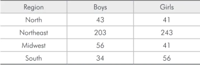
This descriptive study was designed as a cross-sectional research project. The sample was taken from the patient digital iles of six Oral Radiologi-cal Clinics from the North, Northeast, Midwest and South regions of the country. A total of 717 subjects (381 girls and 336 boys) aged 7.0 to 15.9 years were selected (Table 1). Ethical approval was obtained from the Piracicaba Dental School Committee after the Ethical Principles in Research Program exami-nation.
Radiographic images taken between June 2000 and April 2008 were selected. Because the radio-graphs were selected from the digital iles of Oral Radiological Clinics, no information about the pa-tients was available. Thus, papa-tients were included if they fulilled the following criteria: (1) Subjects had a Brazilian ethnic origin, regardless of ethnic-racial classiication, (2) the radiographs presented high quality and allowed good visualization of anatomi-cal structures, speciianatomi-cally the third and fourth cer-vical vertebral bodies, (3) all lateral cephalometric radiographs and hand-wrist radiographs were taken at the same time.
A cervical vertebral computerized analysis was created in the Radiocef Studio 2 computer software for digital cephalometric analysis (Radio Memory Ltda., Belo Horizonte, MG, Brazil). On the digital lateral cephalograms, anatomical landmarks were marked on the third and fourth cervical vertebrae (Figure 1):
C4ai, C4pi: the most inferior points of the ante-rior and posteante-rior borders of the body of C4.
C4as, C4ps: the most superior points of the an-terior and posan-terior borders of the body of C4.
C4ai’: the intersection of the base line (C4ai-C4pi) with a perpendicular line passing through
•
•
• Table 1 - Patient distribution according to geographic
re-gion.
Region Boys Girls
North 43 41
Northeast 203 243
Midwest 56 41
C4as.
C4pi’: the intersection of the base line (C4ai-C4pi) with a perpendicular line passing through C4ps.
C4mi: middle point of the base line (C4ai-C4pi).
C4ms: the intersection of the base line (C4ai-C4pi) with a perpendicular line passing through C4mi. It is marked on the superior border of the body of C4.
C4am: middle point of the C4as-C4ai line.
C4pm: the intersection of the C4as-C4ai line with a perpendicular line passing through C4am. It is marked on the posterior border of the body of C4.
The name and deinition of each landmark were indicated on screen. Image improvement resources such as brightness control, inversion, pseudo-color-ing, and zoom could be used to make it easier to ind each point. With the aid of these landmarks, the fol-lowing measurements were automatically obtained:
Anterior vertebral body height (AH): distance from C4as to C4ai’.
Vertebral body height (H): distance from
•
• •
• •
•
•
C4mi to C4ms.
Posterior vertebral body height (PH): distance from C4ps to C4pi’.
Anteroposterior vertebral body height (AP):
distance from C4am to C4pm.
Cervical vertebral bone age was automatically calculated using the formulas developed by Caldas
et al.17 (2007).
•
•
= 1.4892 + 11.3736 × AH3 + 4.8726×
AP3
H4 AP4 Male cervical
vertebral bone age
= 1.3523 + 6.7691 × AH3 + 8.6408×
AP3
AH4 AP4 Female cervical
vertebral bone age
Hand-wrist bone age was evaluated by the TW3 method, which assessed speciic ossiication centers of the hand and wrist (radius, ulna, and selected metacarpals and phalanges), leading to their classii-cation into one of several stages. Scores were derived from each bone stage and calculated to compute the skeletal age.
Intra-operator error was calculated according
to Dahlberg’s formula18 (1940) using 10
cephalo-metric radiographs selected randomly; these were measured with the computer software, and the same radiographs were measured again 10 days later. The formula revealed values below 1.0, indicating
suf-icient accuracy of the measurements.
Analysis of variance (ANOVA) was used to com-pare cervical vertebral bone age, hand-wrist bone
age and chronological age. Follow up Tukey tests were used to identify speciic differences. All analy-ses were performed with a signiicant level of 5%.
Results
Statistical analysis showed no signiicant dif-ference between cervical vertebral bone age and chronological age in all regions studied (Tables 2-9).
Table 2 - North region: Mean and standard deviation (SD) of cervical vertebral bone age (CVBA), hand-wrist bone age (HWBA) and chronological age (CA) for the girls’ sample.
Group Mean SD
CVBA 11.9088 b 1.9162
HWBA 12.3078 a 2.3322
CA 12.1934 ab 2.1838
Means followed by different lowercase letters differ statistically, with a signifi-cant p-value of 5% by the Tukey test. (aplicable to tables 2 through 9)
Table 3 - North Region: Mean and standard deviation of cervical vertebral bone age (CVBA), hand-wrist bone age (HWBA) and chronological age (CA) for the boys’ sample.
Group Mean SD
CVBA 13.2272 b 1.9133
HWBA 13.8446 a 1.9618
CA 13.3995 b 1.8370
Table 4 - Northeast region: Mean and standard deviation of cervical vertebral bone age (CVBA), hand-wrist bone age (HWBA) and chronological age (CA) for the girls’ sample.
Group Mean SD
CVBA 12.4083 b 2.0596
HWBA 12.6495 a 2.4617
CA 12.3849 b 2.3326
Table 5 - Northeast region: Mean and standard deviation of cervical vertebral bone age (CVBA), hand-wrist bone age (HWBA) and chronological age (CA) for the boys’ sample.
Group Mean SD
CVBA 12.7582 b 2.2395
HWBA 13.0756 a 2.5405
CA 12.7987 ab 2.3484
Table 6 - Midwest region: Mean and standard deviation of cervical vertebral bone age (CVBA), hand-wrist bone age (HWBA) and chronological age (CA) for the girls’ sample.
Group Mean SD
CVBA 11.9239 a 2.0430
HWBA 12.2836 a 2.5567
CA 12.0366 a 2.2363
Table 7 - Midwest region: Mean and standard deviation of cervical vertebral bone age (CVBA), hand-wrist bone age (HWBA) and chronological age (CA) for the boys’ sample.
Group Mean SD
CVBA 12.4582 b 1.8105
HWBA 12.9093 a 1.9336
CA 12.7030 ab 1.9911
Table 8 - South region: Mean and standard deviation of cervical vertebral bone age (CVBA), hand-wrist bone age (HWBA) and chronological age (CA) for the girls’ sample.
Group Mean SD
CVBA 11.3530 a 2.0549
HWBA 11.5673 a 2.0646
CA 12.3252 a 2.1217
Table 9 - South region: Mean and standard deviation of cervical vertebral bone age (CVBA), hand-wrist bone age (HWBA) and chronological age (CA) for the boys’ sample.
Group Mean SD
CVBA 11.5912 a 1.3357
HWBA 11.7382 a 1.5833
When analyzing bone age, it was possible to observe a statistically signiicant difference between cervi-cal vertebral bone age and hand-wrist bone age for female and male subjects in the North and North-east regions, as well as for male subjects in the Mid-west region. However, the differences encountered were no more than 0.61, 0.31 and 0.45 years in the North, Northeast and Midwest regions, respectively (Tables 2, 3, 4, 5, and 7).
No signiicant difference was observed between bone age and chronological age in all regions except for male subjects in the North and female subjects in the Northeast, which were 0.44 and 0.26 years, respectively (Tables 3 and 4).
Discussion
The hand-wrist radiograph has classically been used to determine the maturation level of a child. To avoid taking additional radiographs, the cervical ver-tebrae investigation method has become more popu-lar in recent years. Almost all authors found statisti-cally signiicant correlations between hand-wrist and skeletal maturation of the cervical vertebrae.5,13,14,16
The present study found a statistically signiicant difference between cervical vertebral bone age and hand-wrist bone age for female and male subjects in the North and Northeast regions, as well as for male subjects in the Midwest region. Even though the dif-ferences between cervical vertebral bone age and hand-wrist bone age were statistically signiicant, they were very low in both sex groups. Thus, these results question the necessity of taking hand-wrist radiographs when lateral cephalograms are routinely used for orthodontic documentation.
Similarly to this study, Soegiharto et al.19 (2008)
compared the cervical vertebral maturation (CVM) index and the skeletal maturation index (SMI) in both Indonesians and white subjects and found a statistically signiicant difference between both in-dexes. We agree that caution must be taken when interpreting the results of the present study because it was based on cross-sectional data, which have limitations for analyzing growth. Ideally, studies of this type should be longitudinal, but the dificul-ties involved in obtaining large sample sizes and the associated increase in the number of radiographic
exposures tend to rule out the application of this methodology. Still, another factor to be considered in this study is that different sample sizes were used in each region studied, although the overall sample size was satisfactory. Even though the patient iles of the Oral Radiological Clinics were large, it was very dificult to ind lateral cephalometric radiographs and hand-wrist radiographs taken on the same day.
It has long been recognized that an individual’s chronologic age does not necessarily correlate well with his maturation age. Skeletally, one may be re-tarded or advanced in various degrees of deviation from the actual chronological age.20,21 In this study,
no signiicant difference was observed between bone age and chronological age in all regions except for male subjects in the North and female subjects in the Northeast, which were 0.44 and 0.26 years, respec-tively. However, Hunter reported that the difference between bone age and chronological age is consid-ered normal when it does not exceed one year.22
For the present study, radiographs were select-ed from the patient iles of six Oral Radiological Clinics, presenting good visualization of the cervi-cal vertebrae. As to the methodology employed in this work, masking of patient identiication was a very important factor in preventing the inluence of knowing the chronological age on the determina-tion of the stage in which subjects were posidetermina-tioned in the growth curve. The randomized choice of ra-diographs pertaining to patients ranging from 7 to 15.9 years was intended to include all the matura-tion stages in the period during which patients seek orthodontic treatment and with the possibility of taking advantage of the craniofacial growth.
Almost all previous evaluations based on cervi-cal vertebrae on cephalometric radiographs used the method reported by Lamparski13 (1972) and
modi-ied by Hassel, Farman15 (1995). This method takes
into account morphological characteristics of the cervical vertebrae, such as concavity of the lower border and height and shape of the vertebral bod-ies. However, cervical vertebrae have been used to evaluate growth in a subjective manner. Özer et al.23
this reason, our study used the method of Caldas et al.17 (2007) instead of the classiication of
Lampar-sky13 (1972), which is of great importance because it
allows skeletal age to be calculated in an objective manner.
Many investigators have suggested that the size and shape of the cervical vertebrae change from birth to full maturity at each level of skeletal devel-opment.6 Baccetti et al.1 (2002) showed that only
the shape changes of C2, C3, and C4 were enough to show skeletal maturation. However, C2 is very dificult to measure and it shows little morphologi-cal change. In this study, we measured the third and fourth cervical vertebral bodies because the cervical vertebras lower than C4 cannot be observed when a thyroid protective collar is worn during radiation exposure.
Brazil is the largest country in South America. It is divided into ive geographic regions, each with its own distinct characteristics: the North, North-east, Midwest, SouthNorth-east, and South. Many studies have been performed on skeletal maturation and its capacity to predict and identify the adolescence growth spurt. However, all of them were conducted in speciic states of the country and, even though they were performed with Brazilian people, the obtained results present inherent limitations for interpretation, as they do not represent the overall population of the country. In this study, we selected individuals from the North, Northeast, Midwest, and South regions, since the Southeast region had
already been studied by Caldas et al.17 (2007).
Cephalometric analysis is an important tool in orthodontic diagnosis, treatment planning, evalua-tion of treatment results, and predicevalua-tion of growth. Rapid advances in computer science have led to its wide application in cephalometry. As a result, in re-cent years, digital cephalometric analysis has gained popularity in orthodontic practices. The use of modern cephalometric software requires importing digital cephalograms or digital capture of analogue data.24 Based on the fact that no study has assessed
cervical vertebral maturation using a computer software for digital cephalometric analysis, in this research we developed a cervical vertebral comput-erized analysis for objectively evaluating skeletal maturation on cephalometric radiographs.
Conclusions
In determining the relationship among chrono-logical age, cervical vertebrae and hand-wrist skel-etal maturation, the results suggest that the meth-od develop by Caldas et al.17 (2007) for objectively
evaluating skeletal maturation on cephalometric radiographs can be used to determine bone age in Brazilian subjects.
The computerized analysis created to automati-cally calculate cervical vertebral bone age is reliable and can be used to increase objectivity. The comput-er software used in the present study was found to be suitable for daily orthodontic diagnostics when treating Brazilian patients.
References
1. Baccetti T, Franchi L, McNamara JA Jr. An improved ver-sion of the cervical vertebral maturation (CVM) method for the assessment of mandibular growth. Angle Orthod. 2002;72(4):316-23.
2. Franchi L, Baccetti T, De Toffol L, Polimeni A, Cozza P. Phases of the dentition for the assessment of skeletal maturity: a diag-nostic performance study. Am J Orthod Dentofacial Orthop. 2008;133(3):395-400.
3. Santos EC, Bertoz FA, Arantes F de M, Reis PM, de Bertoz AP. Skeletal maturation analysis by morphological evaluation of the cervical vertebrae. J Clin Pediatr Dent. 2006;30(3):265-70.
4. Soegiharto BM, Cunningham SJ, Moles DR. Skeletal matu-ration in Indonesian and white children assessed with
hand-wrist and cervical vertebrae methods. Am J Orthod Dentofa-cial Orthop. 2008;134(2):217-26.
5. Garcia-Fernandes P, Torre H, Flores L, Rea J. The cervi-cal vertebrae as maturational indicators. J Clin Orthod. 1998;32(4):221-5.
6. Kucukkeles N, Acar A, Biren S, Arun T. Comparisons between cervical vertebrae and hand-wrist maturation for the assess-ment of skeletal maturity. J Clin Pediatr Dent. 1999;24(1):47-52.
7. Björk A, Helm S. Prediction of the age of maximum puberal growth in body height. Angle Orthod. 1967;37(2):134-43. 8. Grave KC, Brown T. Skeletal ossification and the adolescent
9. Hägg U, Taranger J. Maturation indicators and the pubertal growth spurt. Am J Orthod. 1982;82(4):299-309.
10. Mitani H, Sato K. Comparison of mandibular growth with other variables during puberty. Angle Orthod. 1992;62(3):217-22.
11. Holderbaum RM, Veeck EB, Oliveira HW, Silva CL, Fer-nandes A. Comparison among dental, skeletal and chrono-logical development in HIV-positive children: a radiographic study. Braz Oral Res. 2005;10(3):209-15.
12. Flores-Mir C, Burgess CA, Champney M, Jensen RJ, Pitcher MR, Major PW. Correlation of skeletal maturation stages determined by cervical vertebrae and hand-wrist evaluations. Angle Orthod. 2006;76(1):1-5.
13. Lamparski DG. Skeletal age assessment utilizing cervical ver-tebrae [Thesis]. Pittsburgh: University of Pittsburgh; 1972. 14. O’Reilly M, Yanniello G. Mandibular growth changes and
maturation of cervical vertebrae - a longitudinal cephalometric study. Angle Orthod. 1988;58(2):179-84.
15. Hassel B, Farman AG. Skeletal maturation evaluation us-ing cervical vertebrae. Am J Orthod Dentofacial Orthop. 1995;107(1):58-66.
16. Hellsing E. Cervical vertebral dimensions in 8-11, and 15-year-old children. Acta Odontol Scand. 1991;49(4):207-13.
17. Caldas M de P, Ambrosano GM, Haiter Neto F. New formula to objectively evaluate skeletal maturation using lateral cepha-lometric radiographs. Braz Oral Res. 2007;21(4):330-5. 18. Dahlberg G. Statistical methods for medical and biological
students. London: George Allen and Unwin; 1940.
19. Soegiharto BM, Moles DR, Cunningham SJ. Discriminatory ability of the skeletal maturation index and the cervical ver-tebrae maturation index in detecting peak pubertal growth in Indonesian and white subjects with receiver operating characteristics analysis. Am J Orthod Dentofacial Orthop. 2008;134(2):227-37.
20. Bambha JK. Longitudinal cephalometric roentgenographic study of face and cranium in relation to body height. J Am Dent Assoc. 1961;63:776-99.
21. Fishman LS. Chronological versus skeletal age, an evaluation of craniofacial growth. Angle Orthod. 1979;49(3):181-9. 22. Hunter CJ. The correlation of facial growth with body height
and skeletal maturation at adolescence. Angle Orthod. 1966;36(1):44-54.
23. Özer T, Kama JD, Ozer SY. A practical method for determin-ing pubertal growth spurt. Am J Orthod Dentofacial Orthop. 2006;130(2):131.e1-6.