118
Bone mineralization in Brazilian adolescents:
the years of maximum bone mass incorporation
Carla C. Silva, Tamara B. L. Goldberg, Altamir S. Teixeira, José C. Dalmas
Paulo State University (UNESP), São Paulo, Brazil., Londrina State University-UEL
SUMMARY. Puberty is the fundamental period for bone mass (BM) acquisition. In this period mineralization is found to increase with levels of high bone formation. The critical years of intense bone anabolism deserve special attention, as adequate gain could minimize fracture risk in later years. The objective of this work was to study bone mineral content (BMC) and bone mineral density (BMD) in male adolescents with age bracket and maturation level. Sixty-one healthy male 10 to 19 year-olds were evaluated for calcium intake, weight, stature, BMI, puberty stage and BMC and BMD in the lumbar spine and femur. BM was measured by bone densitometry (DXA). Calcium intake was calculated by recording 3 days diet. Puberty stage was defined as per Tanner. Descriptive statistics was used with means and standard deviations, linear correlation, and analysis of variance for comparison between age groups, and the Tukey test (p<0.05). Linear correlation was positive and indicated body weight as the main correlation variable with BMD in both studied locations (p<0.01). BMC and BMD increased with age, differences were significant from 14 to 15 years, and when adolescents reached Tanner stage G4. These results showed a pronounced increase in bone mineralization, with the years after 14 to 15 being critical for BM acquisition in Brazilian adolescents.
Key words: Adolescence, pubertal events, bone mass, bone mineral density, calcium intake.
RESUMEN. Mineralización ósea en adolescentes brasileños: años de máxima incorporación de masa ósea. La pubertad es un momento fundamental para la adquisición de la masa ósea (MO). En este período, la mineralización se encuentra en aumento en los niveles de formación ósea. Los años críticos de intenso anabolismo óseo necesitan de atención, porque el adecuado aumento de la masa ósea podría minimizar el riesgo de fracturas en los años posteriores. El objetivo de esta investigación fue estudiar el contenido mineral óseo (CMO) y la densidad mineral ósea (DMO) en adolescentes según el grupo de edad y el nível de maduración. Sesenta y un adolescentes saludables de 10 a 19 años fueron evaluados cuanto a la ingesta de calcio, peso, estatura, índice de masa corpórea (IMC), etapa puberal, CMO y DMO en la columna lumbar y en el fémur. La MO fue medida por densitometría ósea (DEXA). La ingesta de calcio fue calculada mediante un recordatorio de ingesta de tres días. La etapa puberal fue definida por los criterios de Tanner. Estadística descriptiva fue utilizada con media y desviación estándar, correlación linear y análisis de varianza para comparar los grupos y test de Tukey (p<0,05). Correlación linear fue positiva e indicó que el peso corporal fue la principal variable correlacionada con la DMO en los dos sitios estudiados (p<0,01). CMO y DMO aumentaron con la edad y las diferencias fueron considerables de los 14 a los 15 años, cuando los adolescentes alcanzaban la etapa G4 de Tanner. Estos resultados muestran un aumento pronunciado en la mineralización ósea; entre los 14 y 15 años fueron críticos para la adquisición de MO en adolescentes brasileños.
Palabras clave: Adolescencia, eventos puberales, masa ósea, densi-dad mineral ósea, ingesta de calcio.
INTRODUCTION
The bone mineralization process begins in the fetus, extending throughout infancy, and peaks in adolescence. These years are the fundamental period for bone mass acquisition. Several researchers consider that infancy and adolescence have highest bone mineral capital increase for both sexes(1-6).
International research has been developed showing the relationship between adolescence and bone health. These studies are based on the cyclic principle involving bone mass deposition throughout life. Infancy and adolescence are marked by a very important bone formation rate with predominance of formation over reabsorption. In adulthood both processes stabilize and from 45 to 50 years, especially for females, there is a predominance of bone reabsorption.
However, bone reabsorption is not exclusive to females as both osteopenia and osteoporosis have significantly increased in males (7).
Expressive longitudinal growth during puberty has three distinct phenomena which occur sequentially. They are: growth spurt lasting about 2 to 3 years, characterized by a reduced growth velocity prepuberal phase, an accelerated growth velocity known as Peak Height Velocity (PHV), and a growth cessation phase which contributes to over 20% of final adult stature; a rapid acquisition of bone mineral content known as bone mass peak, and the skeletal maturation process which ends with epiphyseal closure (1,2,8,9).
has shown that when bone mass accumulation is potentialized during puberty and maintained in adulthood we can minimize reductions from advancing age, thus helping to prevent osteopenia/osteoporosis and consequent fractures (11).
Osteoporosis is a heterogenous disorder considered a severe public heath problem which can in part be due to inadequate bone gain during infancy and adolescence (12). Nutritional factors such as adequate calcium supplement, according to age bracket and gender [13] and physical exercise, especially with high impact show protective effects related to healthy bone tissue maintenance independent to the time in life when these measures are adopted; however they should be put into practice early in infancy and adolescence (11,14,15).
In recent years, methods have been developed to accurately evaluate bone mass allowing us to better understand bone tissue dynamics. Dual energy x-ray absorptiometry (DXA) allows very precise analysis with low radiation exposure; this is suitable for evaluating children and adolescents (16,17).
Variations in bone density during infancy and adolescence have been seen in epidemiological studies in different countries. In Brazil however, there have been few investigations on bone mineralization in healthy children and adolescents (18). The objective of this study was to determine the behavior of bone mineral content (BMC) and bone mineral density (BMD) in male adolescents in relation to age bracket and sexual maturity levels.
SUBJECTS AND METHODS
This study included 10 to 19-year-old healthy volunteer students from a private school (Associação Brasileira de Educadores Lassalistas, Colégio La Salle, Botucatu, São Paulo State). The research was approved by the Research Ethics Committee of Botucatu School of Medicine – UNESP, and the volunteers and their parents/guardians gave informed written consent.
Inclusion criteria were weight between the 10 and 90 percentiles, and height between the 10 and 97.5 percentiles for each age bracket (19), with adequate body mass index (BMI) for their age (20), and with daily dairy product intake. They had to be non-smokers and non-drinkers, could not be involved in any extra-curricular sporting activity, only the school’s physical education classes. Control of normal physical activity was not necessary as investigations indicate that it is programmed sporting activities that produce higher increases in bone mass (1,2,15).
Exclusion criteria were: history of prematurity or low birth weight, prolonged corticoid therapy, or calcium or iron supplement in the twelve months prior to research. Other exclusion criteria were: diabetes mellitus, acute or chronic malnutrition, congenital or acquired bone diseases, gastrointenstinal diseases followed by malabsorption, history of nephropathy with or without chronic renal insufficiency,
endocrinopathies, precocious and delay puberty, chronic drug consumption, cystic fibrosis, celiac disease, and use of drugs negatively affecting bone metabolism such as anticonvulsants and antacids with aluminum. Exclusions related to diet were: vegetarianism, high fiber, caffeine, or soft drinks consumption, and no daily dairy product intake.
Data collection started at school; randomly selected students without any dysfunction or disorder exclusions were invited for weight and height measurements. When weights and heights were within the proposed limits, they were asked about drinking and smoking habits. Those fulfilling the criteria were then invited to participate in the study. Their parents/guardians were then contacted to explain the methods used and seek consent. Students or parents could withdraw from the study at any time.
From 497 students, 61 who fitted the inclusion criteria participated in all evaluations. A private school was chosen because it represents a socially privileged population thus ensuring the most favorable conditions to achieve full bone gain potential. As far the ethnic question is concerned, the extent of miscegenation in Brazil is very high, however none of our adolescents were children of exclusively African or Asian origin parents.
Volunteers fitting the criteria were then invited with their parents to attend the Adolescent Outpatient Clinic at Botucatu University Hospital School of Medicine – UNESP where they were interviewed with their parents and submitted to a general and specific physical examination to detect any physical alterations. Secondary Sexual Characters were evaluated, and compared to Tanner Criteria (21). To assess the impact of puberty stage on bone mineralization, maturation level by visual inspection of genitals was compared to BMC and BMD results from dual energy x-ray absorptiometry (DXA). Skeletal maturity (bone age) was obtained by the GP method (22), where hand and wrist x-rays are compared with the Atlas.
Diet characterization then followed using a three-day dietary record completed by participants and analyzed by the authors to obtain information on food intake, preferences, refusals, the main meals involving calcium and any other factors that could possibly interfere in the bio-availability of this mineral (23). Centesimal quantification of food data was by a computerized nutritional analysis system developed by São Paulo University School of Public Health Nutrition Department (24).
BMC (g) and BMD (g/cm2) were determined for each adolescent by a DXA with Hologic QDR 2000-Plus densitometer. Bone mass evaluation was performed on the lumbar spine between L1-L4 and the femural neck.
and standard deviations were used to characterize weight, height, BMI, and three-day mean calcium intake. Pearson simple linear correlation coefficients were calculated between bone mass and morphological aspects, and puberty stage (p<0.01). Analysis of variance was used to compare all ABs and maturation levels with BMC and BMD, and the Tukey test was used to locate significant differences (p<0.05).
RESULTS
Table 1 shows general characteristics: body weight, height, BMI, and mean daily calcium intake calculated by recording diet over three days for each age bracket.
We observed increased body weight, height, and BMI with advancing age; these were significant from 14 to 15 yrs (AB 3) on (Table 1).
TABLE 1
General characteristics of the adolescents studied: body weight (kg), stature (m), BMI (kg/m2), and total calcium intake (mg/day)
Age Bracket Weight (kg) Stature (m) BMI (kg/m2) Calcium (mg/day)
Age (years) X ± SD X ± SD X ± SD X ± SD
(AB1)10-11 (n=14) 35.75 ± 4.57 c 1.432 ± 0.07 bc 17.39 ± 1.43 783 ± 236 (AB2)12-13 (n=14) 43.49 ± 8.47 ac 1.550 ± 0.08 ac 17.91 ± 1.63 740 ± 198 (AB3)14-15 (n=14) 57.71 ± 7.53 ab 1.710 ± 0.06 ab 19.67 ± 1.83 a 887 ± 228 (AB4)16-17 (n=12) 62.71 ± 7.48 ab 1.731 ± 0.07 ab 20.91 ± 1.99 ab 894 ± 275 (AB 5) 18-19 (n=7) 70.34 ± 3.20 abc 1.803 ± 0.05 ab 21.65 ± 1.16 ab 1073 ± 434 b ANOVA analysis of variance and Tukey’s test for differences between age groups.
Letters show the differences between age groups (p<0.05). a Difference between AB1 and the other groups.
b Difference between AB2 and the other groups. c Difference between AB3 and the other groups.
BMI= body mass index; AB= Age brackets; SD= standard deviations
After analysis of the dietary diaries records completed by the adolescents, we observed that calcium intake for the different age groups were from 740
±
198 mg/day to 1,073±
434 mg/day, with an average of 863±
280 mg/day.Pearson’s simple linear correlation coefficient was used
to investigate the impact of body dimension and nutritional changes related to genital maturation stage classification over bone mass. Table 2 shows significant and positive differences for simple linear correlation between all variables; significance level was less than 1%.
TABLE 2
Simple correlation coefficients between variables related to bone mass, morphological aspects, and secondary sexual characteristics
Bone Mass Age Bone Age Weight Stature BMI Genitals
More precise analysis reveals that, from all the studied indicators, BMI had the poorest correlation to adolescent bone mass. The highest correlation was between BMC and body weight; the score for lumbar spine was r= 0.88, and for femural neck was r=0.91. The highest correlation for BMD was also to body weight; the score for lumbar spine was r=0.85, and femural neck r=0.80. The correlation between skeletal and
sexual maturation indicators was greater than 0.70, showing a strong participation from biological maturation in relation to bone mass increase for these adolescents.
Table 3 shows BMC and BMD values for lumbar spine and femural neck with adolescent age. Significant differences (p<0.05) were seen from 14 to 15 yrs (AB 3) for both BMC and BMD in these regions.
TABLE 3
Mean and standard deviations of bone mineral content and bone mineral density in the lumbar spine and femoral neck for age groups
Age (Years) BMC- Spine BMD-Spine BMC-Femur BMD-Femur
(grams) (g/cm2) (grams) (g/cm2)
(AB-1)10–11 28.71± 3.95 c 0.618 ± 0.049 c 26.01 ± 3.69 c 0.800 ± 0.07 c (AB-2)12–13 36.08 ± 10.16 c 0.708 ± 0.116 c 32.85 ± 9.73 c 0.844 ± 0.08 c (AB-3)14–15 51.54 ± 13.28 ab 0.836
± 0.132 ab 48.87
± 9.40 ab 0.978
± 0.14 ab (AB-4)16–17 59.26 ± 11.38 ab 0.937 ± 0.113 ab 54.54 ± 11.65 ab 1.095 ± 0.17 ab (AB-5)18–19 76.72 ± 10.46 abc 1.094 ± 0.132 abc 62.15 ± 7.35 abc 1.205 ± 0.11 abc Total (n=61) 47.16 ± 18.50 0.806 ± 0.187 42.59 ± 15.54 0.956 ± 0.18 ANOVA analysis of variance and Tukey’s test for differences between age groups
Letters show differences between age groups (p<0.05). a Difference between AB1 and the other groups. b Difference between AB2 and the other groups. c Difference between AB3 and the other groups.
BMC= bone mineral content; BMD= bone mineral density
Bone mineralization parameters were compared with sexual maturation level, particularly genital development, to see which stages of puberty had the highest increase in bone
mass (Table 4). Significant differences (p<0.05) were seen in G4 and G5 for both BMC and BMD; there were no significant alterations in mineralization parameters between G1 and G3.
TABLE 4
Mean and standard deviations of bone mineral content and bone mineral density in the lumbar spine and femoral neck according to sexual maturity levels
Genital BMC- Spine BMD-Spine BMC-Femur BMD-Femur
Development (grams) (g/cm2) (grams) (g/cm2)
G 1 (n=6) 25.98 ± 2.86 0.584 ± 0.045 24.53 ± 3.43 0.786 ± 0.089 G2 (n=12) 29.61 ± 4.26 0.639 ± 0.053 26.48 ± 3.71 0.806 ± 0.066 G3 (n=8) 34.48 ± 6.77 0.694 ± 0.102 33.13 ± 3.93 0.842 ± 0.062 G4 (n=15) 51.55 ± 13.46 ab 0.844 ± 0.121 ab 50.00 ± 13.35 abc 0.999 ± 0.178 b G5 (n=20) 65.03 ± 13.51 abc 0.989 ± 0.138 abc 55.90 ± 8.97 abc 1.109 ± 0.151 abc ANOVA analysis of variance and Tukey’s test for differences between age groups
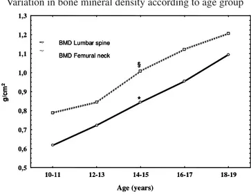
Figure 1 shows BMD variation in ABs. Increased growth can be seen from 10 to 19 yrs, with significant differences in femur neck and lumbar spine BMD between 14 and 15 yrs. Femural neck values are higher than lumbar spine at all ages.
FIGURE 1
Variation in bone mineral density according to age group
Bone mineralization behavior was similar and increased with sexual maturation (Figure 2), indicating significant differences in G4 and G5 for BMD in both regions. However in G3, we can clearly see pronounced increases in BMD.
FIGURE 2
Variation in bone mineral density according to sexual maturity levels
DISCUSSION
Bone mineralization is a complex process with several factors affecting bone mass acquisition, the most important being genetic factors; body dimension, weight, and height alterations; hormonal profile which leads to sexual and skeletal maturity; physical exercise; and an adequate calcium intake at this age which is reflected in strong bone mineralization (7,12,25,26).
This study showed mean nutrition indicator values for body weight, stature and BMI for each age bracket similar to those presented by the National Center for Health Statistics (NCHS) data, as inclusion criteria were similar to the methods chapter (19,20).
In this group, the calcium supplement intake did not reach minimum recommended levels. Intake ranged from 713 ± 292 to 1451 ± 334mg/day, when ideal intake for adolescents of either sex should be 1300mg/day (13). Literature shows that maximum intake should not exceed 2500mg/day; this was not reached by any adolescent in this study. However values in this study were higher than in other Brazilian studies for the same age bracket (27-29). Transverse studies on children and adolescents indicate the beneficial effects of adequate calcium intake on bone mass peak (12). Apparently, low calcium intake during child and adolescent growth results in lower bone mineralization than with same age bracket individuals who had adequate intake (30).
According to Abrams et al. (31) there is no doubt that adolescents may adapt to very low calcium intakes (<500mg/ day) by increasing fractional absorption and decreasing both urinary and endogenous fecal calcium excretion, but the exact contributions to “calcium economy” are not known at this time. They suggest that those on very low calcium intakes are at substantial risk of low calcium retention. However, a double blinded controlled study on calcium carbonate supplementation over 13 months in 143 male adolescents showed a significant increase in spinal (+2.5%), proximal femur (+2.3%), and whole body (+1.3%) bone mineral content. The authors also emphasized that bone mass potentialization is associated with an increase in stature (0.4%) equivalent to 7mm (32).
The mean for calcium intake in our study was 863±280mg/ day, values under the dietary intake references for calcium (DRI) (13), but superior to those considered as very low calcium intake (31).
The beneficial impact of calcium intake on child and adolescent bone mineralization was reported in a longitudinal study by Lee et al. (33). Calcium carbonate (800mg/day) was given for 18 months to children of both sexes with a mean age of 8.5 years. The results showed a significant increase in lumbar spine BMC in relation to controls.
The potential benefits of a calcium rich diet and systematic
* > significant in BMD – Lumbar spine (p<0.05). § > significant in BMD – Femural neck (p<0.05).
Age (years) § * g/c m 2 0,5 0,6 0,7 0,8 0,9 1,0 1,1 1,2 1,3
10-11 12-13 14-15 16-17 18-19
BMD Lumbar spine
BMD Femural neck
* > significant in BMD – Lumbar spine (p<0.05). § > significant in BMD – Femural neck (p<0.05).
Age (years) § * g/c m 2 0,5 0,6 0,7 0,8 0,9 1,0 1,1 1,2 1,3
10-11 12-13 14-15 16-17 18-19
BMD Lumbar spine
BMD Femural neck BMD Lumbar spine
BMD Femural neck
Sexual maturity level §
§
* > significant in BMD – Lumbar spine (p<0.05). § > significant in BMD – Femural neck (p<0.05).
* * 0,5 0,6 0,7 0,8 0,9 1,0 1,1 1,2 1,3
G1 G2 G3 G4 G5
g/
c
m
2
BMD Lumbar spine
BMD Femural neck
Sexual maturity level §
§
* > significant in BMD – Lumbar spine (p<0.05). § > significant in BMD – Femural neck (p<0.05).
* * 0,5 0,6 0,7 0,8 0,9 1,0 1,1 1,2 1,3
G1 G2 G3 G4 G5
g/
c
m
2
BMD Lumbar spine
BMD Femural neck BMD Lumbar spine
physical exercise during infancy and adolescence have been reported by several authors (2,4,11). These behaviors are the basis of a healthy lifestyle related to bone mass. Literature shows that adequate habits started in pediatric populations tend to last throughout adult life and minimize the risk of fractures later on (2,12,15).
The results in Table 2 are similar to other investigations correlating BMD with anthropometric variables such as body weight, height, and alterations in sexual and bone maturation (12,18,29).
In a Brazilian study, Pessoa et al. (34) evaluated BMC and BMD in pre-pubescent children, 7–8 years old, and found high and positive correlations between lumbar spine BMC and BMD and bone age, body weight, and height. They suggest that in-terpretation of bone mass in pre-pubescence should be linked to body weight and bone age variation (34). Similarly, Klein reported a significant correlation between total body BMD and chronological and bone age in children of approximately 10 years old and of both sexes (35).
In relation to age, this study shows significant differences from 14 to 15 years, both in BMC and BMD in the lumbar spine and femural neck regions. Rubin evaluated BMD in 299 children and adolescents of both sexes between 6 and 18 years old (12). The results indicated a major acceleration in lumbar spine BMD from 13 years in males, which stabilized around 15 to 16 years, similar to our study. In a study with 207 Caucasian children and adolescents of both sexes between 9 and 17 years, there was a pronounced difference in males in both lumbar spine and femural neck between 13 and 17 years [36]. According to Theintz, the period between 13 and 17 years was fundamental for BMD increase in the lumbar spine and femural neck (37).
Everything indicates that the period from 14 to 16 years old is critical for bone mineralization. These data are in agreement with several studies where there a linear increase was seen in bone mass during infancy, with an exponential increase during puberty in several bone sites (12,18,38).
Although age is a major temporal indicator for alterations occurring in adolescence, it is limited in relation to the con-stant modifications occurring in puberty due to maturation level variability in individuals of the same age. More recently re-searchers have reported that puberty stage and bone age should be considered when interpreting bone mass measurements (26). Male adolescents significantly increase bone mass between the ages of 14 and 15, and between G4 and G5 maturation levels; this is reflected in the bone mineral content and bone mineral density gains seen in our study in the lumbar spine and femoral neck regions. Therefore, to be between 14 and 15 years old and over the G3 maturation stage, corresponds to a critical gain in bone mass acquisition which impacts high mineralization rate.
This study shows that the increase in bone mineralization during puberty occurs at the same time as significant increases in body dimensions and is related to secondary sexual characters.
It is important to emphasize that the adolescents in this study come from a socially differentiated stratum with adequate weight and height, and higher calcium intake than other Brazilian studies (27-29). The results indicate variations in BMC and BMD in healthy adolescents. Currently, this is the only study that considers rigorous inclusion and exclusion criteria for the Brazilian population. When compared with international published data they indicate great similarity.
ACKNOWLEDGEMENTS
To Fundação de Amparo à Pesquisa do Estado de São Paulo for financing part of this research. FAPESP- Process 04/07007-1.
REFERENCES
1. Pettersson U, Nordströnm P, Alfredson H, Henriksson-Larsén K, Lorentzon R. Effect of high impact activity on bone mass and size in adolescent female: a comparative study between two different types of sports. Calcif Tissue Int 2000; 67: 207-214.
2. Lima F, Falco V, Baima J, Carazzato JG, Pereira RMR. Effect of impact load and active load on bone metabolism and body composition of adolescent athletes. Med Sci Sports Exerc 2001; 33: 1318-1323.
3. Jones G, Riley MD, Whiting S. Association between urinary potassium, urinary sodium, current diet, and bone density in prepuberal children. Am J Clin Nutr 2001; 73: 839-844. 4. Outila TA, Kärkkäinen MUM, Lamberg-Allardt CJE. Vitamin
D status affects serum parathyroid hormone concentrations during winter in female adolescents: associations with forearm bone mineral density. Am J Clin Nutr 2001; 74: 206-210. 5. Crawford PB, Wang MC, Sabry ZI, Hudes M, VanLoan M,
Bachach LK. Adolescent diet is predictive of peak bone mass. Am J Clin Nutr 2002; 75: 356S.
6. Chinn DJ, Fordham JN, Kibirige MS, Crabtree NJ, Venables J, Bates J, Pitcher O. Bone density at the os calcis: reference values, reproducibility, and effects of fracture history and physical activity. Arch Dis Child 2005; 90: 30-35.
7. Lorentzon M, Lorentzon R, Bäckström T, Nordstöm P. Estro-gen receptor Estro-gene polymorphism, but not estradiol levels, is related to bone density in health adolescents boys: A cross-sectional and longitudinal study. J Clin Endocrinol Metab 1999; 84: 4597-4601.
8. Rubin K. Pubertal development and bone. Curr Opin Endocrinolol Diab 2000; 7: 65-70.
9. Phillip M, Lazar L. The regulatory effect of hormones and growth factors on the pubertal growth spurt. Endocrinology 2003;13: 465-469.
reflects teenage sports-exercise patterns but not teenage cal-cium intake. Am Academy Pediatrics 2000; 106:40-44. 11. Badenhop-Stevens N, Matkovic V. Calcium needs in children.
Orthopaedic Nursing 2004; 23(4): 228-232.
12. Rubin K, Schirduan V, Gendreau P, Sarfarazi M, Mendola R, Daisky G. Predictors of axial and peripheral bone mineral density in health children and adolescents, with special attention to the role of puberty. J Pediatr 1993; 123: 863-870.
13. Institute of Medicine (US). Dietary references intakes for cal-cium, phosphorus, magnesium, vitamin D and fluoride. Wash-ington (DC): National Academy Press; 1998.
14. Kohrt WM, Bloomfield SA, Little KD, Nelson ME, Yingling VR. Physical activity and bone health. Med Sci Sports Exerc; Special Communications 1985-1996; 2004.
15. Mackelvie KJ, Khan KM, Mckay HA. Is there a critical period for bone response to weight-bearing exercise in children and adolescents? A systematic review. Br J Sports Med 2002; 36:250-7.
16. Bonjour JP. Peak bone mass, calcium, and protein intakes. Feed Toddlers Adolesc 1996; 37: 31-43.
17. Boot AM, Bouquet J, Ridder MAJ, Krenning EP, Keizer-Schrama SMPFM. Determinants of body composition measures by dual-energy x-ray absorptiometry in Dutch children and adolescents. Am J Clin Nutr 1997; 66: 232-238.
18. Fonseca ASM, Szejnfeld VL, Terreri MT, Goldenberg J, Ferraz MB, Hilário MOE. Bone mineralization density of the lumbar spine of Brazilian children and adolescents aged 6 to 14 years. Braz J Med Biol Res 2001; 34: 347-352.
19. Hamill PVV, Drizd TA, Johnson CL, Reed RB, Roche AF, Moore WM. Physical growth: National Center for Health Sta-tistics Percentiles. Am J Clin Nutr 1979; 32: 607-629. 20. Prevalence of overweight among adolescents-United States,
1988-91. MMWR Morb Wkly Rep 1994; 43: 818-821. 21. Marshall WA, Tanner JM. Variation in the pattern of pubertal
changes in boy. Arch Dis Child 1970; 45: 13.
22. Greulich WW, Pyle SI. Radiographic atlas of skeletal development of the hand and wrist, 2nd. ed. Standford: University Press; 1959.
23. Cintra IP, Von Der Heydy MED, Schitz BAS, Francheschini SCC, Taddei JAAC, Sigulen DM. Métodos de inquéritos dietéticos. Cad Nutr 1997; 13: 11-23.
24. Philiphi ST, Szarfarc SC, Lattrza AR. Virtual Nutri. Versão 1.0 for Windows. Departamento de Nutrição: Faculdade de Saúde Pública Universidade de São Paulo; 1996.
25. Blanchet C, Giguère Y, Prud’homme D, Dumont M, Rousseau F, Dodin S. Association of physical activity and bone: influence of vitamin D receptor genotype. Med Sci Sport Exerc 2002; 34: 24-31.
26. Ahmed SF, Weaver LT. Letter to the editor. J Pediatr Gastroenterol Nutr 2005; 40: 99-103.
27. Albuquerque MFM, Monteiro AM. Ingestão de alimentos e adequação de nutrientes no final da infância. Rev Nutr PUCCAMP 2002; 15: 291-99.
28. Garcia GCB, Gambardella AMD, Frutuoso MFP. Estado nutricional e consumo alimentar de adolescentes de um centro de juventude da cidade de São Paulo. Rev Nutr PUCCAMP 2003;16: 41-50.
29. Vargas DM, Rigotti T, Gütz CNRM, Lobe MCS, Fernandes JA. Mineralização óssea em crianças e adolescentes com diabetes melito tipo 1. J Pediatr 2003; 79: 253-258. 30. Jackman LA, Millane SS, Martin BR, Wood OB, McCabe GP,
Peacock M et al. Calcium retention in relation to calcium intake and postmenarcheal age in adolescent females. Am J Clin Nutr 1997; 66: 327-333.
31. Abrams SA, Griffin IJ, Hicks PD, Gunn SK. Pubertal girls only partially adapt to low dietary calcium intakes. J Bone Miner Res 2004;19:759 –763.
32. Prentice A, Ginty F, Stear SJ, Jones SC, Laskey MA, Cole TJ. Calcium supplementation increases stature and bone mineral mass of 16- to 18-year-old boys. J Clin Endocrinol Metab 2005; 90: 3153–3161.
33. Lee WTK, Leung SSF, Leung DMY, Cheng JCY. A follow-up study on the effects of calcium-supplement withdrawal and puberty on bone acquisition of children. Am J Clin Nutr 1996; 64: 71-77.
34. Pessoa JHL, Lewin S, Longui CA, Mendonça BB, Bianco AC. Densidade mineral óssea: correlação com peso corporal, estatura, idade óssea e fator de crescimento símile à insulina. J Pediatr 1997; 73: 259-264.
35. Klein KO, Larmore KA, Lancey E, Brown JM, Considine RV, Hassink SG. Effect of obesity on estradiol level, and its rela-tionship to leptine, bone maturation, and bone mineral density in children. J Clin Endocrinol Metab 1998; 83: 3469-3475. 36. Bonjour JP, Theintz G, Buchs B, Sloman D, Rizzoli R. Critical
years and stages of puberty for spinal and femoral bone mass accumulation during adolescence. J Clin Endocrinol Metab 1991;73: 555-563.
37. Theintz G, Buchs B, Rizzoli R, Sloman D, Clavien H, Sizonenko PC et al. Longitudinal monitoring of bone mass accumulation in health adolescents: evidence for marked re-duction after 16 years of age at the levels of lumbar spine and femoral neck in female subjects. J Clin Endocrinol Metab 1992; 75: 1060-1065.
38. Hui SL, Dimeglio LA, Longcope C, Peacock M, McClintock R, Perkins AJ et al. Difference in bone mass between black and white American children: Attributable to body build, sex hormone levels, or bone turnover? J Clin Endocrinol Met 2003; 88:642-49.