Katherine Cooper(a) Vincent Bennani(a) Andrew Tawse-Smith(b) Malcolm Reid(c)
Claudine Stirling(c) George Dias(d)
(a) Department of Oral Rehabilitation, School of Dentistry, Univ of Otago, Dunedin, New Zealand.
(b) Deans Department, School of Dentistry, Univ of Otago, Dunedin, New Zealand. (c)Department of Chemistry, Centre for Trace
Element Analysis, Univ of Otago, Dunedin, New Zealand.
(d) Department of Anatomy, Otago School of Medical Sciences, Univ of Otago, Dunedin, New Zealand.
Corresponding Author:
Vincent Bennani
E-mail: [email protected]
Effect of a cordless retraction paste
on titanium surface: a topographic,
chemical and biocompatibility
evaluation
Abstract: Good exposure of the preparation margins and haemostasis in the sulcular gingiva are necessary for accurate impressions to produce precise restorations. The use of cordless retraction paste material in im-plant dentistry is a relatively novel application. However, few studies have been conducted on the use of retraction pastes and their possible in-teraction with implant surfaces. Recent literature has described remnants on titanium implant surfaces and expressed the need for an assessment of the biocompatibility of the exposed surface (Chang et al.). This in vitro
study evaluated the effect of a cordless gingival retraction paste on sterile titanium disks. Surface chemistry was determined using energy-disper-sive X-ray spectroscopy (EDS), and further investigated using laser abla-tion inductively coupled plasma mass spectrometry (LA-ICP-MS). After exposure to retraction paste, surface chemistry alterations were identi-ied. A ibroblast cell line (L929) was exposed to the disks and the live/ dead viability/cytotoxicity assay was used to determine any effects on the proliferation and health of cells. The disks exposed to the retraction paste showed fewer dead cells compared to the unexposed disks. This was statistically signiicant.
Descriptors: Dental Impression Materials; Dental Impression Technique; Dental Implants.
Introduction
The use of gingival retraction paste material in implant dentistry ap-pears to have considerable potential. Although some studies have been conducted on this innovative application, there is a need for further in-vestigation into the biocompatibility of the implant surfaces after they have been exposed to a retraction paste material.
Gingival sulcus opening and haemostasis around an abutment is re-quired to obtain an accurate impression of the preparation site.1 Most of
the time, a gingival retraction cord soaked in an astringent medicament, such as aluminium chloride (AlCl3), is used for tissue retraction and
hae-mostasis before impression taking for cement-retained implant restorations. AlCl3 is the least irritating among the chemicals used for gingival retraction
and has the longest lasting retraction effect after its removal from the sul-cus. Displacing forces exerted on peri-implant tissue during the cord
pack-Declaration of Interests: The authors certify that they have no commercial or associative interest that represents a conflict of interest in connection with the manuscript.
Submitted: Nov 14, 2012
ing procedure could cause irreversible trauma. Using an injectable matrix containing AlCl3 as an
alterna-tive for gingival retraction is an atraumatic procedure with reduced risk of trauma to peri-implant tissue.2,3
The surface of dental implants is a key contribu-tor to successful osseointegration.3 For titanium
im-plants, oxidation of its surface results in an excess of free radicals and oxygenated derivatives which form a titanium dioxide layer. In order to make the bone/ implant interface highly dynamic, some ions, such as calcium and phosphorus, also present in bone, are incorporated within this titanium oxide layer. Alteration or damage to the oxide layer leads to a pathological loss of osseointegration called peri-im-plantitis.4 The factors contributing to
peri-implanti-tis fall into four categories:
• lesions of peri-implant attachment,
• presence of aggressive bacterial strains,
• excessive mechanical stress and
• corrosion.3
If the implant surface topography and chemistry is altered during the gingival retraction process, im-plant biocompatibility may be affected.
According to Chang et al.5 traces of retraction
paste remain on implant surfaces even after two rinses with distilled water. Further research is re-quired to quantify the amount and effect of these remnants on implant surface biocompatibility. Some tests have used cell cultures of ibroblasts or osteo-blast-like cells to investigate biocompatibility on a titanium surface.6,7 Imaging techniques have been
used to analyze cell adhesion, proliferation and dif-ferentiation of cells cultured on these surfaces, in-cluding scanning electron microscopy.8
ISO categories are designed to evaluate the bio-compatibility of medical devices used in dentistry9,10
by assessing cell damage through morphologic anal-ysis, the measuring of cell growth/proliferation and the investigation of speciic aspects of cell metabo-lism. The most reliable method of assessment is a standardised analysis of cell morphology by a histo-morphologist. Culture and quantitative measure-ment of human diploid ibroblasts are an appropri-ate model for identifying the early cytotoxic effects of dental materials.11
In this in vitro study, we evaluated the effect of a cordless gingival retraction paste on sterile titanium disks. The surface chemistry was irst determined using energy-dispersive X-ray spectroscopy (EDS), and further investigated using laser ablation induc-tively coupled plasma mass spectrometry (LA-ICP-MS). Cell cytotoxicity was determined using the live/dead viability/cytotoxicity assay.12 The aim of
this investigation was to determine whether the ap-plication of a gingival retraction paste to a titanium surface alters surface chemistry, and, if so, whether this would have a detrimental effect on cells, hence on biocompatibility.
Methodology
Disk samples and gingival retraction paste
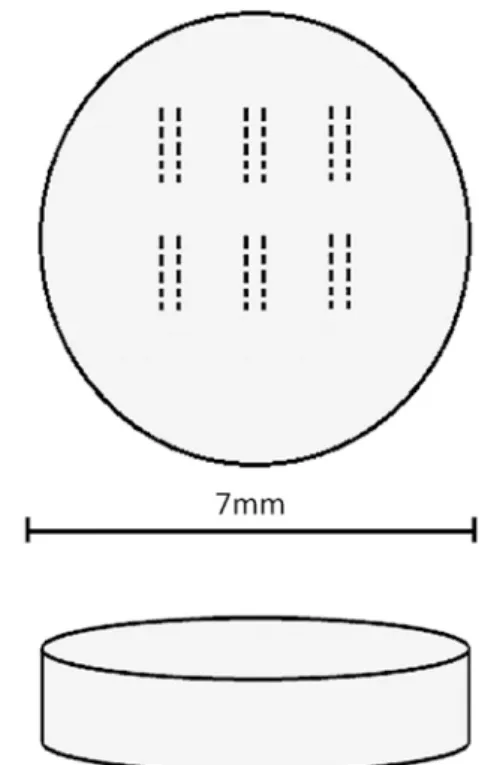
Seventy-two 7 mm × 2 mm thick sterile titanium disks from Southern Implant (Southern Implant, Irene, South Africa; ASTM-F67-95 Grade 4 pure ti-tanium) were used. The disks had standardised en-hanced moderately rough surfaces (Sa = 1.43 nm)
with the same surface topography and chemistry as those of Southern implants. Table 1 demonstrates how the disks were assigned to the different meth-ods.
A gingival retraction paste (Expasyl; Acteon, Bordeaux, France) was applied on the surface of the disks using a handgun applicator. Expasyl (Acte-on) is a kaolin-based material with 15% aluminium chloride.
Exposure protocol
Half of a cartridge (0.30 cm³) of Expasyl gin-gival retraction paste (Acteon) was applied onto the whole surface of each disk for 10 minutes to simulate possible incomplete removal of the paste after rinsing (the manufacturer’s recommenda-tion is one to two minutes). To achieve the same pressure generated during placement of Expasyl
Table 1 - Study outline specifying the number of disks used for each test.
SEM EDS LA-ICP-MS Cell culture
Control 1 5 6 24
were analysed (Table 1), followed by instrument calibration. For each disk, laser ablation data were acquired in ixed positions across the surface of the disk as twelve tracks, each 600 µm in length. Aver-age Al/Ti and Si/Ti count ratios were obtained and then scaled according to known ratios for the NIST calibration glasses to derive concentration ratios.
Cell vitality tests
Viability tests were carried out on a L929 ibro-blast cell line.11 Disks were seeded at a density of
4 × 10³ cells/cm², and allowed to adhere for 30
min-utes at 37°C. Cells were then incubated in α-MEM supplemented with 10% fetal bovine serum (FBS; Invitrogen, Carlsbad, USA) for 48 hours at 37°C. The media contained L-glutamine, and no antibi-otics were used. Cells were grown in 5% CO2 in a
humidiied cell culture incubator (Galaxy mini CO2
incubator; New Brunswick Scientiic, Enield, USA). The relative numbers of live and dead cells were measured using the live/dead viability/cytotoxicity assay.12
A confocal microscope was used to detect the (Acteon) material in vivo (143 kPa),13 a weight
of 225 g was applied to the paste during contact with the disk. Following the protocol established by Chang et al.5, each disk was rinsed twice with
distilled water and spray set at 50 psi for two min-utes. All these procedures were carried out in a sterile environment, with sterile plastic equipment to avoid any contamination of the disks.
Surface topography and chemistry
A scanning electron microscope (SEM; JEOL 6700 Field Emission SEM, Tokyo, Japan) was used to capture images of the surface before and after ap-plication of Expasyl (Acteon). An accelerating volt-age of 15 kV was selected for analysis with the SEM. Local compositional differences across the surface were observed in the backscattered electron mode (BSE).
Survey electron-dispersive spectroscopy (EDS) elemental data were acquired under an accelerating voltage of 25 kV. SEM and semi-quantitative EDS analysis was carried out on the disk surfaces before and after contact with the gingival retraction paste.
Furthermore, the composition of Al and Si (main components of Expasyl) relative to Ti for each sam-ple was determined at the Centre for Trace Element Analysis, University of Otago, using LA-ICP-MS high sensitivity analysis.14 Speciically, a quadrupole
7500 cs ICP-MS instrument (Agilent Technologies, Chicago, USA) was coupled to a NewWave UP-213 Nd:YAG deep UV (213 nm) laser ablation system (Electro Scientiic Industries, Portland, USA) for the in situ analysis of elemental composition at the micron scale. Ablation was performed under a pure helium atmosphere whilst the laser was operated in continuous mode using a spot size of 50 µm, a repe-tition rate of 5 Hz, and a scan rate of 5 µm/s (Figure 1). The ablated aerosol was entrained into the mass spectrometer by an argon carrier gas low. Immedi-ately after each sample analysis, the instrument was calibrated with NIST (National Institute of Stan-dards and Technology) glass stanStan-dards (NIST 610 and NIST 612).
Analyses by LA-ICP-MS were carried out on the disk surface before and after exposure to Expasyl (Acteon). A total of twelve disks (Southern Implant)
wavelengths produced in the assay. Three images were taken randomly per disk by the same operator, and quantitative analysis of the images was carried out using ImageJ software (Version 1.45S, National Institutes of Health, Baltimore, USA). The colour channels for each image were split, and the green and red considered separately. The threshold for each image was set to 90–255 and the area itting that intensity was measured with ImageJ software.
Statistical analysis
The data obtained from each disk was analysed making an educated assumption of a normal distri-bution pattern. The exposure of disks was the as-signed variable for comparison. The student t-test was used for the chemical data. p < 0.05 was consid-ered statistically signiicant. A non-parametric Wil-coxon T test was used for the biocompatibility data.
Results
Surface topography using SEM
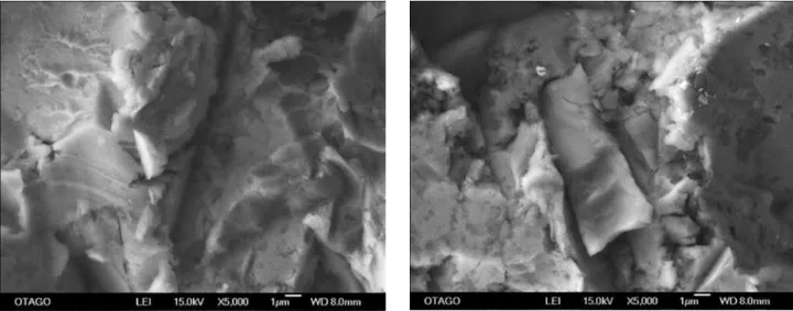
Under low magniication (×500), no difference in surface topography could be seen between exposed and control disks. The surface was rough with sharp, deined valleys and tops, and variable lank length. At higher magniication (×5000), minute particles were visible, particularly in the back-scat-ter electron (BSE) detector view (Figure 2). These particles measured 0.7 µm at most.
Surface chemistry
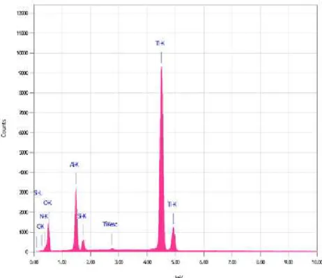
EDS results showed that traces (less than 1%) of oxygen and silicon were present on the disks exposed to Expasyl (Acteon). The silicon compo-nent in the exposed and control groups was signii-cantly different. Both the exposed and control disks (Southern Implant) were similar with regard to alu-minium and titanium, in that both of these elements yielded the highest readings among the elements considered (Table 2).
LA-ICP-MS was carried out to quantify the trace amount of silicon and aluminium detected with EDS (Figure 3). The results showed a statistically signii-cant difference in the ratio of Si/Ti between control and exposed disks, in that this ratio was higher in the exposed samples. In contrast, higher amounts of alu-minium were detected in the control samples, but this difference was not statistically signiicant (Table 3).
Figure 2 - SEM image at ×5000 magnification of the unpolished disk surface, control (left) and exposed (right). BSE shows deposits of retraction paste on the surface of the exposed disk.
Table 2 - EDS results for control and exposed disks.
C* Al2O3*/** SiO2* TiO2**
Control 2 2 t 26 25 23 t 0 0 72 71 74
1 1 1 30 24 22 0 0 0 67 74 75
Exposed 1 3 2 27 25 21 t 2 3 71 71 74
1 1 1 22 19 20 1 t t 74 79 77
p > 0.05 p < 0.05
*Elements originating From Expasyl (Acteon, Bordeaux, France);
**ele-ments originating from the Southern-Implants disk (Southern Implant,
Cell vitality tests
The area, thus number, of live cells (those which take up calcein) was greater in the exposed group than in the control group (Figure 4). The area of dead cells was higher in the control disks than in those exposed to Expasyl (Acteon). The remaining percentage corresponded to black space, or to areas where the lorescence was less than 90, thus did not it the threshold parameters (Table 4).
Discussion
Packing a gingival retraction cord is the usu-al method applied in implantodontics and ixed prosthodontics.1 The Expasyl retraction paste
(Ac-teon) provides adequate gingival crevicular opening to achieve an accurate impression of the inishing margins in ixed prosthodontics.
Minimal alterations to surface morphology after contact with Expasyl (Acteon) were observed in SEM images, which partially supports the indings by Chang et al.5 The only change was observed at
×5000 magniication, where “white” particles were visible on the surface. However, this was not repli-cated in all images of the exposed disks (Southern Implant). The particles could be residual Expasyl (Acteon) material on the surface, as the EDS results
showed a statistically signiicant difference in the silicon component (Table 2). The occasional appear-ance of white particles would explain the increased variability in the hi-spatial resolution LA-ICP-MS signals, even if not evident on a low spatial resolu-tion technique like EDS. This observaresolu-tion could plain why Al/Ti signals were more variable in the ex-posed counts compared to those of the control.
After exposure to Expasyl (Acteon), EDS mea-surements indicated that silicon was present, but only at levels near the instrumental detection limit, an observation consistent with the indings of Chang
et al.5 Because the EDS silicon data was only con-Table 3 - LA-ICP-MS results, for control and exposed disks, as a ratio relative to titanium.
Ratio Si/Ti Al/Ti
Control
Mean (SD) 1.75 × 10−05 (0.02) 9.12 × 10−05 (1.85)
Max 5.10 × 10−05 1.60 × 10−04
Min 8.32 × 10−06 6.10 × 10−05
Exposed
Mean (SD) 2.60 × 10−05 (0.06) 8.90 × 10−05 (7.41)
Max 5.98 × 10−05 1.90 × 10−04
Min 9.30 × 10−06 4.71 × 10−05 Si/Ti, p < 0.05.
sidered to be indicative, LA-ICP-MS was employed to fully quantify elemental abundances owing to its sensitivity and spatial resolution.14
The EDS showed that titanium and some alumin-ium were present before the disk (Southern Implant) was exposed. Titanium was present because the disk was grade 4 titanium. The presence of aluminium was accounted for by its addition used to modify the surface to enhance osseointegration.3,5
LA-ICP-MS analysis of the disks showed a predominance of titanium and aluminium counts before treatment with Expasyl (Acteon). Aluminium, which is pres-ent in both the unexposed disk surfaces and in the Expasyl paste (Acteon), varied within and between sample groups (exposed and control). The variation
in the control disks meant that a baseline for alu-minium could not be accurately determined. Hence, the anticipated increase after exposure could not be accurately measured.
One limitation of the LA-ICP-MS method is that the depth of ablation is dificult to control. Further research on this aspect is needed.
The cell viability assay results indicated that there was a difference in biocompatibility between the surfaces. L929 cells are well characterised and widely used in research. They have a high prolif-eration rate, and are relatively resilient. Experimen-tally, the cells were reaching 80% conluence at 48 hours when initially seeded with 2 × 10³ cells/well in
24-well plates. This was calculated using an ATCC L929 data sheet protocol, and is an appropriate number for analysis and to ensure healthy cells.
When seeding the cells, it was noticed that the cells spread better on the exposed disks as compared to the control disk. This could imply an increased biocompatibility. The results showed less than 2% dead cells in both disk groups. The exposed disks achieved higher areas of live cells (98% in the ex-posed versus 95.32% in the control). It is possible that an increased wettability of the surface caused the slight difference in cell viability. This difference
Figure 4 - Confocal images of live/dead viability tests. The control surface (left) shows more red dots than the exposed disk (right). The black spaces represent areas that are not occupied by cells; they are less abundant in the exposed disk (right).
Table 4 - Mean area of all images taken with respect to green and red channels.
Control Exposed
Green 17.54% 24.47%
Red 0.87% 0.33% p < 0.05
Black 81.60% 75.21%
Total % surface with cells
present (area not black) 18.41% 24.80%
was not statistically signiicant, therefore with neg-ligible clinical implications; however, it could be an area for further research.
Furthermore, the aforementioned result was complemented by analysing the black area in the images. This area relects the area not covered with live or dying cells, and was lower in the exposed disk group.
Another inding that could support this statement is that, when using the proteolysis enzyme trypsin to detach cells during the 3-(4,5-dimethylthiazol-2- yl)-5-(3-carboxymethoxyphenyl)-2-(4-sulfophenyl)-2H-tetrazolium (MTS) assay (as described in ISO-10993)10 to determine cell viability, the cells would
not detach from the exposed disk surface. Only after applying trypsin for twice the amount of time rec-ommended (10 minutes) did the cells detach, which is detrimental to the cells. Thus, the live/dead viabili-ty cytotoxiciviabili-ty test, that does not require cell detach-ment, was used.
Conclusion
1. Chemical analysis indicated the presence of sili-con after application of Expasyl (Acteon).
2. Exposing the titanium disks to Expasyl (Acte-on) did not affect their viability.
Acknowledgements
The authors thank Pierre-Roland, a division of the Acteon Group (Bordeaux, France), for providing inancial support for this research, and appreciate the use of the testing facilities at the University of Otago, New Zealand, and the Sir John Walsh Re-search Institute for their support. The authors also wish to thank Professor Peter Herbison, for his assis-tance with the statistical analysis; Keppel Kooman, for his help with the cell culture, and Liz Girvan, for the SEM work involved in this study. Claudine Stirling and Malcom Reid gratefully acknowledge the Community Trust of Otago for initial inancial support to the Centre for Trace Element Analysis.
References
1. Shannon A. Expanded clinical uses of a novel tis-sue retraction material. Compend Contin Educ Dent. 2002 Jan;23(1Suppl):3-6.
2. Bennani V, Schwass D, Chandler N. Gingival retraction techniques for implants versus teeth. J Am Dent Assoc. 2008 Oct;139(10):1354-63.
3. Dohan Ehrenfest DM, Coelho PG, Kang B-S, Sul Y-T, Al-brektsson T. Classification of osseointegrated implant surfac-es: materials, chemistry and topography. Trends Biotechnol. 2010 Apr; 28(4):198-206.
4. Mouhyi J, Ehrenfest DD, Albrektsson T. The peri-implantitis: implant surfaces, microstructure and physicochemical aspects. Clin Implant Dent Relat Res. 2012 Apr;14(2):170-83. 5. Chang Y-S, Bennani V, Tawse-Smith A, Girvan L. Effect of a
cordless retraction paste material on implant surfaces: an in vitro study. Braz Oral Res. 2011 Nov-Dec;25 (6):492-9. 6. Groessner-Schreiber B, Neubert A, Muller A-D, Hopp M,
Griepentrog M, Lange K-P. Fibroblast growth on surface-modified dental implants: an in vitro study. J Biomed Mater Res. 2003;64:591-9.
7. Pae A, Kim S, Kim H, Woo Y. Osteoblast-like cell attach-ment and proliferation on turned, blasted and anodized ti-tanium surfaces. Int J Oral Maxillofac Implants. 2011 May-Jun;26(3):475-81.
8. Mustafa K, Wennerberg A, Wroblewski J, Hultenby K, Lo-pez B, Arvidson K. Determining optimal surface roughness
of aTiO2 blasted titanium implant material for attachment, proliferation and differentiation of cells derived from hu-man hu-mandibular alveolar bone. Clin Oral Implant Res. 2001 Oct;12(6):515-25.
9. International Organization for Standardization. ISO-7405: dentistry – evaluation of biocompatibility of medical devices used in dentistry. Geneva: ISO; 2008. 7 p.
10. International Organization for Standardization. ISO-10993-5: biological evaluation of medical devices – part 5: tests for in vitro cytotoxicity. Geneve: ISO; 2009. 34 p.
11. Willerhausen B, Marroquin B, Schaefer D, Schulze R. Cyto-toxicity of root canal illing materials to three different human cell lines. J Endo. 2000 Dec;26(12):703-7.
12. Viability/Cytotoxicity Kit for mammalian cells. Molecular Probes. Invitrogen Detection technologies.Eugene: Product information; 2005.7 p.
13. Bennani V, Aarts J, He L. A comparison of the pressure gener-ated by cordless gingival displacement techniques. J Prosthe Dent. 2012 Jun;107(6):388-92.