Submitted 15 December 2014 Accepted 18 June 2015 Published9 July 2015 Corresponding author
Andrew R. Cuff, andrew.cuff@ucl.ac.uk
Academic editor
Mark Young
Additional Information and Declarations can be found on page 17
DOI10.7717/peerj.1093 Copyright
2015 Cuffand Rayfield
Distributed under
Creative Commons CC-BY 4.0 OPEN ACCESS
Retrodeformation and muscular
reconstruction of ornithomimosaurian
dinosaur crania
Andrew R. Cuff∗and Emily J. Rayfield
School of Earth Sciences, University of Bristol, Bristol, United Kingdom
∗Current affiliation: Department of Genetics, Environment and Evolution, University College
London, London, United Kingdom; Structure and Motion Laboratory, Royal Veterinary College, London, United Kingdom
ABSTRACT
Ornithomimosaur dinosaurs evolved lightweight, edentulous skulls that possessed keratinous rhamphothecae. Understanding the anatomy of these taxa allows for a greater understanding of “ostrich-mimic” dinosaurs and character change during theropod dinosaur evolution. However, taphonomic processes during fossilisation often distort fossil remains. Retrodeformation offers a means by which to recover a hypothesis of the original anatomy of the specimen, and 3D scanning technologies present a way to constrain and document the retrodeformation process. Using computed tomography (CT) scan data, specimen specific retrodeformations were performed on three-dimensionally preserved but taphonomically distorted skulls of the deinocheiridGarudimimus brevipesBarsbold, 1981and the ornithomimids Struthiomimus altusLambe, 1902andOrnithomimus edmontonicusSternberg, 1933. This allowed for a reconstruction of the adductor musculature, which was then mapped onto the crania, from which muscle mechanical advantage and bite forces were calculated pre- and post-retrodeformation. The extent of the rhamphotheca was varied in each taxon to represent morphologies found within modern Aves. Well constrained retrodeformation allows for increased confidence in anatomical and functional analysis of fossil specimens and offers an opportunity to more fully understand the soft tissue anatomy of extinct taxa.
Subjects Paleontology
Keywords Skull, Rhamphotheca, Retrodeformation, Myology, Ornithomimosaurs, Bite forces
INTRODUCTION
Fossil skulls can offer insights into many aspects of vertebrate ecology and evolution. The cranium hosts the major sensory systems and, along with the mandible and hyolingual apparatus, is responsible for the ingestion of food items. Three-dimensionally preserved skulls provide even greater insight by allowing studies of endocranial morphology (Brochu, 2000;Sanders & Smith, 2005;Witmer & Ridgely, 2009), reconstruction of soft tissues (e.g., rhamphothecae and musculature:Holliday, 2009;Lautenschlager, 2013;
(both pre- and post-burial) can lead to the disarticulation or distortion of skeletal remains. As such, reconstructing and retrodeforming fossil remains can correct for taphonomic damage and is important for furthering our understanding of extinct taxa (Tschopp, Russo & Dzemski, 2013;Williams, 1990).
Various methods have been used to retrodeform fossil taxa. Methods particularly applicable to fossils preserved on a 2D bedding plane range from rescaling drawings (Rushton & Smith, 1993) to the determination of the strain ellipse (Cooper, 1990;Hughes & Jell, 1992) or other ways of deducing tectonic deformation (Motani, 1997). Digital techniques lend themselves to retrodeformation of 3D preserved fossils, including employing 3D computer models for user manipulation of individual disarticulated bones (Lautenschlager, 2013;Porro, Rayfield & Clack, 2015), modifying digital models by reference to closely related extant taxa (Zollikofer et al., 2005;Gunz et al., 2009) or by using landmarks (Molnar et al., 2012;Tallman et al., 2014) and geometric morphometrics (Angielczyk & Sheets, 2007;Hedrick & Dodson, 2013). The efficacies of these methods may be debated, but ultimately they are limited by the quality of preserved material (including brittle and plastic deformation) and perception of what the original specimen should look like, whether informed by symmetry or informed by closely related extant or extinct taxa.
Ornithomimosauria are a clade of coelurosaurian theropod dinosaurs that are com-monly known as “ostrich-mimicking” dinosaurs due to their cranial and postcranial con-vergences with palaeognathous birds. The convergence is seen in their lightweight skulls, with relatively large orbits and edentate jaw margins that bear rhamphotheca (Makovicky, Kobayashi & Currie, 2004). The most primitive members of Ornithomimosauria (Nqwebasaurus thwaziDe Klerk et al., 2000, andPelecanimimus polyodonPerez-Moreno et al., 1994) possess numerous tiny teeth in the premaxillae, maxillae and mandibles. More derived members of the group lose their upper dentition, maintaining a reduced dentition on the mandible (Harpymimus okladnikoviBarsbold & Perle, 1984; andShenzhousaurus orientalisJi et al., 2003), before becoming fully edentate (as in deinocheirids (Lee et al., 2014) and ornithomimids (Makovicky et al., 2010)). Where teeth are lost, ornithomimids possess beaks, inferred from the presence of foramina on the lateral surfaces the premaxilla, maxilla and mandible and the preservation of remnants of keratinous rhamphothecae in two specimens, theOrnithomimusspecimen used in this study, RTMP 1995.110.0001, andGallimimus bullatusOsm´olska, Roniewicz & Barsbold, 1972, specimen GIN100/1133 (Norell, Makovicky & Currie, 2001). The posterior extent of the beak is subject to debate, yet important for functional considerations as it provides a food capture and manipulation surface and plays a role in the reduction of feeding-related bony stress (Lautenschlager et al., 2013).
Rayfield et al., 2001;Holliday, 2009;Bates & Falkingham, 2012;Lautenschlager, 2013). The studies range from simple identification and line drawings based on osteological correlates (e.g.,Haas, 1969), to clay modelling of the muscles (Rayfield et al., 2001), to digital reconstructions (e.g.,Lautenschlager, 2013). The increased sophistication of adductor reconstruction has permitted more accurate estimation of not just the size of individual muscles, and therefore the force they can potentially generate, but their spatial relations to each other and effects of muscle bulging during contractions.
The aim of this paper is to document the process and consequences of retrodeformation of the crania of three ornithomimosaur theropod dinosaurs. Then using our hypotheses of retrodeformed morphology we reconstruct the comparative adductor muscle anatomy and calculate and compare the relative differences between adductor mechanical advantage and the resulting estimated bite force along the jaw. We do this for skulls pre- and post-retrodeformation, to deduce, in the context of the specimens presented here, the influence of retrodeformation on our predictions of function. This allows characterisation of bite forces arising during the evolution of edentulism between the ornithomimids and deinocheirids and more broadly within the ornithomimosaurs, one of at least three clades of coelurosaurian theropods that diverge from hypercarnivory (Zanno & Makovicky, 2011). We compare our predicted bite forces to the only other estimate from a herbivorous theropod,Erlikosaurus andrewsiiPerle, 1981, a therizinosaur (Lautenschlager et al., 2013). Given that the three ornithomimosaurians andE. andrewsiihave similar sized skulls, we test for congruence in bite force magnitudes between these putatively herbivorous taxa.
METHODS
Specimens
Few well preserved, three-dimensional ornithomimosaur skulls are known. Here we focus on crania from three taxa:Garudimimus brevipes,Struthiomimus altusandOrnithomimus edmontonicus.Garudimimusis known from only a single specimen. Our chosen specimens ofS. altusandO. edmontonicusrepresent the best prepared material for either taxon. There are other cranial remains, but most are badly crushed, encased within matrix prohibiting detailed observation, or remain taxonomically contentious. A number of specimens were examined first hand (seeAppendix S1) and information from the published literature on the well preserved skulls ofGallimimus(Osm´olska, Roniewicz & Barsbold, 1972), Deinocheirus(Lee et al., 2014), andSinornithomimus(Kobayashi & L¨u, 2003), was used for comparison where possible and inform on the retrodeformation process.
The specimen ofGarudimimus brevipes(GIN 100/13, described byBarsbold (1981)
andKobayashi & Barsbold (2005)) was scanned at the University of Texas using a P250D scanner at 419 kV, 1.8 mA, aluminum filter, slice thickness=0.5 mm, total slices=517.
TheOrnithomimus edmontonicusspecimen (RTMP 1995.110.0001) was scanned along
Struthiomimusthe scans are of relatively low quality. To provide better detail, the scans were upsampled inAvizo7.0 (FEI Visualization Sciences Group, USA). This process creates interpolations between each of the original CT slices to provide twice the number of slices in every axis for smoother reconstructions, but not providing any further resolution. The GarudimimusCT dataset was not resampled.
Reconstructions
The CT datasets were loaded into the visualisation and analysis packageAvizo 7.0. Segmentation and isolation of each individual cranial bone was performed, as far as the deformed, and in some places incomplete, datasets permitted. As all of the specimens suffered deformation, it was necessary to undertake retrodeformation to provide a complete undeformed skull for each species on which the soft tissue reconstructions could be based. Notably, the nature and magnitude of deformation differed in each taxon, and hence specimen-specific retrodeformation processes were applied to each specimen. Furthermore, there is no known undistorted skull for any of the taxa studied. The process of deformation was therefore informed by the topographic relationships of the individual cranial elements in the 3D dataset, evidence of breakage and cracks revealed from direct observation of specimens and the CT scan data, and information gathered from related ornithomimosaur material from museum collections and the literature (as outlined above, and seeAppendix S1). Where possible, a set of criteria were employed to perform and constrain the process. As outlined inArbour & Currie (2012), the shape of the orbit was used a proxy to determine the degree of deformation. Orbital retrodeformation was therefore employed to reconstruct the arrangement of the surrounding facial bones. In all studied ornithomimosaurs, both actual specimens and literature study, the pattern of breakage and deformation to the bones of the orbital region suggest that the orbits in undeformed taxa should be approximately circular. As such, this was the first correction applied to theGarudimimusandStruthiomimusskulls. InGarudimimus, the individual bones were segmented from the CT scan datasets and the bones surrounding the orbit were rotated into position using the editing tools inAvizo(sensuLautenschlager et al., 2013;Button, Rayfield & Barrett, 2014). This process was sequentially repeated with bones further from the orbits, until all of the right side of the specimen was reconstructed with the original material (no obvious plastic deformation was seen in this specimen except for the posterior margin of the maxilla). In theStruthiomimusskull, the orbital region was dorsally shifted by translating the bones within the “edit label field” function inAvizountil a circular orbit was restored. This process was continued anteriorly and posteriorly until a smooth cranial roof was created. The orbit was then measured in anteroposterior and dorsoventral axes with the “measure” tool withinAvizoto check whether a near circular structure has been achieved via the retrodeformation process.
the mediolateral dimensions and required expansion of the skulls. ForOrnithomimus the palatal bones were separated and aligned, and the remainder of the skull expanded mediolaterally to fit the palate. The palatal morphology of observed and well preserved specimens in the literature was used to inform on this procedure.
For the remaining bones it was possible to determine whether cortical bone had collapsed or was damaged using the CT scan data, so that the surface topography of the bones could be reconstructed using the paintbrush region-selecting tool within Avizoto match that seen in other specimens or ornithomimosaur taxa (e.g., the jugals inOrnithomimus). In some places bone was so badly damaged that full reconstruction required material from the other scans and digital manipulation using the paintbrush tools to create “new bone”. This was always informed by the individuals studied here as well as other specimens and taxa from museum collections and the literature. For example, in the anterior portion of the jugal inGarudimimus, the bone is broken and partially missing, but should overlap the posterior ramus of the maxilla and contact the lacrimal. The maxillary ramus was therefore ventrally displaced to bring it into alignment with the preserved remains of the jugal (as described for the orbit ofStruthiomimus), and the jugal was extended using the paintbrush tool in three dimensions to provide the required contact whilst maintaining the shape seen in the other scanned and observed ornithomimosaurs. InGarudimimusthe right side of the skull was better preserved than the left, whilst the opposite was true inStruthiomimus. Bones of the better preserved sides, once aligned and reconstructed, were mirrored about the sagittal midline of the skull, using the mirror function inAvizo(Lautenschlager, 2013).
Ornithomimosaur myology
Following methods ofHolliday (2009),Lautenschlager (2013)andButton, Rayfield & Barrett (2014), the individual insertions and origination sites for the adductor muscles were digitally mapped onto the 3D ornithomimosaur skull reconstructions. Where there was a lack of osteological correlates on the bones in either the CT scans or the actual specimens, phylogenetic bracketing was used to ascertain likely insertion and origination locations. These originations and insertions were demarcated on the skull and mandible. As there was no scannedGarudimimusjaw, theStruthiomimusjaw was used (scaled and rotated into place) to ascertain muscle orientation as it was the closest in morphology of the two ornithomimids.
2013;Button, Rayfield & Barrett, 2014). This process was repeated for all adductor muscles. All of the fleshed out muscles were then enlarged until they occupied the maximum amount of space within the chambers without intersecting in three-dimensional space, whichAvizocan prevent. The expanded muscle bodies were then digitally smoothed using tools inAvizo.
Muscle forces were estimated using the dry skull method (Thomason, 1991), where force (Fmus) equals the cross-sectional area (CSA) multiplied by the isometric muscle stress (σ
here taken as 0.3 N mm−2:Weijs & Hillen, 1985;Thomason, 1991):
Fmus=CSA×σ.
The CSA is calculated inAvizo, using the ‘clipping plane’ tool to define the cross section and the ‘material statistics’ module which calculates the surface area. This was done for each muscle at its widest location to give the maximum CSA and thus maximum estimated force. As this method fails to take into account pennation angle of muscle fibres, the forces were multiplied by a scale factor (calculated from experimental comparisons between modelled and actual data (Thomason, 1991)) of 1.5 to compensate. Given the arrangement of muscle bodies, the total muscle force is the resultant of anteroposterior, dorsoventral and mediolateral force components. Mediolaterally orientated muscle force has limited influence on jaw closing due to the almost vertical orientation of the muscle lines of action. As such the dorsoventral component is studied for bite force lever mechanics (as inLautenschlager, 2013). The force of each muscle (Fmus:Table 2) can be multiplied by
the perpendicular distance of the muscle centroid from the jaw joint (measured in Avizo) to provide a muscle moment:
Fin=Fmus×perpendicular distance from joint.
The sum of each of the muscle input moments can then be used to calculate bite forces (Fbf) at individual locations along the skull (Table 2):
TotalFin=Σ(Fmus×perpendicular distance from joint)
TotalFin=TotalFout
TotalFout=Fbf×perpendicular distance from joint.
Rhamphothecae
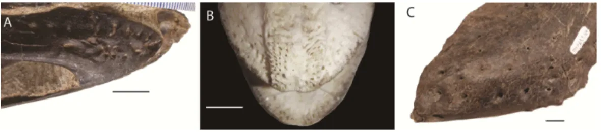
Figure 1 Foramina and rugosities in the rostra of certain taxa. (A) Anterior, right mandible of Struthiomimus altus (RTMP 1990.026.0001); (B) Dorsal view of anterior premaxilla of ostrich and mandible (ROM R1080); (C) Anterior dentary of a tyrannosaur (Daspletosaurus?) RTMP (1967.009.0164). Scale bars=1 cm.
In non-avian theropods, the picture is more complicated. Ornithomimosaurs, oviraptorids, therizinosaurs, andLimusaurus(a ceratosaur) underwent tooth loss leading to partial edentulism and inferences of rhamphothecae (Zanno et al., 2009;Zanno & Makovicky, 2011). These taxa bear regular foramina across the lateral surface of edentulous regions of the premaxilla and dentary. There are also grooves on the mandible of Erlikosaurus(a therizinosaur) that appear to demarcate a keratinous rhamphotheca/beak (Lautenschlager, 2013;Lautenschlager et al., 2013). However, neurovascular foramina are also present in large theropods (e.g., tyrannosaurs:Fig. 1; spinosaurs:Dal Sasso et al., 2005;
Morhardt, 2009) where teeth are present and keratinous beaks are not inferred.
Table 1 Selection of measurements pre- and post-retrodeformation for each skull.Length is measured from the centre of the quadrate condyle to the tip of premaxilla; width is measured as the distance between the centres of each quadrate condyle; orbit height is measured as the dorsoventral height of the centre of the orbit. All measures are in millimetres.
Garudimimus Struthiomimus Ornithomimus
Pre- Post- Pre- Post- Pre-
Post-Length 226 225 183 183 185 185
Width 34a 46 64a 56 26 42
Orbit height 59.5 61 35 54 68 68
Notes.
aWhere there is an anterior–posterior offset resulting in a shear, inflating the measure.
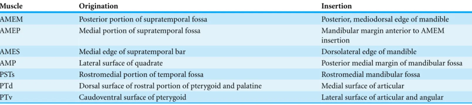
Table 2 Reconstructed muscle originations and insertions for the ornithomimosaurs studied here (see text for muscle abbreviations).
Muscle Origination Insertion
AMEM Posterior portion of supratemporal fossa Posterior, mediodorsal edge of mandible AMEP Medial portion of supratemporal fossa Mandibular margin anterior to AMEM
insertion
AMES Medial edge of supratemporal bar Dorsolateral edge of mandible
AMP Lateral surface of quadrate Posterior medial margin of mandibular fossa PSTs Rostromedial portion of temporal fossa Rostromedial mandibular fossa
PTd Dorsal surface of rostral portion of pterygoid and palatine Medial surface of articular
PTv Caudoventral surface of pterygoid Lateral surface of articular and angular
nares ofErlikosaurus(Lautenschlager et al., 2013). As the lower jaw was not used in any functional studies, beaks were not reconstructed for the mandibles.
RESULTS
The cranial reconstructions are shown inFigs. 2–4. No new gross anatomical descriptive information is revealed but the overall dimensions of the skull are modified by retrodefor-mation (Table 1). The width of the skull is modified in all taxa post-retrodeforretrodefor-mation, as are the dimensions of the orbit inGarudimimusandStruthiomimus. The few areas where cranial material was digitally added compared to original bone can be seen inFig. 5.
Figure 2 Garudimimus brevipesreconstruction (GIN 100/13).(A), (C), (E), original skull, (B), (D), (F), retrodeformed skulls. (A), (B), right lateral; (C), (D) dorsal; (E), (F), ventral views. Scale bar=5 cm.
SeeVideo S1andVideo S2showing video of the skull before and after retrodeformation.
Figure 3 Struthiomimus altusreconstruction (RTMP 1990.026.0001).Note the dorsoventral expansion of the skull after retrodeformation, particularly of the orbital region. (A), (C), (E), original skull, (B), (D), (F), retrodeformed skulls. (A),(B), right lateral; (C), (D) dorsal; (E), (F), ventral views. Scale bar=5 cm.
Figure 4 Ornithomimus edmontonicusreconstruction (RTMP 1995.110.0001) showing the effect of the mediolateral expansion after separating the taphonomically deformed bones of the palate.(A), (C), (E), original skull, (B), (D), (F), retrodeformed skulls. (A), (B), right lateral; (C), (D) dorsal; (E), (F), ventral views. Scale bar=5 cm. SeeVideos S5andS6showing video of the skull before and after
retrodeformation.
specimen (Figs. 2Eand2F) as this is one of the better preserved and prepared skulls available to study.
Figure 5 Reconstructions showing the regions where material was added using thepaintbrush region-selecting tool withinAvizo.Regions in red showing the areas where new material was added. (A)–(C) Garudimimus, (D)–(F)Struthiomimus, (G)–(I)Ornithomimus.
Myology
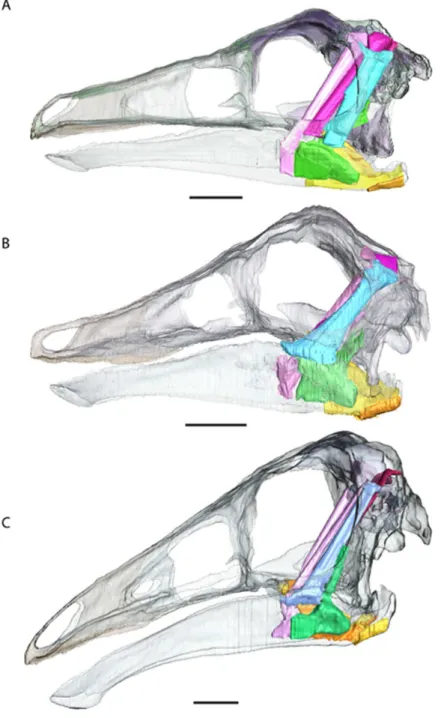
The reconstructions do not find any major differences between insertions and originations of the ornithomimosaurian myology and other dinosaurs (Fig. 6andTable 2), except that we could not reliably restore the M. pseudotemporalis profundus. This muscle usually attaches on the epipterygoid in extant sauropsids and has been identified in other dinosaurs (Holliday, 2009). Because none of the specimens had an identifiable epipterygoid attachment visible on the quadrate (as in birds:Holliday & Witmer, 2007) the muscle was not reconstructed. It is possible the muscle occupies some of the space used here in the reconstruction of the M. adductor mandibulae posterior.
Figure 6 Full cranial reconstruction including musculature of the jaw. (A) Garudimimus, (B) Struthiomimus, (C)Ornithomimus.Scale bars=5 cm. Pink, PSTs; purple, AMEp; red, AMEm; blue,
AMEs; green, AMP; yellow, PTd; orange, PTv.
forces at any of the positions along the jaw, whilstOrnithomimusproduces the lowest. The presence of a rhamphotheca marginally reduces estimated bite forces.
DISCUSSION
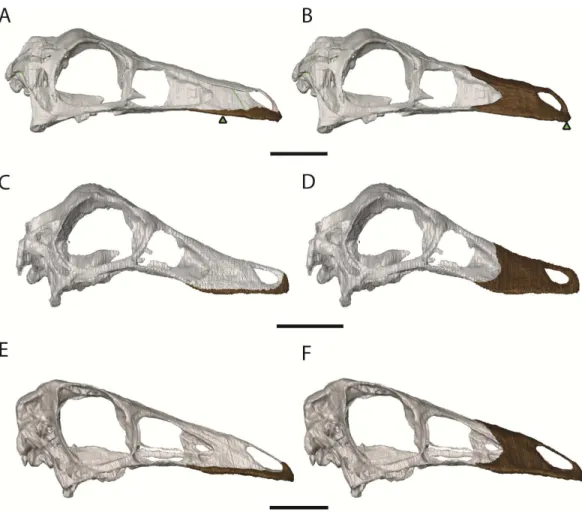
Figure 7 Ornithomimosaur beaks.(A) Small and (B) big beak morphs onGarudimimus; (C) small and (D) big beak morphs onOrnithomimus; (E) small and (F) big beak morphs onStruthiomimus. Scale bars
=5 cm. Triangles represent bite locations for mid-beak and tip of the beak bites (Table 6).
Table 3 Muscle moment arms and mechanical advantages for the specimens prior to retrodeforma-tion.The mechanical advantage out-lever was calculated as the distance from the jaw joint to the anterior tip of the premaxilla with no rhamphothecae:Garudimimus= 226 mm;Struthiomimus=183 mm;
Ornithomimus=185 mm.
Moment arm distances (mm)
Mechanical advantage (jaw tip-joint)
Garudi. Struthio. Ornitho. Garudi. Struthio. Ornitho.
AMEm 33.4 27.1 30.2 0.120 0.148 0.163
AMEp 49.0 30.5 26.0 0.135 0.166 0.141
AMEs 31.8 30.4 30.7 0.135 0.166 0.166
AMP 32.8 20.2 20.8 0.089 0.110 0.112
PSTs 50.8 33.3 37.1 0.147 0.182 0.200
PTd 27.0 7.90 13.8 0.035 0.043 0.075
PTv 14.7 13.1 9.3 0.058 0.072 0.050
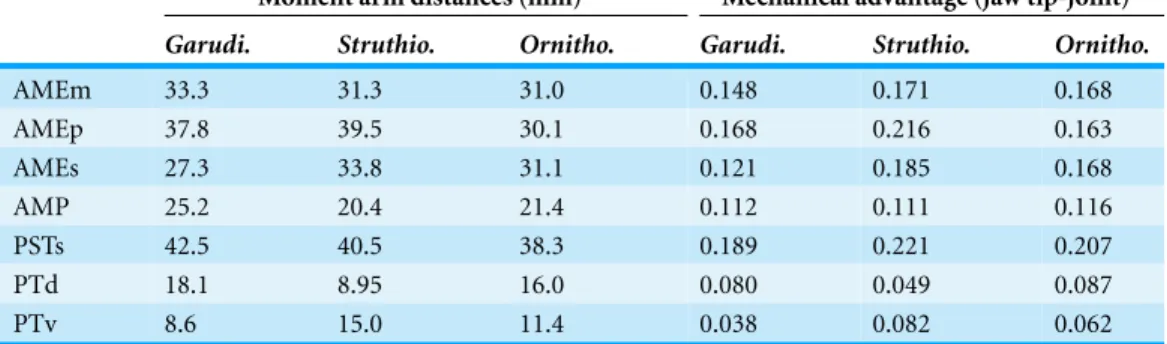
Table 4 Muscle moment arms and mechanical advantages for the specimens after retrodeforma-tion.The mechanical advantage out-lever was calculated as the distance from the jaw joint to the anterior tip of the premaxilla with no rhamphothecae:Garudimimus= 225 mm;Struthiomimus=183 mm;
Ornithomimus=185 mm.
Moment arm distances (mm) Mechanical advantage (jaw tip-joint)
Garudi. Struthio. Ornitho. Garudi. Struthio. Ornitho.
AMEm 33.3 31.3 31.0 0.148 0.171 0.168
AMEp 37.8 39.5 30.1 0.168 0.216 0.163
AMEs 27.3 33.8 31.1 0.121 0.185 0.168
AMP 25.2 20.4 21.4 0.112 0.111 0.116
PSTs 42.5 40.5 38.3 0.189 0.221 0.207
PTd 18.1 8.95 16.0 0.080 0.049 0.087
PTv 8.6 15.0 11.4 0.038 0.082 0.062
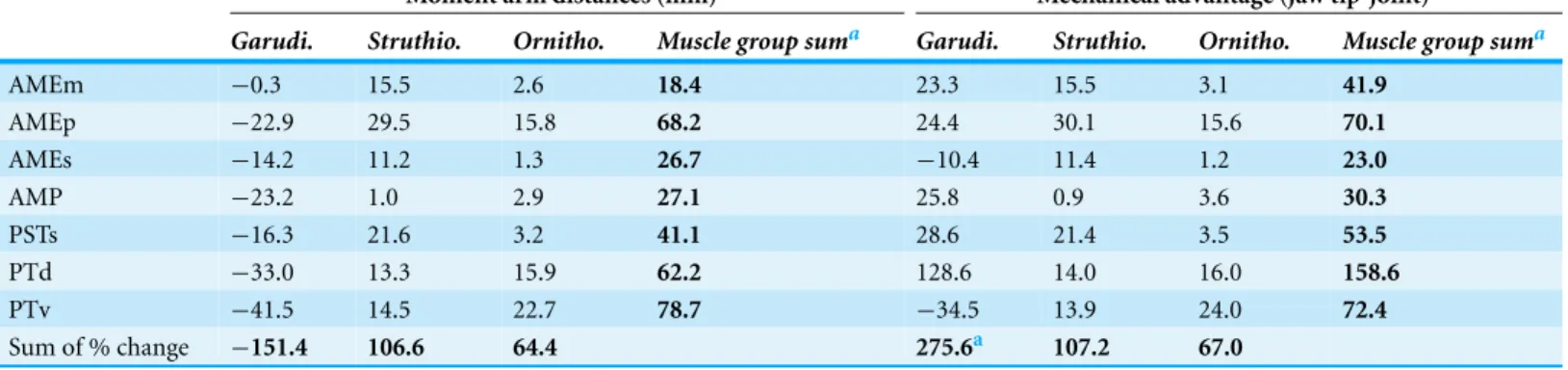
Table 5 Percentage change in muscle moment arms and mechanical advantage after retrodeformation.
Moment arm distances (mm) Mechanical advantage (jaw tip-joint)
Garudi. Struthio. Ornitho. Muscle group suma Garudi. Struthio. Ornitho. Muscle group suma
AMEm −0.3 15.5 2.6 18.4 23.3 15.5 3.1 41.9
AMEp −22.9 29.5 15.8 68.2 24.4 30.1 15.6 70.1
AMEs −14.2 11.2 1.3 26.7 −10.4 11.4 1.2 23.0
AMP −23.2 1.0 2.9 27.1 25.8 0.9 3.6 30.3
PSTs −16.3 21.6 3.2 41.1 28.6 21.4 3.5 53.5
PTd −33.0 13.3 15.9 62.2 128.6 14.0 16.0 158.6
PTv −41.5 14.5 22.7 78.7 −34.5 13.9 24.0 72.4
Sum of % change −151.4 106.6 64.4 275.6a 107.2 67.0
Notes.
aSum of absolute percentage change.
Table 6 Muscle loads and bite forces as calculated from muscle reconstructions for each ornithomi-mosaur.All forces in Newtons. Positions for mid beak (half the distance from the rostral to distal margins of the rhamphothecae) and tip of beak bites are shown inFig. 7.
Garudimimus Ornithomimus Struthiomimus
AMEm 14.1 8.69 24.1
AMEp 29.0 12.9 28.3
AMEs 17.2 10.5 31.7
AMP 14.3 15.0 13.2
PSTs 23.7 10.4 30.7
PTd 3.17 17.1 40.4
PTv 8.56 7.08 35.3
Tip of beak 19.0 22.0 57.6
Mid beak 23.9 28.6 75.2
(Lee et al., 2014). This large, derived (almost hadrosaurid-like) skull has relatively small temporal fenestrae so may have had small adductor muscles (Lee et al., 2014). This, combined with the long rostrum, suggests it too had a relatively small bite force despite its large size. This likely has a consequence on its diet:Deinocheirusis known to have consumed small fish based on stomach contents, but is also believed to have consumed plant matter, as hypothesized for other ornithomimosaurs.
Ornithomimosaur bite forces are the lowest reported to date for any non-avian theropod and are lower than those found in another putatively herbivorous theropod (Zanno et al., 2009;Zanno & Makovicky, 2011),Erlikosaurus(Lautenschlager, 2013). In that study, it was suggested that such low bite forces (43–134 N depending on location of the bite along the jaw) combined with a keratinous rhamphotheca, could be used to help hold plant material, whilst neck musculature (Rayfield, 2004;Snively & Russell, 2007) provided a ventrocaudal force to strip vegetation (Lautenschlager, 2013;Lautenschlager et al., 2013;Button, Rayfield & Barrett, 2014). This may be a valid method of food acquisition in ornithomimosaurs but further study is required. There are few estimates of bite force in other herbivorous dinosaur taxa. For Sauropoda, estimates of between 235–324 N and 982–1859 N have been calculated forDiplodocusandCamarasaurusrespectively (Button, Rayfield & Barrett, 2014). The bite force ofStegosaurus stenops(USNM 4934) has been estimated at between 140 and 275 N depending on the bite position along the jaw, modelled as sufficient to bite through smaller braches and leaves (Reichel, 2010). Further investigation of individual taxa will contribute to a broader picture of cranial evolution within Dinosauria.
CONCLUSION
The retrodeformation of three ornithomimosaurian skulls has allowed for greater insight into ornithomimosaur cranial anatomy and function than was possible with deformed skulls, particularly the reconstruction of the myology and rhamphothecae. The reconstructions and functional interpretations presented here should be treated as biologically informed hypotheses of musculoskeletal anatomy that can inform on future myological, endocranial and biomechanical studies.
Institution abbreviations
GIN Mongolian Academy of Sciences, Ulan Bator, Mongolia
RTMP Royal Tyrrell Museum of Palaeontology, Drumheller, Alberta, Canada
Myological abbreviations
AMEm adductor mandibulae externus medialis AMEp adductor mandibulae externus profundus AMEs adductor mandibulae externus superficialis AMP adductor mandibulae posterior
PTd pterygoideus dorsalis PTv pterygoideus ventralis
ACKNOWLEDGEMENTS
We would like to thank Hans Larsson and Yoshi Kobayashi for providing the CT scans used in the study. Thanks also to Stephan Lautenschlager and Jen Bright for help withAvizo. In addition, thanks go to Kevin Seymour (ROM), Brandon Strilisky (RTMP), Chinzorig Tsogtbataar (GIN), Xu Xing (IVPP) for allowing access to the museum collections. We also thank Eric Snively, Victoria Arbour and an anonymous reviewer for comments and suggestions that have improved the manuscript.
ADDITIONAL INFORMATION AND DECLARATIONS
Funding
This work was carried out as part of a self-funded PhD. The funders had no role in study design, data collection and analysis, decision to publish, or preparation of the manuscript.
Competing Interests
The authors declare there are no competing interests.
Author Contributions
• Andrew R. Cuff performed the experiments, analyzed the data, wrote the paper,
prepared figures and/or tables, reviewed drafts of the paper.
• Emily J. Rayfield conceived and designed the experiments, contributed
reagents/materials/analysis tools, wrote the paper, prepared figures and/or tables, reviewed drafts of the paper.
Data Deposition
The following information was supplied regarding the deposition of related data: Ornithomimus
http://phenome10k.org/ornithomimus-edmontonicus-2/ http://phenome10k.org/ornithomimus-edmontonicus/ Struthiomimus
http://phenome10k.org/struthiomimus-altus/ http://phenome10k.org/struthiomimus-altus-2/ Garudimimus
http://phenome10k.org/garudimimus-brevipes-2/ http://phenome10k.org/garudimimus-brevipes/
Supplemental Information
REFERENCES
Adams LA. 1919.A memoir of the phylogeny of the jaw muscles in recent and fossil vertebrates.
Annals of the New York Academy of Science58:51–166.
Angielczyk KD, Sheets HD. 2007.Investigation of simulated tectonic deformation in fossils using geometric morphometrics.Paleobiology33:125–148DOI 10.1666/06007.1.
Arbour VM, Currie PJ. 2012.Analyzing taphonomic deformation of ankylosaur skulls using retrodeformation and finite element analysis.PLoS ONE7(6):e39323
DOI 10.1371/journal.pone.0039323.
Barsbold R. 1981. Toothless carnivorous dinosaurs of Mongolia. Transactions, Joint Soviet-Mongolian Palaeontological Expedition15:28–39.
Barsbold R, Perle A. 1984.On first new find of a primitive ornithomimosaur from the Cretaceous of the MPR.Paleontologicheskii Zhurnal2:121–123.
Bates KT, Falkingham PL. 2012.Estimating maximum bite performance in Tyrannosaurus rex using multi-body dynamics.Biology Letters8:660–664DOI 10.1098/rsbl.2012.0056. Bell PR, Snively E, Shychoski L. 2009.A comparison of the jaw mechanics in hadrosaurid and
ceratopsid dinosaurs using finite element analysis.The Anatomical Record292:1338–1351 DOI 10.1002/ar.20978.
Brochu CA. 2000.A digitally-rendered endocast forTyrannosaurus rex.Journal of Vertebrate Paleontology20:1–6DOI 10.1671/0272-4634(2000)020[0001:ADREFT]2.0.CO;2.
Button DJ, Rayfield EJ, Barrett PM. 2014.Cranial biomechanics underpins high sauropod diversity in resource-poor environments.Proceedings of the Royal Society B281:20142114 DOI 10.1098/rspb.2014.2114.
Cooper RA. 1990.Interpretation of tectonically deformed fossils.New Zealand Journal of Geology and Geophysics33:321–332DOI 10.1080/00288306.1990.10425690.
Curtis N, Kupczik K, O’Higgins P, Moazen M, Fagan MJ. 2008. Predicting skull loading: applying multibody dynamics analysis to a macaque skull.Anatomical Record291:491–501 DOI 10.1002/ar.20689.
Dal Sasso C, Maganuco S, Buffetaut E, Mendez MA. 2005.New information on the skull of the enigmatic theropodSpinosaurus, with remarks on its sizes and affinities.Journal of Vertebrate Paleontology25:888–896DOI 10.1671/0272-4634(2005)025[0888:NIOTSO]2.0.CO;2. Davies SJJF. 2003.Struthioniformes (Tinamous and Ratites). In: Hutchins M, Jackson A, Bock
WJ, Olendorf D, eds.Grzimek’s animal life encyclopedia. 8 birds I tinamous and ratites to hoatzin. 2nd edition. Farmington Hills: Gale Group, 75–77.
De Klerk WJ, Forster CA, Sampson SD, Chinsamy A, Ross CF. 2000.A new coelurosaurian dinosaur from the Early Cretaceous of South Africa. Journal of Vertebrate Paleontology
2:324–332DOI 10.1671/0272-4634(2000)020[0324:ANCDFT]2.0.CO;2.
Fairman JE. 1999.Prosauropod and iguanid jaw musculature: a study on the evolution of form and function. Unpublished M.A. thesis, Johns Hopkins University.
Gunz P, Mitteroecker P, Neubauer S, Weber GW, Bookstein FL. 2009.Principles for the virtual reconstruction of hominin crania.Journal of Human Evolution57:48–62
DOI 10.1016/j.jhevol.2009.04.004.
Haas G. 1955.The jaw musculature inProtoceratopsand in other ceratopsians.American Museum Novitates1729:1–24.
Hedrick BP, Dodson P. 2013.Lujiatun psitacosaurids: understanding individual and taphonomic variation using 3D geometric morphometrics. PLoS ONE8(8):e69265 DOI 10.1371/journal.pone.0069265.
Hieronymus TL, Witmer LM. 2010. Homology and evolution of avian compound rhamphothecae.The Auk127:590–604DOI 10.1525/auk.2010.09122.
Holliday CM. 2009.New insights into the dinosaur jaw muscle anatomy.The Anatomical Record
292:1246–1265DOI 10.1002/ar.20982.
Holliday CM, Witmer LM. 2007.Archosaur adductor chamber evolution: Integration of musculoskeletal and topological criteria in jaw muscle homology.Journal of Morphology
268:457–484DOI 10.1002/jmor.10524.
Hughes NC, Jell PA. 1992.A statistical/computer-graphic technique for assessing variation in tectonically deformed fossils and its application to Cambrian trilobites from Kashmir.Lethaia
25:317–330DOI 10.1111/j.1502-3931.1992.tb01401.x.
Ji Q, Norell M, Makovicky PJ, Gao K, Ji S, Yuan C. 2003. An early ostrich dinosaur and implications for ornithomimosaur phylogeny.American Museum Novitates 3420:1–19 DOI 10.1206/0003-0082(2003)420<0001:AEODAI>2.0.CO;2.
Kobayashi Y. 2004.Asian ornithomimosaurs. PhD Thesis, Southern Methodist University. Kobayashi Y, Barsbold R. 2005.Anatomy ofHarpymimus okladnikoviBarsbold and Perle 1984
(Dinosauria; Theropoda) of Mongolia. In: Carpenter K, ed.The carnivorous dinosaurs. Bloomington: Indiana University Press, 97–126.
Kobayashi Y, L¨u J-C. 2003.A new ornithomimid dinosaur with gregarious habits from the Late Cretaceous of China.Acta Palaeontologica Polonica48:235–259.
Lambe LM. 1902.On Vertebrata of the mid-Cretaceous of the North-west Territory. 2. New genera and species from the Belly River Series (mid-Cretaceous).Contributions to Canadian Palaeontology3:25–81.
Lautenschlager S. 2013.Cranial myology and bite force performance ofErlikosaurus andrewsi: a novel approach for digital muscle reconstructions.Journal of Anatomy 222:260–272 DOI 10.1111/joa.12000.
Lautenschlager S, Witmer LM, Altangerel P, Rayfield EJ. 2013. Edentulism, beaks and
biomechanical innovations in the evolution of theropod dinosaurs.Proceedings of the National Academy of Sciences of the United States of America110:20657–20662
DOI 10.1073/pnas.1310711110.
Lee YN, Barsbold R, Currie PJ, Kobayashi Y, Lee HJ, Godefroit P, Escuilli´e FO, Chinzorig T. 2014.Resolving the long-standing enigmas of a giant ornithomimosaurDeinocheirus mirificus.
Nature515:257–260DOI 10.1038/nature13874.
Makovicky PJ, Kobayashi Y, Currie PJ. 2004.Ornithomimosauria. In: Weishampel DB, Dodson P, Osm ´olska H, eds.The dinosauria. 2nd edition. Berkeley: University of California Press, 137–150.
Makovicky PJ, Li D, Gao K-Q, Lewin M, Erickson GM, Norell MA. 2010. A giant
ornithomimosaur from the Early Cretaceous of China.Proceedings of the Royal Society B
277:191–198DOI 10.1098/rspb.2009.0236.
Molnar JL, Pierce SE, Clack JA, Hutchinson JR. 2012.Idealized landmark-based geometric reconstructions of poorly preserved fossil material: a case study of an early tetrapod vertebra.
Morhardt AC. 2009.Dinosaur smoles: do the texture and morphology of the premaxilla, maxilla, and dentary bones of sauropsids provide the osteological correlates for inferring extra-oral structures reliably in dinosaurs? MSc Thesis, Western Illinois University.
Motani R. 1997.New technique for retrodeforming tectonically deformed fossils, with an example for ichthyosaurian specimens.Lethaia30:221–228DOI 10.1111/j.1502-3931.1997.tb00464.x. Norell MA, Makovicky P, Currie PJ. 2001.The beaks of ostrich dinosaurs.Nature412:873–874
DOI 10.1038/35091139.
Osm ´olska H, Roniewicz E, Barsbold R. 1972.A new dinosaur,Gallimimus bullatusn. gen., n. sp. (Ornithomimidae) from the Upper Cretaceous of Mongolia.Palaeontologia Polonica
27:103–143.
Perez-Moreno BP, Sanz JL, Buscalioni AD, Moratella JJ, Ortega F, Raskin-Gutman D. 1994. A unique multitoothed ornithomimosaur from the Lower Cretaceous of Spain.Nature
370:363–367DOI 10.1038/370363a0.
Perle A. 1981.A new segnosaurid from the Upper Cretaceous of Mongolia.Transactions, Joint Soviet-Mongolian Palaeontological Expedition15:50–59 (in Russian).
Porro LB, Rayfield EJ, Clack JA. 2015.Descriptive anatomy and three-dimensional reconstruction of the skull of the early tetrapodAcanthostega gunnariJarvik, 1952.PLoS ONE10(3):e0118882 DOI 10.1371/journal.pone.0118882.
Rayfield EJ. 2004.Cranial mechanics and feeding inTyrannosaurus rex.Proceedings of the Royal Society B271:1451–1455DOI 10.1098/rspb.2004.2755.
Rayfield EJ, Milner AC, Xuan VB, Young PG. 2007.Functional morphology of spinosaur ‘crocodile-mimic’ dinosaurs.Journal of Vertebrate Paleontology27:892–901
DOI 10.1671/0272-4634(2007)27[892:FMOSCD]2.0.CO;2.
Rayfield EJ, Norman DB, Horner CC, Horner JR, Smith PM, Thomason JJ, Upchurch P. 2001.Cranial design and function in a large theropod dinosaur.Nature409:1033–1037 DOI 10.1038/35059070.
Reichel M. 2010.A model for the bite mechanics in the herbivorous dinosaur Stegosaurus (Ornithischia, Stegosauridae).Swiss Journal of Geosciences103:235–240
DOI 10.1007/s00015-010-0025-1.
Rushton AWA, Smith M. 1993.Retrodeformation of fossils-a simple technique.Palaeontology
36:927–930.
Sanders RK, Smith DK. 2005.The endocranium of the theropod dinosaurCeratosaurusstudied with computed tomography.Acta Palaeontologica Polonica50:601–616.
Seki Y, Bodde SG, Meyers MA. 2010.Toucan and hornbill beaks: a comparative study.Acta Biomaterialia6:331–343DOI 10.1016/j.actbio.2009.08.026.
Sereno PC, Zhao X-T, Tan L. 2010.A new psittacosaur from Inner Mongolia and the parrot-like structure and function of the psittacosaur skull.Proceedings of the Royal Society of London B
277:199–209DOI 10.1098/rspb.2009.0691.
Snively E, Russell AP. 2007.Functional variation of neck muscles and their relation to feeding style in Tyrannosauridae and other large theropod dinosaurs.The Anatomical Record290:934–957 DOI 10.1002/ar.20563.
Sternberg CM. 1933.A newOrnithomimuswith complete abdominal cuirass.The Canadian Field Naturalist47:79–83.
Tahara R, Larsson HCE. 2011.Cranial pneumatic anatomy ofOrnithomimus edmontonicus
Tallman M, Amenta N, Delson E, Frost SR, Deboshmita G, Klukkert ZS, Morrow A, Sawyer GJ. 2014.Evaluation of a new method of fossil retrodeformation by algorithmic symmetrization: Crania of Papionins (Primates, Cercopithecidae) as a test case.PLoS ONE9(7):e100833 DOI 10.1371/journal.pone.0100833.
Thomason JJ. 1991.Cranial strength in relation to estimated biting forces in some mammals.
Canadian Journal of Zoology69:2326–2333DOI 10.1139/z91-327.
Tschopp E, Russo J, Dzemski G. 2013.Retrodeformation as a test for the validity of phylogenetic characters: an example from diplodocid sauropod vertebrae.Palaeontologia Electronica16(1): 2T, 23p.
Weijs WA, Hillen B. 1985.Cross-sectional areas and estimated intrinsic strength of the human jaw muscles.Acta Morphologica Neerlando-Scandinavica23:267–274.
Williams SH. 1990.Computer-assisted graptolite studies. In: Bruton DL, Harper DAT, eds.
Microcomputers in palaeontology,Contributions from the Paleontological Museum,370. Oslo: University of Oslo, 446–455.
Witmer LM, Ridgely RC. 2009.New insights into the brain, braincase, and ear region of tyrannosaurs (Dinosauria, Theropoda), with implications for sensory organization and behavior.The Anatomical Record292:1266–1296DOI 10.1002/ar.20983.
Young MT, Rayfield EJ, Holliday CM, Witmer LM, Button DJ, Upchurch P, Barrett PM. 2012.Cranial biomechanics ofDiplodocus(Dinosauria, Sauropoda): testing hypotheses of feeding behaviour in an extinct megaherbivore.Naturwissenschaften99:637–643 DOI 10.1007/s00114-012-0944-y.
Zanno LE, Gillette DD, Albright LB, Titus AL. 2009.A new North American therizinosaurid and the role of herbivory in ‘predatory’ dinosaur evolution.Proceedings of the Royal Society B
276:3505–3511DOI 10.1098/rspb.2009.1029.
Zanno LE, Makovicky PJ. 2011.Herbivorous ecomorphology and specialization patterns in theropod dinosaur evolution.Proceedings of the National Academy of Sciences of the United States of America108:232–237DOI 10.1073/pnas.1011924108.
Zanno LE, Makovicky PJ. 2013. No evidence for directional evolution of body mass in herbivorous theropod dinosaurs.Proceedings of the Royal Society B: Biological Sciences
280:201225226DOI 10.1098/rspb.2012.2526.
Zollikofer CPE, Ponce de Le ´on MS, Lieberman DE, Guy F, Pilbeam D, Likius A, Mackaye HT, Vignaud P, Brunet M. 2005.Virtual cranial reconstruction ofSahelanthropus tchadensis.Nature