Renal Cancer Cell Lines
Takashi Okabe1*, Megumi Kumagai2,3, Yoshihiro Nakajima4, Suguru Shirotake1, Kiichiro Kodaira1,
Masafumi Oyama1, Munehisa Ueno1, Masaaki Ikeda2,3
1Department of Uro-oncology, Saitama Medical University International Medical Center, Saitama, Japan,2Department of Physiology, Saitama Medical University, Saitama, Japan,3Molecular Clock Project, Project Research Division, Research Center for Genomic Medicine, Saitama Medical University, Saitama, Japan,4Health Research Institute, National Institute of Advanced Industrial Science and Technology (AIST), Kagawa, Japan
Abstract
In mammals, the circadian rhythm central generator consists of interactions among clock genes, includingPer1/2/3,Cry1/2,
Bmal1, andClock. Circadian rhythm disruption may lead to increased risk of cancer in humans, and deregulation of clock genes has been implicated in many types of cancers. Among these genes,Per2is reported to have tumor suppressor properties, but little is known about the correlation betweenPer2and HIF, which is the main target of renal cell carcinoma (RCC) therapy. In this study, the rhythmic expression of thePer2gene was not detectable in renal cancer cell lines, with the exception of Caki-2 cells. In Caki-2 cells, HIF1aincreased the amplitude ofPer2oscillation by directly binding to the HIF-binding site located on the Per2promoter. These results indicate that HIF1a may enhance the amplitude of the Per2 circadian rhythm.
Citation:Okabe T, Kumagai M, Nakajima Y, Shirotake S, Kodaira K, et al. (2014) The Impact of HIF1aon thePer2Circadian Rhythm in Renal Cancer Cell Lines. PLoS ONE 9(10): e109693. doi:10.1371/journal.pone.0109693
Editor:Shin Yamazaki, University of Texas Southwestern Medical Center, United States of America
ReceivedJanuary 9, 2014;AcceptedSeptember 12, 2014;PublishedOctober 21, 2014
Copyright:ß2014 Okabe et al. This is an open-access article distributed under the terms of the Creative Commons Attribution License, which permits unrestricted use, distribution, and reproduction in any medium, provided the original author and source are credited.
Funding:The authors have no support or funding to report.
Competing Interests:The authors have declared that no competing interests exist.
* Email: [email protected]
Introduction
Renal cell carcinoma (RCC) is the most common malignancy of the adult kidney, which accounts for approximately 2% of cancers worldwide [1]. A somatic mutation of the Von Hippel–Lindau (VHL) gene is the most frequent genetic change observed in RCC [2], and recent efforts have targeted the VHL–hypoxia inducible factor (HIF)-mediated hypoxia-induced gene pathway for RCC therapy [3]. HIFs are heterodimeric transcription factors with two structurally related subunits: an oxygen-sensitive HIFa subunit
and a constitutively expressed HIFß or aryl hydrocarbon receptor nuclear translocator (ARNT) subunit [4]. In normoxia, HIFa
molecules are subjected to a regulatory process involving enzymatic hydroxylation of conserved prolyl and asparaginyl residues, leading to rapid VHL protein-mediated ubiquitination and proteasomal degradation [5]. Hypoxia or mutations in the VHL gene inactivate this pathway. Increased HIFa activity
upregulates genes involved in many aspects of cancer progression, including metabolic adaptation, apoptotic resistance, and angio-genesis [3]. In RCC, intense tumor vascular networks can be attributed to the inappropriate accumulation of HIFa leading to
angiogenic gene induction. Vascular endothelial growth factor (VEGF) is one of the most potent pro-angiogenic factors, whose expression is transactivated by HIF1a/ARNT through binding to
the hypoxia-response element (HRE) in theVegfpromoter [6,7]. Increased expression of VEGF is also associated with malignant progression and a poor treatment outcome [8]. Therefore, suppressing the HIF-mediated gene pathway may be an important therapeutic strategy for the treatment of RCC [3].
Many physiological, biochemical, and behavioral processes are under circadian regulation, which is generated by an internal time-keeping mechanism referred to as the biological clock in almost all organisms from bacteria to mammals [9,10]. Circadian rhythms are controlled by genetically determined networks of transcrip-tion–translation feedback loops involving clock genes, including Per1/2/3, Cry 1/2, Bmal1, and Clock [11]. A common theme underlying circadian rhythmicity is that oscillations of clock gene transcripts are the consequence of intracellular transcriptional– translational feedback loops. For example, in mammals, the transcription factors CLOCK and BMAL1 heterodimerize and activate the expression of threePer genes and twoCrygenes by binding to E-box elements in their promoters. The protein products of these genes multimerize and translocate to the nucleus, where PER and CRY proteins repress the transcriptional activity of the CLOCK–BMAL1 dimer [12,13].
Among these clock genes, Per2 is responsible for setting the period of oscillation [14]. Furthermore,Per2has tumor-suppres-sor properties and is often mutated or downregulated in human breast cancers [15,16]. In renal cancer, altered expression of the Per2gene is reportedly involved in disease onset and progression, but the molecular mechanism responsible remains unclear [17].
In this study, we measured the levels ofPer2promoter activity and mRNA in eight renal cancer cell lines after dexamethasone treatment. ThePer2promoter activity and mRNA level oscillated over an approximately 24-h cycle in Caki-2 cells, which contain BMAL1, CLOCK, and HIF1a proteins. We also found that
HIF1aincreased the amplitude of oscillation by directly binding to
show that HIF1a may affect the amplitude of Per2 circadian
rhythms in renal cancer cell lines.
Materials and Methods
Cells and cell cultures, chemicals, and enzymes
Established human RCC cell lines (A704, ACHN, 786-O, A498, 769-P, and Caki-2) were obtained from the American Type Culture Collection (ATCC; Manassas, VA, USA). RCC4+vector alone and RCC4+VHL were obtained from Sigma (St. Louis, MO, USA). These renal cell lines were maintained in Roswell Park Memorial Institute (RPMI)-1640 medium (Kojin Bio, Tokyo, Japan) supplemented with 10% fetal bovine serum (FBS; Life Technologies, Carlsbad, CA, USA), 24 U/mL penicillin, and 25mg/mL streptomycin (Gibco, Grand Island, NY, USA) in a standard humidified incubator at 37uC in an atmosphere of 5% CO2. We also used the mouse fibroblast NIH3T3 and human osteosarcoma U2OS cell models of the autonomous circadian clock [18,19]. These cell lines were also obtained from ATCC, and were maintained in Dulbecco’s modified Eagle’s medium (DMEM), supplemented with 10% FBS, penicillin (24 U/mL), and streptomycin (25mg/mL). Chrysin was purchased from Sigma, and its purity exceeded 96%. A stock solution of chrysin was prepared in dimethyl sulfoxide (DMSO). Chrysin was dissolved in DMSO at three different concentrations (1, 10, and 100 mM) and added each 2mL to 2 mL culture media (final concentration; 1, 10, 100mM). Cells were treated with culture media containing 1, 10, 100mM chrysin or same concentration of DMSO as control for 2 hours.
Plasmid construction
To construct reporter vectors carrying the mPer2promoter, the mPer2promoter fragment (2279 to+112 bp, where+1 indicates the putative transcription start site) was polymerase chain reaction (PCR)-amplified from the C57BL/6J mouse genome, and cloned into the NheI/XhoI site of pGL3 Basic (Promega, Madison, WI, USA). Firefly luciferase (FLuc) was replaced with the NcoI and XbaI fragment of pSV40-dFLuc, resulting in mPer2-dFLuc. The HRE-mutant mPer2 promoter reporter was generated with inverse PCR using a KOD-Plus-Mutagenesis Kit (Toyobo, Osaka, Japan).
Real-time reporting of circadian-regulated gene expression using luciferase bioluminescence
All cells were seeded (56104per dish) in a 35-mm dish 2 days before transfection, and the reporter plasmid was transfected using Lipofectamine 2000 (Invitrogen, Carlsbad, CA, USA) according to the manufacturer’s instructions. The appropriate amount of reporter plasmid for each cell line was determined according to differences in transfection efficiency among the cell lines. One day after transfection, cells were treated with 100 nM dexamethasone (Nakalai Tesque, Kyoto, Japan) for 2 h, and the medium was replaced with medium in the absence of phenol red supplemented with 10% FBS and 100mM D-luciferin (Toyobo). Biolumines-cence was measured at 37uC under a 5% CO2atmosphere and integrated for 1 min at intervals of 10 min using a dish-type luminometer, AB-2550 Kronos Dio (ATTO, Tokyo, Japan) [20,21]. Bioluminescence activity was expressed as relative light units (RLUs). Each experiment was repeated at least four times. The cells were cultured in the luminometer for at least 4 days while the instrument counted their bioluminescence. The obtained crude data (10-min bins) were smoothed by a 10-point moving average method and detrended by subtracting a 12-h moving average from the smoothed data [21].
Analysis of circadian rhythms using bioluminescence To test the significance of the circadian rhythmicity and to calculate circadian parameters (i.e., period, amplitude, and acrophase), we performed computerized data analysis in the Cosinor software downloaded from the Circadian Rhythm Laboratory (Walterboro, SC, USA) software home page (http:// www.circadian.org/software.html) [22,23]. Circadian parameters were calculated using data from 1–5 days after dexamethasone treatment.
Automated image capture and analysis
NIH3T3 cells were seeded (56104per well) on 6-well plates 1 day before transfection, and the expression plasmid was transfect-ed using Lipofectamine 2000 (Invitrogen) according to the manufacturer’s instructions. One day after transfection, cells were treated with 100 nM dexamethasone (Nakalai Tesque) for 2 h, and the medium was replaced with medium in the absence of phenol red supplemented with 10% FBS. Cells were stained with 0.1mg/mL Hoechst 33342 (Invitrogen) for 1 hour and analyzed using ArrayScan XTI (Thermo Scientific, Waltham, MA, USA).
Quantification of mRNA by real-time RT-PCR
All cells were harvested at 4-h intervals from six plates at each time point beginning 24 h after treatment with dexamethasone. Total RNA from these cells was extracted using ISOGEN (Nippon Gene, Tokyo, Japan) and reverse transcribed. Per2 and Gapdh transcripts were quantified using an ABI Prism 7300 (Applied Biosystems, Foster City, CA, USA). PCR was performed using the One Step SYBR PrimeScript RT-PCR Kit (Takara Bio, Kyoto, Japan) with the following thermal cycling parameters: 94uC for 5 min followed by 40 cycles at 94uC for 20 s and 62uC for 1 min. The Gapdh transcript was used to normalize the expression of each transcript. Circadian rhythmicity significance was analyzed using the Cosinor software (Circadian Rhythm Laboratory) [22,23]. Primers for each gene were designed based on the information available from the National Center for Biotechnology Information (NCBI). The PCR primer sequences were as follows: Per2(GenBank accession no., NM_022817; amplicon, 85 bp): sense primer 59-CACACACAGAAGGAGGAGCA-39 and anti-sense primer 59-AGTAATGGCAGTGGGACTGG-39.
Gapdh (GenBank accession no., M33197; amplicon, 185 bp): sense primer 59-GAGTCAACGGATTTGGTCGT-39 and anti-sense primer 59-GACAAGCTTCCCGTTCTCAG-39.
Luciferase assay
Transfected NIH3T3 cells were used for luciferase assays. One day before transfection, cells were seeded (56104per well) on 24-well plates containing DMEM supplemented with 10% FBS, penicillin (24 U/mL), and streptomycin (25mg/mL). Cells were transfected using Lipofectamine 2000 (Invitrogen). For each sample, transfected DNA was added to each well. Twenty-four hours after transfection, cells were washed in phosphate-buffered saline (PBS) and disrupted with 100mL of passive lysis buffer (Promega). Luciferase activity was determined using a Dual-Luciferase Reporter Assay System (Promega) and an Ascent FS II luminometer (Thermo Scientific).
Western blotting
All cells were synchronized by 100 nM dexamethasone treatment for 2 h. Then, the medium was replaced with fresh medium. After 24-h incubation, these cells were lysed in Cell Lytic-MT (Sigma). The cell lysates were centrifuged at 15,000 rpm at 4uC for 10 min. The supernatants were stored as whole cell
extracts at280uC until use. For Western blotting, 20-mg protein were resolved on 7.5% sodium dodecyl sulfate polyacrylamide (SDS-PAA) gels and transferred onto a nitrocellulose membrane (Bio-Rad, Hercules, CA, USA). The membranes were blocked with Tris-buffered saline (TBS)-Tween containing 5% non-fat dried milk. Proteins were detected using antibodies against HIF1a
(dilution, 1: 500; BD Transduction Laboratories, Franklin Lakes, NJ, USA), PER2 (dilution, 1:1000; Santa Cruz Biotechnology, Santa Cruz, CA, USA), CRY1 (dilution, 1:2000; Santa Cruz Biotechnology), CLOCK (dilution, 1:1000; Thermo Scientific), GAPDH (dilution, 1:10000; Sigma), and BMAL1 (dilution, 1:100; mouse monoclonal antibody generated in our lab). We performed four replicate Western blots; a representative blot is shown.
ChIP assay
ChIP experiments were performed using a commercially available kit according to the manufacturer’s instructions (Magna-ChIP; Millipore, Bedford, MA, USA). Briefly, Caki-2 cells were plated in 100-mm diameter dishes (56104cells per dish); after 24 h, cells were incubated with formaldehyde (final concentration, 1%) for 10 min at 37uC to cross-link proteins to
DNA. Unreacted formaldehyde was quenched with 1 mL of 106 glycine. The plate was washed twice with ice-cold PBS, and the pellets were harvested in 1 mL PBS with protease inhibitor cocktail and pooled together in a 1.5-mL tube. The cross-linked chromatin was sheared by sonication 20 times for 1 min each time with 1 min of cooling on ice between pulses using a Branson 2510 Ultrasonic Cleaner (Branson, Danbury, CT, USA). Immunopre-cipitation (IP) was performed with 5mg of either anti-HIF1a
(dilution, 1: 500; Novus Biologicals Inc., Littleton, CO, USA) or anti-IgG antibody (Millipore) as a negative control. Washes and elution of the IP DNA were performed according to the Magna-ChIP protocol (Millipore). Ten percent (10%) of the original sheared chromatin DNA was similarly reverse cross-linked and purified, and the recovered DNA was used as an input control. PCR was performed with specific primers flanking the HRE-like sequence within the promoter region of the human Per2 gene (2476 to2284 bp, sense: 59- ACGCCGGAAGTGGATGAGAC -39and antisense: 59 CGACTCCGTCTCATCTGCATACAT -39) with the following thermal cycling parameters: 94uC for 3 min, followed by 40 cycles at 94uC for 20 s, annealing at 59uC for 30 s, and extension at 72uC for 30 s.
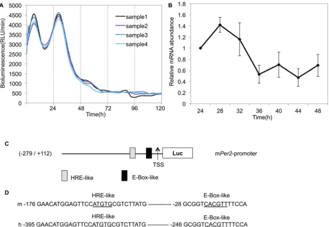
Figure 1. Rhythmic expression ofPer2in Caki-2 cells.(A) All renal cancer cell lines were transfected with the Per2 promoter reporter (2mg) and the bioluminescence was then measured using a real-time monitoring assay. Real-time monitoring of luciferase activity of thePer2promoter showed that activity oscillated over an approximately 24-h cycle. The luciferase activities of four replicate samples are shown. These cultures showed significant circadian rhythms (Table 1). (B) mRNA levels ofPer2were determined by real-time PCR for six plates at each time point. Total RNA was extracted every 4 h, beginning 24 h after treatment with dexamethasone for one 24-h cycle, andPer2transcripts were quantified. Error bars indicate the standard errors of the mean values (n= 6). The data from a single 24 hours after dexamethasone treatment were analyzed using the Cosinor software for rhythmicity (Table 1). (C) The structure of thePer2promoter and an analysis of the potential transcription factor-binding motifs in this region. The 2,994-bp region contains one E-box-like sequence (CACGTT) and one HRE-like sequence (ATGTG), similar to the consensus HRE sequence (ACGTG) located upstream of the transcription start site (TSS). (D) Sequence comparisons: upper line, mouse sequence; lower line, human sequence. The nucleotide sequence of potential transcription factor-binding motifs for E-box-like sequence and HRE-like sequence are 100% conserved between mouse and human.
Statistical analysis
Each experiment was repeated at least four times. Data are expressed as means 6 standard errors. To evaluate the significance of differences, Student’s t-test was performed. We used a one-way analysis of variance (ANOVA) for comparisons among the drug concentration groups, followed by application of Tukey’s post hoc tests. For all analyses, the significance level was set at P,0.05. The Cosinor software (Circadian Rhythm Laboratory) [22,23] was used to analyze circadian rhythmicity.
Results
Circadian expression of thePer2gene in renal cancer cell lines
To explore the transcriptional oscillation ofPer2, all cell lines were transfected with a luciferase reporter gene driven by thePer2 promoter, and a real-time monitoring assay was performed using
Kronos Dio (AB-2550; ATTO). A luciferase-bound promoter in Caki-2 cells displayed circadian rhythms after 2 h dexamethasone treatment (Fig. 1A, Table 1), but rhythmicity was not detected in the other cell lines (Fig. S1). Each experiment was repeated four times and these results were consistent. 24 h after dexamethasone treatment,Per2mRNA levels had a circadian rhythm in Caki-2 cells (Fig. 1B, Table 1). These results showed that the circadian rhythmicity of thePer2gene was not detectable in renal cancer cell lines, excluding Caki-2 cells.
Analysis of thePer2 promoter region
A previous study demonstrated that an E-box-like sequence (CACGTT) and its downstream region are essential for transcrip-tional oscillation ofPer2, a crucial component of molecular clocks [24]. We focused on this E-box-like region and HRE. The transcription factor-binding motifs located on thePer2promoter in mice and humans were analyzed using MatInspector software Table 1.Circadian parameters ofPer2promoter activities and mRNA in Caki-2 cells.
period (h) amplitude acrophase (h) P value
Promoter activitiy 24.1860.05 442.24616.72 6.1760.11 ,0.000001
mRNA N/A 0.4223 4.95 ,0.05
Promoter activity and mRNA levels ofPer2showed significant circadian rhythms (p,0.000001,p,0.05, by Cosinor). doi:10.1371/journal.pone.0109693.t001
Figure 2. Western blot analysis of the indicated proteins. All cell lines were lysed and harvested 24 h after synchronization by 2-h dexamethasone treatment. We performed four replicate Western blots; a representative blot is shown. (A) Western blots of renal cancer whole-cell extracts (20mg) with BMAL1, CLOCK, PER2, CRY1 and GAPDH antibodies are shown. Full-length blots of PER2 and BMAL1, in addition to a positive control, are presented in Figure S2, 3. (B) Western blots of renal cancer whole-cell extracts (20mg) with HIF1aand GAPDH antibodies are shown. doi:10.1371/journal.pone.0109693.g002
Figure 3. The impact of HIF1a/ARNT onPer2transcriptional activity.(A) NIH3T3 cells were co-transfected with thePer2promoter reporter
(400 ng) and the indicated expression plasmids (300 ng) for HIF1a/ARNT or empty vector pcDNA3 (600 ng) as a control. Bioluminescence was then measured using a real-time monitoring assay. Control, transfected with empty vector pcDNA3 (uncloned-vector control);+HIF1a/ARNT, transfected with the expression plasmids. Luciferase activities of four replicate samples are shown. (B) Detrended bioluminescence is shown. Period, amplitude, and acrophase of the oscillations were measured on days 2 to 5 using the Cosinor software (Circadian Rhythm Laboratory). Amplitude significantly increased (mean6SEM,n= 4) compared to the control (p,0.01, Student’st-test). See Table 2. (C) U2OS cells were co-transfected with thePer2 promoter reporter (400 ng) and the indicated expression plasmids (300 ng) for HIF1a/ARNT or empty vector pcDNA3 (600 ng) as a control. Bioluminescence was then measured using a real-time monitoring assay. Control, transfected with empty vector pcDNA3 (uncloned-vector control);+ HIF1a/ARNT, transfected with the expression plasmids. Luciferase activities of four replicate samples are shown. (D) Detrended bioluminescence is shown. Period, amplitude, and acrophase of the oscillations were measured on days 2 to 5 using the Cosinor software (Circadian Rhythm Laboratory). Amplitude significantly increased (mean6SEM,n= 4) compared to the control (p,0.01, Student’st-test). See Table 2. (E) Caki-2 cells were co-transfected with thePer2promoter reporter (2mg) and the indicated expression plasmids (1.5mg) for HIF1a/ARNT or empty vector pcDNA3 (3mg) as a control. The bioluminescence was then measured using a real-time monitoring assay. Control, transfected with empty vector pcDNA3 (uncloned-vector control);+HIF1a/ARNT, transfected with the expression plasmids. The luciferase activities of four replicate samples are shown. (F) Detrended bioluminescence is shown. Period, amplitude, and acrophase of the oscillations were measured from days 2 to 5 using the Cosinor software (Circadian Rhythm Laboratory). Amplitude significantly increased (mean6SEM,n= 4) compared to the control (p,0.01, Student’st-test). See Table 2. doi:10.1371/journal.pone.0109693.g003
Table 2.Circadian parameters ofPer2promoter activities based on four days of data.
period amplitude acrophase P value
Caki-2 control 24.1860.025 191.1665.11 5.1860.06 ,0.0001
+HIF1a/ARNT 24.1860.025 325.9762.91** 5.3360.04 ,0.0001
NIH3T3 control 24.4360.05 7930.866442.12 2.9060.05 ,0.000001
+HIF1a/ARNT 24.2360.08 41876.88633.40** 3.1160.07 ,0.000001
U2OS control 23.8360.13 220.63631.95 7.5260.76 ,0.000001
+HIF1a/ARNT 24.0560.02 1121.36658.72** 6.0260.09 ,0.000001
Period, amplitude, and acrophase of the oscillations were measured on days 2 to 5 using the Cosinor software (Circadian Rhythm Laboratory). Amplitude significantly increased (mean6SEM,n= 4) compared to the control (**p,0.01, Student’st-test).
(Genomatix, Munich, Germany). Sequence analysis of thePer2 promoter region revealed high homology between mice and humans. Sequence analysis also revealed one E-box-like sequence (CACGTT) and one HRE-like sequence (ATGTG), similar to the consensus HRE sequence (ACGTG) [25] located upstream of the transcription start site (TSS) (Fig. 1C). These sequences were 100% conserved between mice and humans (Fig. 1D). A real-time monitoring assay (Fig. 1A) indicated that the promoter region we cloned is sufficient to produce circadian transcriptional oscillation in human cell lines.
Expression of clock genes in renal cancer cell lines To examine the difference between Caki-2 and other cell lines, we examined the expression of BMAL1, CLOCK, PER2, and CRY1 proteins. Caki-2, 786-O, and A498 cells expressed BMAL1 protein. All renal cancer cell lines expressed CLOCK and CRY1 protein, but did not express PER2 protein (Fig. 2A). Full-length blots of PER2 and BMAL1, in addition to a positive control, are presented in Figure S2, S3.
Expression of HIFaprotein under normoxic conditions in
renal cancer cell lines
Since the HIF1a protein may be generally overexpressed in
RCC, we also examined the expression of HIF1aprotein. In
Caki-2 and RCC4+vector alone, HIF1a protein was overexpressed
(Fig. 2B). Considering the results that thePer2circadian rhythm was shown only in Caki-2 cells, which contained BMAL1, CLOCK, and HIF1aprotein, it is possible that HIF1ais related
to thePer2circadian rhythm in renal cancer cell lines.
The impact of HIF1aonPer2 transcriptional activity To examine the impact of HIF1a on Per2 transcriptional
activity, NIH3T3 and U2OS cells were transfected with a luciferase reporter gene driven by the Per2 promoter and co-transfected withHif1aand Arntexpression vector.
Co-transfec-tion with HIF1a/ARNT increased the amplitude of oscillation
and had no influence on the period or acrophase of oscillation in
these cell lines (Fig. 3A–D, Table 2). The same results were observed in Caki-2 cells (Fig. 3E, F, Table 2).
HIF1ahas no effect on number of NIH3T3 cells
To determine whether HIF1a enhance the amplitude of
oscillation ofPer2 promoter activities by increasing the number of cells, we performed cell count by using ArrayScan XTI (Thermo Scientific). Co-transfection with HIF1a/ARNT had no
influence on the number of NIH3T3 cells (Fig. 4). This indicates that HIF1a/ARNT increased the bioluminescence of Per2
promoter activities not affecting the number of cells.
HIF1adirectly binds to the HRE-like sequence within the Per2promoter
To determine whether HIF1a affected Per2 transcription
through the HRE-like element, a putative HIF1a-binding
sequence, we examined the effects of HIF1a/ARNT on Per2
expression using a luciferase assay in NIH3T3 cells. HIF1a/
ARNT increasedPer2 transcriptional activity, but had no effect on HRE-mutant Per2 promoters (Fig. 5A, B). CoCl2 treatment induces HIF1a expression by binding to the PAS domain,
resulting in blockage of HIF1a-pVHL binding and thereby HIF1a
stability [26,27]. To investigate the effect of CoCl2-induced HIF1a overexpression onPer2 transcriptional activity, cells were treated with CoCl2. CoCl2 upregulated Per2 transcription but had no effect on the HRE-mutantPer2promoter (Fig. 5C). These results suggest that the HRE-like sequence in the Per2 promoter we cloned responded to HIF1a overexpression. To investigate
whether HIF1a directly binds to the HRE-like sequence in the Per2promoterin vivo, a ChIP assay was performed. Cross-linked Caki-2 cells were immunoprecipitated with rabbit anti-HIF1a
antibody or normal rabbit IgG. The resulting immunoprecipitates were analyzed by PCR assays using primers flanking the HRE-like sequences (2476 to2284 bp) of thePer2promoter. A noticeable increase in the intensity of the DNA band was observed for the rabbit anti-HIF1aantibody (Fig. 5D, lane 3) but not for normal
rabbit IgG (Fig. 5D, lane 2). These results indicated that HIF1a
may increasePer2 transcriptional activity by directly binding to Figure 4. The effects of HIF1a/ARNT on the number of NIH3T3 cells.NIH3T3 cells were co-transfected with the Per2 promoter reporter
(400 ng) and the indicated expression plasmids (300 ng) for HIF1a/ARNT or empty vector pcDNA3 (600 ng) as a control. Plates were read on the ArrayScan XTI (Thermo Scientific) for cell count indicated time after dexamethasone treatment. Numbers of viable cells were not affected by HIF1a/ ARNT at all time points (mean6SEM,n= 6, Student’st-test).
doi:10.1371/journal.pone.0109693.g004
the HRE-like element and enhance the amplitude of oscillation of Per2promoter activities.
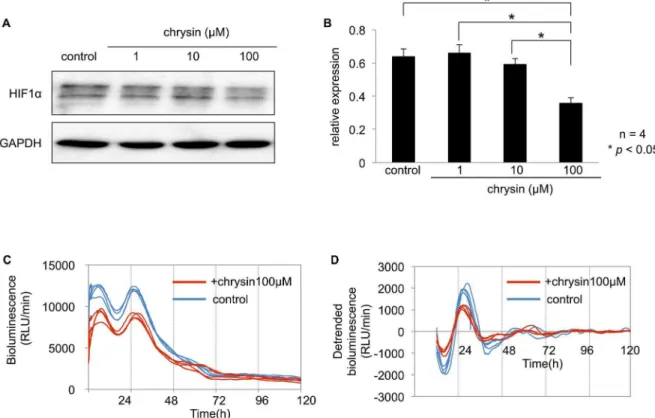
The effect of inhibiting HIF1aon the Per2circadian
rhythm
Chrysin is a natural flavonoid, which is known to inhibit HIF1a
expression by reducing protein synthesis and thereby decreases HIFastability without affecting cell viability [28]. To examine the
effect of inhibiting HIF1aprotein onPer2circadian rhythm, the
cells were pretreated with different chrysin concentrations. The expression of HIF1aprotein was significantly suppressed after a
2-h incubation wit2-h 100mM chrysin in Caki-2 cells (Fig. 6A, B). The amplitude of the circadian rhythm of thePer2promoter activity significantly decreased after a 2-h incubation with 100mM chrysin in Caki-2 cells (Fig. 6C, D, Table 3). Based on these results, HIF1a may enhance the circadian rhythms of Per2 at the
promoter level.
Discussion
In this study, rhythmic expression of the Per2 gene was observed in Caki-2 cells. However,Per2promoter activities and mRNA levels did not have circadian rhythms in any other cell lines. Some differences may exist between Caki-2 and other renal cancer cell lines. BecausePer2gene transcription is activated by the heterodimerized transcription factor BMAL1/CLOCK by binding to the E-box-like sequence [24], we examined the expression of BMAL1 and CLOCK protein in these cell lines. Caki-2, 786-O, and A498 cells expressed BMAL1, and all cell lines contained CLOCK protein. Furthermore, all renal cancer cell lines expressed CRY1 protein, but did not express PER2 protein. In Caki-2 cells, we also examined the expression of PER2 protein at 4-h intervals beginning 24 h after treatment with dexametha-sone. However, no PER2 protein was detected at all time points (Fig. S4). No definitive mechanism to account for the discrepancy between the lack of PER2 protein expression and positive mRNA expression in Caki-2 cells has yet been elucidated. It is reported that the protein kinase CK2 specifically binds and phosphorylates Figure 5. The effects of HIF1a/ARNT on thePer2promoter.(A) Schematic representation of the mousePer2promoter. The upper area
represents the wild-type mousePer2promoter and the lower area represents the HRE-mutantPer2promoter. (B) HIF1a/ARNT potently inducedPer2 promoter activity. ThePer2promoter and the HRE-mutantPer2promoter reporter (60 ng) were co-transfected with the indicated expression plasmids (+; 50 ng).Per2promoter activities were significantly increased (mean6SEM,n= 4,p,0.01, Student’sttest) compared to the control (without the expression plasmid), but the HRE-mutantPer2promoter was not affected. (C) Twenty-four hours after treatment with CoCl2(10, 30, 100mM for 6 h), luciferase activity was measured.Per2promoter activities were significantly increased (mean6SEM,n= 4,p,0.05, one-way ANOVA followed by Tukey’s post hoc tests) concentration-dependently compared to the control, but the HRE-mutant Per2 promoter was not affected. (D) HIF1a specifically interacts with the HRE-like sequence within thePer2promoter. Caki-2 cells were cross-linked, lysed, and immunoprecipitated with anti-HIF1aantibody or normal rabbit IgG (negative control). The precipitated DNA was subjected to PCR with primers specific for the target region (2 476/2284). One aliquot of input DNA was used as a positive control. PCR product was observed in the anti-HIF1aChIP (lane 3) and 10% Input DNA (lane 4). Substantially less was detected in the no antibody ChIP (lane 1) and normal rabbit IgG ChIP (lane 2) lanes.
PER2 and interacts with the protein kinase CKIe to promote
PER2 degradation [29]. Furthermore, the expression levels and activities of CK2 are reported to be increased in many tumors and tumor cell lines [30]. Thus, in this case, there is a possibility that posttranslational modification by CK2 causes the loss of PER2 protein. However, further studies will be needed to elucidate the detailed mechanism underlying CK2-mediated enhancement of CKIe-dependent PER2 degradation.
Upregulation of HIF1amay be common in RCC sinceVHLis
often mutated [31]. Thus, we examined the expression of HIF1a
protein in these cell lines. We found that Caki-2 and RCC4+vector alone cells contained HIF1a protein. Taken together, rhythmic
expression of the Per2 gene was observed in Caki-2 containing BMAL1, CLOCK, and HIF1aprotein. This suggested that both
BMAL1/CLOCK and HIF1a may be related to the circadian
expression of Per2 in renal cancer cell lines. Thus, we hypothesized that HIF1a may affect circadian expression of the Per2gene. In the present study, HIF1aincreased the amplitude of
thePer2circadian rhythm in Caki-2 cells, as well as mouse and human models. Sequence analysis of thePer2 promoter region revealed high homology between mice and humans. These results suggest that the promoter region we cloned is suitable for use in experiments on human cell lines. We also examined the difference between the wild-type mPer2 promoter and the HRE-mutant mPer2promoter in NIH3T3 cells without HIF1aor CoCl2. The
transcriptional activity and amplitude of the HRE-mutant mPer2 promoter was higher than those of the wild-type mPer2promoter (Fig. S5 A–C). It is possible that the factor which has negative
Table 3.Circadian parameters ofPer2promoter activities based on four days of data in Caki-2 cells.
period (h) amplitude acrophase (h) P value
control 24.360.04 1045.57641.99 5.4960.10 ,0.000001
+chrysin100mM 24.260.04 766.08623.95* 5.7460.11 ,0.000001
Period, amplitude, and acrophase of the oscillations were measured on days 2 to 5 using the Cosinor software (Circadian Rhythm Laboratory). Amplitude significantly decreased (mean6SEM,n= 4) compared to the control (*p,0.05).
doi:10.1371/journal.pone.0109693.t003
Figure 6. The effect of HIF1ainhibition onPer2circadian rhythm in Caki-2 cells.(A) Caki-2 cells were cultured to 60–70% confluence. The
cells were treated with DMSO as a control, or different chrysin concentrations (1, 10, 100mM) for 2 h. (B) HIF1aprotein levels were measured in optical density values normalized to their respective GAPDH loading control, then averaged 6 SEM, and graphed (relative expression) to semiquantitatively compare protein levels (n= 4). HIF1aexpression was significantly suppressed by 100 nM chrysin compared to the control incubated with DMSO (p,0.05, one-way ANOVA followed by Tukey’s post hoc test). (C) Caki-2 cells were transfected with thePer2promoter reporter (2mg). Twenty-four hours after the transfection, cells were incubated with 100mM chrysin or DMSO for 2 h. Bioluminescence was then measured using a real-time monitoring assay. The luciferase activities of four replicate samples are shown. (D) Detrended bioluminescence is shown. Period, amplitude, and acrophase of the oscillations were measured on days 2 to 5 using the Cosinor software (Circadian Rhythm Laboratory). Amplitude significantly decreased (mean6SEM,n= 4) compared to the control (p,0.05). See Table 3.
doi:10.1371/journal.pone.0109693.g006
effects on the HRE-like sequence can not bind to the mutated HRE-like sequence, hence the transcriptional activity and amplitude of the HRE-mutant mPer2promoter was higher than those of the wild-type mPer2promoter. Moreover, another factor might upregulate thePer2transcriptional activity and amplitude of oscillation by binding to the mutated HRE-like sequence. We are investigating these factors, however the detailed mechanism remains unclear. This will be addressed in future studies. Based on our luciferase assay with HIF1aand CoCl2, the HRE-like element
we identified may respond to overexpression of HIF1a.
Further-more, ChIP assay revealed the direct binding of HIF1a to the
HRE-like element. These results indicate that HIF1amay activate Per2 transcription and increase the amplitude of the Per2 circadian rhythm by directly binding to the HRE-like element within thePer2promoter.
As shown in Fig. 6, inhibition of HIF1a decreased the
amplitude of the circadian rhythm of thePer2promoter activities. These data support that HIF1a plays a role in the circadian
expression ofPer2in renal cancer cell lines.
We hypothesized thatPer2 circadian rhythms in Caki-2 cells were produced by BMAL1/CLOCK and HIF1a following our
observation that HIF1amay enhance thePer2circadian rhythms.
However, further investigation is required to determine the molecular mechanisms of HIF1a-induced enhancement ofPer2
circadian rhythms and differences between Caki-2 and other renal cancer cell lines.
Rhythmic expression of thePer2gene was not observed in renal cancer cell lines, excluding Caki-2 cells. Although rhythmicity of Per2 promoter activities was determined using online software, bioluminescence levels in Caki-2 cells were much lower than in NIH3T3 and U2OS cells. Rhythmic expression of theBmal1gene was not observed in any renal cancer cell line (Fig. S6). These results suggest that disruption of circadian rhythms of clock genes may be common in renal cancer cell lines. Previous studies revealed that circadian rhythm disruption in mice is associated with accelerated growth of malignant tumors [32]. Also, Per2 plays an important role in setting the period of oscillation [14] and has tumor-suppressor properties [33]. ThePer2gene functions in tumor suppression by regulating DNA-damage-responsive path-ways, andPer2-deficient mice show signs of premature aging and increased neoplastic tissue development following gamma irradi-ation [34]. Mutirradi-ations in thePer2 gene have been identified in human colorectal and breast cancers17. Moreover, overexpression of Per2 inhibits tumor proliferation in culture and in animals [35,36]. In the present study, HIF1a may activate Per2
transcription and increase the amplitude of the Per2 circadian rhythm by directly binding to thePer2promoter. Considering that Per2is reported to have tumor-suppressor properties, it is possible that HIF1aincreasesPer2transcriptional activity thereby inhibits
tumor proliferation in contrast to the previous finding that HIF-mediated gene pathway is the main risk-factor of tumor growth in renal cancer. However, a recent study showed that, in contrast to previous reports, deficiency in either thePer1orPer2gene alone does not render mice more tumor-prone; moreover, some long-term effects of ionizing radiation inPer2-deficient mice are more reminiscent of accelerated aging rather than a tumor-prone phenotype [37]. Therefore, we should investigate the detailed molecular mechanisms ofPer2as potentially important targets for
renal cancer therapy and clarify the effect of HIF inducedPer2 upregulation on tumor progression in renal cancer in future study. Furthermore, their detailed role that seems to conflict with the role of HIF-mediated angiogenic pathway should be investigated as well.
Supporting Information
Figure S1 Real-time monitoring of luciferase activity of thePer2 promoter in renal cancer cell lines and NIH3T3 cells. Rhythmicity was not detectable in renal cancer cell lines (p.0.05, by Cosinor). The amount of plasmid and Lipofectamine 2000 used in each of the cell lines are shown.
(TIFF)
Figure S2 Full-length blots of BMAL1. (A) Full-length blots of BMAL1 in indicated cell lines including a circadian competent cell line (NIH3T3) as a positive control are presented. (B) The positive controls using a circadian competent cell line (NIH3T3) transfected with the indicated expression plasmids are presented. (TIFF)
Figure S3 Full-length blots of PER2. The positive controls using a circadian competent cell line (NIH3T3) and HeLa cells, which is listed in manufacturer’s datasheet as a positive control, are presented.
(TIFF)
Figure S4 Full-length blots of PER2 in Caki-2 cells at seven time points.
(TIFF)
Figure S5 Difference between the wild-type mPer2 promoter and the HRE-mutant mPer2 promoter in NIH3T3 cells without HIF1a or CoCl2. (A) Difference between the relative luciferase
activities of the wild-type mPer2promoter and the HRE-mutant mPer2 promoter in NIH3T3 cells. (B) Bioluminescence of the wild-type mPer2promoter and the HRE-mutant mPer2promoter in NIH3T3 cells. Four replicate samples are shown. (C) Detrended bioluminescence of the wild-type mPer2promoter and the HRE-mutant mPer2promoter in NIH3T3 cells. Four replicate samples are shown.
(TIFF)
Figure S6 Real-time monitoring of luciferase activity of the Bmal1 promoter in renal cancer cell lines and NIH3T3 cells. Rhythmicity was not detectable in renal cancer cell lines (p.0.05, by Cosinor). The amount of plasmid and Lipofectamine 2000 used in each of the cell lines are shown.
(TIFF)
Acknowledgments
We thank Dr. Yasuhiro Takenaka for advice on the ChIP experiments.
Author Contributions
Conceived and designed the experiments: TO MU MI. Performed the experiments: TO MK. Analyzed the data: TO MU MI. Contributed reagents/materials/analysis tools: YN. Wrote the paper: TO. Served as scientific advisors: SS KK MO.
References
1. McLaughlin JK, Lipworth L (2000) Epidemiologic aspects of renal cell cancer. Semin Oncol 27: 115–123.
2. Talks KL, Turley H, Gatter KC, Maxwell PH, Pugh CW, et al. (2000) The expression and distribution of the hypoxia-inducible factors HIF-1alpha and
HIF-2alpha in normal human tissues, cancers, and tumor-associated macro-phages. Am J Pathol 157: 411–421.
vascular endothelial growth factor expression through HIF-2a. Carcinogenesis 3: 529–536.
4. Jiang BH, Rue E, Wang GL, Roe R, Semenza GL (1996) Dimerization, DNA binding, and transactivation properties of hypoxia-inducible factor 1. J Biol Chem 271: 17771–17778.
5. Pugh CW, Ratcliffe PJ (2003) Regulation of angiogenesis by hypoxia: role of the HIF system. Nat Med 9: 677–684.
6. Semenza GL (2003) Targeting HIF-1 for cancer therapy. Nat Rev Cancer 3: 721–732.
7. Harris AL (2002) Hypoxia—a key regulatory factor in tumour growth. Nat Rev Cancer 2: 38–47.
8. Thelen P, Hemmerlein B, Kugler A, Seiler T, Ozisik R, et al. (1999) Quantification by competitive quantitative RT-PCR of VEGF121 and VEGF165 in renal cell carcinoma. Anticancer Res 19: 1563–1565.
9. Panda S, Hogenesch JB, Kay SA (2002) Circadian rhythms from flies to human. Nature 417: 329–335.
10. Reppert SM, Weaver DR (2002) Coordination of circadian timing in mammals. Nature 418: 935–941.
11. Reppert SM, Weaver DR (2001) Molecular analysis of mammalian circadian rhythms. Annu Rev Physiol 63: 647–676.
12. Gekakis N, Staknis D, Nguyen HB, Davis FC, Wilsbacher LD, et al. (1998) Role of the CLOCK protein in the mammalian circadian mechanism. Science 280: 1564–1569.
13. Griffin EA Jr, Staknis D, Weitz CJ (1999) Light-independent role of CRY1 and CRY2 in the mammalian circadian clock. Science 286: 768–771.
14. Wilkins AK, Barton PI, Tidor B (2007) The Per2 negative feedback loop sets the period in the mammalian circadian clock mechanism. PLOS Comput Biol 3: e242. Available: http://www.ploscompbiol.org/article/info%3Adoi%2F10. 1371%2Fjournal.pcbi.0030242.
15. Chen ST, Choo KB, Hou MF, Yeh KT, Kuo SJ, et al. (2005) Deregulated expression of the PER1, PER2 and PER3 genes in breast cancers. Carcinogenesis 26: 1241–1246.
16. Sjo¨blom T, Jones S, Wood LD, Parsons DW, Lin J, et al. (2006) The consensus coding sequences of human breast and colorectal cancers. Science 314: 268–274. 17. Mazzoccoli G, Piepoli A, Carella M, Panza A, Pazienza V, et al. (2012) Altered expression of the clock gene machinery in kidney cancer patients. Biomed Pharmacother 66: 175–179.
18. Nagoshi E, Saini C, Bauer C, Laroche T, Naef F, et al. (2004) Circadian gene expression in individual fibroblasts: cell-autonomous and self-sustained oscilla-tors pass time to daughter cells. Cell 119: 693–705.
19. Baggs JE, Price TS, DiTacchio L, Panda S, FitzGerald GA, et al. (2009) Network features of the mammalian circadian clock. PLoS Biology 7: e52. Available: http://www.plosbiology.org/article/info%3Adoi%2F10.1371%2Fjournal.pbio. 1000052#pbio-1000052-g005.
20. Noguchi T, Ikeda M, Ohmiya Y, Nakajima Y (2012) A dual-color luciferase assay system reveals circadian resetting of cultured fibroblasts by co-cultured adrenal glands. PLoS One 7: e37093. Available: http://www.plosone.org/ article/info%3Adoi%2F10.1371%2Fjournal.pone.0037093.
21. Yang F, Nakajima Y, Kumagai M, Ohmiya Y, Ikeda M (2009) The molecular mechanism regulating the autonomous circadian expression of Topoisomerase I in NIH3T3 cells. Biochem Biophys Res Commun 380: 22–27.
22. Thomas KA, Burr RL (2008) Circadian research in mothers and infants: how many days of actigraphy data are needed to fit cosinor parameters? J Nurs Meas 16: 201–206.
23. Tamaru T, Hattori M, Ninomiya Y, Kawamura G, Vare`s G, et al. (2013) ROS stress resets circadian clocks to coordinate pro-survival signals. PLoS One 8: e82006. Available: http://www.plosone.org/article/info%3Adoi%2F10.1371% 2Fjournal.pone.0082006#s2.
24. Akashi M, Ichise T, Mamine T, Takumi T (2006) Molecular mechanism of cell-autonomous circadian gene expression of Period2, a crucial regulator of the mammalian circadian clock. Mol Biol Cell 17: 555–565.
25. Kong D, Park EJ, Stephen AG, Calvani M, Cardellina JH, et al. (2005) Echinomycin, a small-molecule inhibitor of hypoxia-inducible factor-1 DNA-binding activity. Cancer Res 65: 9047–9055.
26. Kanaya K, Kamitani T (2003) pVHL-independent ubiquitination of Hif1aand
its stabilization by cobalt ion. Biochem Biophys Res Commun 306: 750–755. 27. Yuan Y, Hilliard G, Ferguson T, Millhorn DE (2003) Cobalt inhibits the
interaction between hypoxia inducible factor-aand von Hippel-Lindau protein
by direct binding to hypoxia inducible factor-a. J Biol Chem 278: 15911–15916.
28. Fu B, Xue J, Li Z, Shi X, Jiang BH, Fang J (2007) Chrysin inhibits expression of hypoxia-inducible factor-1alpha through reducing hypoxia-inducible factor-1 alpha stability and inhibiting its protein synthesis. Mol Cancer Ther 1: 220–226. 29. Tsuchiya Y, Akashi M, Matsuda M, Goto K, Miyata Y, et al. (2009) Involvement of the protein kinase CK2 in the regulation of mammalian circadian rhythms. Sci Signal. 2: ra26.
30. Romieu-Mourez R, Landesman-Bollag E, Seldin DC, Sonenshein GE (2002) Protein kinase CK2 promotes aberrant activation of nuclear factor-kappaB, transformed phenotype, and survival of breast cancer cells. Cancer Res. 62: 6770–6778.
31. Rini BI, Jaeger E, Weinberg V, Sein N, Chew K, et al. (2006) Clinical response to therapy targeted at vascular endothelial growth factor in metastatic renal cell carcinoma: impact of patient characteristics and Von Hippel-Lindau gene status. BJU Int 98: 756–762.
32. Filipski E, King VM, Li X, Granda TG, Mormont MC, et al. (2002) Host circadian clock as a control point in tumor progression. J Natl Cancer Inst 94: 690–697.
33. Fu L, Lee CC (2003) The circadian clock: pacemaker and tumour suppressor. Nat Rev Cancer 3: 350–361.
34. Fu L, Pelicano H, Liu J, Huang P, Lee C (2002) The circadian gene Period2 plays an important role in tumor suppression and DNA damage response in vivo. Cell 111: 41–50.
35. Gery S, Virk RK, Chumakov K, Yu A, Koeffler HP (2007) The clock gene Per2 links the circadian system to the estrogen receptor. Oncogene 26: 7916–7920. 36. Hua H, Wang Y, Wan C, Liu Y, Zhu B, et al. (2007) Inhibition of tumorigenesis
by intratumoral delivery of the circadian gene mPer2 in C57BL/6 mice. Cancer Gene Ther 9: 815–818.
37. Antoch MP, Toshkov I, Kuropatwinski KK, Jackson M (2013) Deficiency in PER proteins has no effect on the rate of spontaneous and radiation-induced carcinogenesis. Cell Cycle 12: 3673–3680.