CASE REPORT
Lowe Syndrome (Oculo-cerebro-renal Syndrome of Lowe): A Case Report from
Eastern India
Dipankar Das 1, Shoumini Chakravarty 1, Jaydeb Ray 2*
1 Department of Radio diagnosis, 2 Department of Pediatrics, Institute of Child Health, Kolkata -700017, West Bengal, India
Abstract:
Lowe syndrome (the oculocerebrorenal syndrome of Lowe, OCRL) is a rare X-linked recessive metabolic disorder that primarily affects eyes, kidneys and brain. It is caused by the deficiency of enzyme phosphatidylinositol 4, 5-bisphosphate 5-phosphatase. The gene coding for this enzyme, OCRL1 and muta-tions in it are responsible to cause Lowe Syndrome. We report a 6 years old boy from Eastern India, with Lowe Syndrome. Diagnosis was suggested by typical features in the MRI of the brain along with other clini-cal feature and investigation.
Keywords: Lowe syndrome; OCRL; Cataract.
Introduction:
Lowe syndrome (the oculocerebrorenal syndrome of Lowe, OCRL) is an uncommon, panethnic, X-linked recessive metabolic disorder described by Lowe and coworkers in 1952 [1]. It is a multisystem disorder which primarily affects the eyes, nervous system, and kidneys. It is characterized by congenital cataracts, infantile glaucoma, neonatal or infantile hypotonia, in-tellectual impairment, and renal tubular dysfunction (Fanconi syndrome). Tentative prevalence is 1 in 500,000 of general population. The mutation of the gene OCRL1 localized at Xq26.1, coding for the en-zyme phosphatidylinositol, bisphosphate 5 phos-phatase, PtdIns P2, in the trans-Golgi network is re-sponsible for the disease. Both enzymatic and mo-lecular testing is available for confirmation of the di-agnosis and for prenatal detection of the disease [2]. Clinical features include congenital cataract, glau-coma, miotic pupils, nystagmus, corneal keloids as ocular involvement, infantile hypotonia, areflexia, sei-zures, mental retardation, behavioral abnormalities as cerebral involvement and fanconi syndrome, renal
tu-bular acidosis and gradual impairment of glomerular function as renal involvement. Although the child was treated by different subspecialist but proper diagno-sis was not predicted in the beginning, as there was no team approach. We should treat a patient as a whole & team approach is needed to diagnose a rare syndrome like Lowe syndrome.
Case Report:
A six years old male patient came thrice in different hospitals at different departments. At one month of age the baby presented for the first time with bilateral congenital cataract and glaucoma which were treated by ophthalmologist.
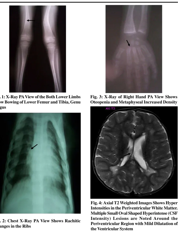
Next time the patient came with lower limb injury for which X-ray was done. X ray showed features of rickets (Fig. 1, 2, 3). Relevant blood examination showed low phosphorus level with raised alkaline phosphatase and normal serum calcium. Urine exami-nation revealed albumin, sugar and phosphate in urine suggesting renal tubular dysfunction. Blood gas analy-sis revealed acidoanaly-sis. USG of abdomen showed bi-lateral small kidneys.
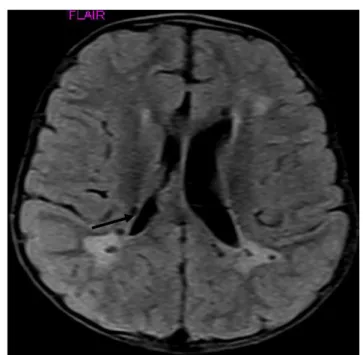
The patient came for the third time and was admitted for convulsion in our hospital at the age of six years. Along with other routine investigations, the patient was referred to our department for MRI of brain. MRI brain was performed in our Magnetom Concerto (Siemens Medical System) which showed periventricular hyperintensities on T2W and FLAIR images with tiny cystic lesions within (Fig. 4, 5, 6, 7). The cysts were hypointense on T1W and FLAIR images (Fig. 7, 8). The cerebellum, basal ganglia and subcortical white matter were not involved.
in-Fig. 3: X-Ray of Right Hand PA View Shows Oteopenia and Metaphyseal Increased Density Fig. 1: X-Ray PA View of the Both Lower Limbs
Show Bowing of Lower Femur and Tibia, Genu Valgus
Fig. 2: Chest X-Ray PA View Shows Rachitic Changes in the Ribs
Fig. 7: Axial FLAIR Image Shows Periventri-cular Hyper Intensities with Multiple Hypo In-tense Cystic Lesions
Fig. 5: Axial T2 Weighted Images Shows Ill-De-fined Hyperintensities in the Region of Centrum Semiovale
Fig. 6: Coronal T2 Weighted Image Shows Hyper Intensities in the Periventricular Region with Multiple Cystic Lesions Within. the Lesions are not Involving Cerebellum and Corpus Callosum
volvement, the cerebral lesions pointed towards the diagnosis of oculo-cerebro-renal syndrome of Lowe. MR spectroscopy could not be done as it was not available in that scanner.
Discussion:
MR imaging findings in Lowe syndrome include patchy areas of high-signal changes on T2W and FLAIR images that are hypointense on T1-weighted images. At the same time there may be small, well-defined, punctate, cyst-like changes, which follow the signal pattern of CSF. The lesions are usually bilat-eral [3, 4, 5, 6]. Cranial MR examination of our pa-tient shows similar bilateral hyper intensities on T2 weighted images with tiny cysts in the periventricular regions.
Past reports attribute that the hyper intensities mostly in the bilateral periventricular regions and centrum semiovale are due to demyelination [7, 4]or gliosis [8] and not necessarily correlated with the severity of clinical manifestations [8]. Recent studies and neuro-pathological data show evidences that these are due to gliosis. Schneider and Sener [5, 4]described cases with proton MR spectroscopy reporting prominent peaks of myoinositol which is a glial marker, suggest-ing the presence of gliosis[9].
In OCRL patients there is deficiency of phosphatidylinositol, bisphosphate 5 phosphatase, located in the Golgi apparatus, which catalyzes the hydrolysis of the 5-position phosphate and regulates cellular levels of phosphatidylinositol 4, 5-biphosphate (PtdIns 4, 5-P2), a metabolite involved in Golgi ve-sicular transport. This enzymatic deficiency leads to accumulation of PtdIns 4, 5-P2 in lysosomal mem-branes and therefore causes increased extracellular release of lysosomal enzymes [10]. This would be responsible for developmental defects in the lens and abnormal renal and neurological functions. In CNS the accumulation of lysosomal products can lead to dilatation of perivascular spaces [5], whereas
extra-cellular release of lysosomal enzymes can lead to toxic gliotic reactions [5].
In our patient, ocular involvement (bilateral cataract and glaucoma) and renal involvement (renal tubular dysfunction, renal rickets) were viewed as isolated entities when attended. However, after the highly sug-gestive MR picture of brain, we reviewed the past history of the patient and detailed work up was done. Although similar tiny cystic lesions and periventricular hyper intensities on long TR sequences can be found in different conditions with dilated perivascular spaces including mucopolysaccharidosis, white matter injury etc. The diagnosis was suggested in the perspective of ocular and renal involvement in the same patient. Any further investigation like genetic mutation, enzy-matic analysis and response to treatment with golgi complex are not possible with this patient because of our socio-economic condition.
Inspite of multisystem involvement, patients with Lowe syndrome survive for about 30-40 years. Renal in-sufficiency [9]is the usual cause of death. Medical management includes correction of electrolytic imbal-ance, supplementation of calcium, vitamin- D, phos-phate, potassium and relation with speech, occupa-tional and physiotherapy. Surgical intervention com-bines ophthalmologic intervention and includes treat-ment of cataract, glaucoma and orthopedic interven-tion including correcinterven-tions of scoliosis and long bone deformities.
Conclusion:
sce-nario can only lead to the correct diagnosis.
Acknowledgement:
Dr. Joydeep Das, Assistant Professor, Department
of Pediatrics, Institute of Child Health, Kolkata, West Bengal, India for his assistance in clinics regarding the case.
References:
1. Lowe CU, Terrey M, MacLachlan EA. Organic-aciduria,
decreased renal ammonia production, hydrophthalmos, and mental retardation; a clinical entity. Am J Dis Child. 1952; 83(2):164-184.
2. Singh AR, Singh JR, Kaur H, Sachdeva GS, Kaur A,
Singh AD. Lowe Syndrome (Oculocerebrorenal Syn-drome of Lowe): A Case Report of Two Brothers from India. Int J Hum Genet 2002; 2(4): 243-249.
3. Demmer LA, Wippold FJ 2nd, Dowton SB. Periventricular
white matter cystic lesions in Lowe (oculocerebrorenal) syndrome. A new MR finding. PediatrRadiol 1992; 22 (1): 76-77.
4. Ono J, Harada K, Mano T, Yamamoto T, Okada S. MR
findings and neurologic manifestations in Lowe
oculocerebrorenal syndrome. Pediatr Neurol 1996;
14(2): 162-164.
5. Schneider JF, Boltshauser E, Neuhaus TJ, Rauscher C,
Martin E. MRI and proton spectroscopy in Lowe syn-drome. Neuropediatrics 2001; 32 (1): 45-48.
6. Van der Knaap MS, Valk J. Magnetic resonance of
my-elin, myelination, and myelin disorders. Springer-Verlag, Heildelberg 1995: 243-245.
7. Carroll WJ, Woodruff WW, Cadman TE. MR findings in
oculocerebrorenal syndrome. AJNR Am J Neuroradiol
1993; 14(2):449-51.
8. Sener RN. Lowe syndrome: proton MR spectroscopy,
and diffusion MR imaging. J Neuroradiol 2004;
31(3):238-240.
9. Loi M. Review Lowe syndrome. Orphanet J Rare Dis
2006; 1: 16.
10. Carvalho-Neto Ad, Ono SE, Cardoso Gde M, Santos ML, Celidonio I. Oculocerebrorenal syndrome of Lowe: magnetic resonance imaging findings in the first six years of life. Arq Neuropsiquiatr 2009; 67(2A):305-7.