772 Letter to the Editor
International Braz J Urol Vol. 34 (6): 772-773, November - December, 2008
Childhood Renal Lymphangiectasia
To the Editor,
An asymptomatic 4 month-old white female had a 2 x 3 cm faint birthmark on her mid-thoracic back consistent with a cutaneous telangiectasia. MRI
RIWKHVSLQHLQFLGHQWDOO\GHPRQVWUDWHGDQLQ¿OWUDWLYH ULJKWUHQDOOHVLRQ8OWUDVRXQGVKRZHGDQLQ¿OWUDWLYH
lesion in the enlarged right kidney and proximal ip-silateral ureter as well as heterogeneously increased renal cortical echogenicity. Abdominal CT scan
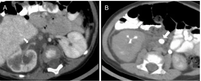
UHYHDOHGDK\SRGHQVHOHVLRQ+RXQV¿HOGXQLWVIURP WRLQ¿OWUDWLQJWKHUHQDOSHOYLVDQGSDUHQFK\PD
circumscribing the right kidney and proximal ureter
)LJXUH$ 'HOD\HG FRQWUDVW LPDJHV VKRZHG FRPSUHVVLRQ RI WKH XUHWHURYHVLFDO MXQFWLRQ ZLWK PHGLDOGLVSODFHPHQWRIWKHULJKWXUHWHU)LJXUH %$EGRPLQDO 05, FKDUDFWHUL]HG WKH OHVLRQ DV
heterogenously hyperintense and hypointense on
7DQG7ZHLJKWHGLPDJHVUHVSHFWLYHO\)LJXUH 5LJKWNLGQH\GLIIHUHQWLDOIXQFWLRQZDVE\ 0$*UHQDOVFDQ7KHLQ¿OWUDWLYHFKDUDFWHURIWKH
OHVLRQZLWKRXWVLJQL¿FDQWHQKDQFHPHQWLVFRQVLVWHQW
with a renal lymphangiectasia. At 2.5 years of follow-up, she remains asymptomatic without radiological change.
Renal lymphangiectasia is a rare, benign
FRQGLWLRQFKDUDFWHUL]HGE\GHYHORSPHQWDOPDOIRUPD -tion of the perirenal lymphatic system. The lymphatic structures that surround the kidney fail to establish normal communication with the rest of the lymphatic system. The physiopathological process is ectasia of
O\PSKDWLFYHVVHOVZLWKRXWREVWUXFWLRQ,WFDQ
be focal, unilateral or bilateral and may be found in pediatric or adult patients. There are reports of familial
SUHGLVSRVLWLRQ/LNHRWKHUO\PSKDWLFOHVLRQVUHQDO
lymphangiectasia can appear suddenly, grow rapidly,
FHDVHJURZWKDEUXSWO\RUHYHQUHJUHVVVSRQWDQHRXVO\ 6LJQVDQGV\PSWRPVPD\YDU\IURPQRQHWRPLFUR
-VFRSLFRUPDFURVFRSLFKHPDWXULDSURWHLQXULDÀDQN
pain, abdominal pain/distension, palpable abdominal
Figure 1 –$$QLQ¿OWUDWLYHK\SRGHQVHOHVLRQLVLGHQWL¿HGLQWKHUHQDOVLQXVSDUHQFK\PDDQGSHULUHQDOVSDFH7KHULJKWUHQDODUWHU\ LVFLUFXPVFULEHGE\WKHOHVLRQDUURZ%'HOD\HG&7LPDJHVKRZVHORQJDWHGDQGGLVWRUWHGFROOHFWLQJV\VWHPE\WKHUHQDOVLQXVPDVV 7KHULJKWXUHWHULVHQODUJHGDQGGLVSODFHGPHGLDOO\DUURZ
773 Letter to the Editor
mass, lower extremity edema and hypertension. Renal
IXQFWLRQLVJHQHUDOO\SUHVHUYHG
5DGLRORJLFDOLPDJLQJPRGDOLWLHVKDYHDLGHG
diagnosis, including renal US, IVP, CT, and MRI. Renal US can show an enlarged and lobulated kidney with increased echogenicity and loss of normal
corti-FRPHGXOODU\GLIIHUHQWLDWLRQ7KH&7VFDQUHYHDOV PXOWLORFXODUF\VWÀXLG¿OOHGPDVVHVZLWKWKLQZDOOV LQWKHSHULUHQDODQGSDUDSHOYLFUHJLRQ7KH05,FDQ
show hyperintensity of the renal parenchyma espe-cially at the cortical region and hypointensity at the medullary region. Also, multiple hyperintense lesions in perirenal spaces on T2-weighted images can be appreciated. Pediatric differential diagnosis includes polycystic renal disease, urinoma, renal lymphoma
ZLWKSHULUHQDOLQYROYHPHQWUHQDOWXPRUVHWF7KH GLDJQRVLVFDQEHFRQ¿UPHGE\QHHGOHDVSLUDWLRQRI FK\ORXV ÀXLG RU E\ UHQDO ELRSV\ EXW PXOWLPRGDO
imaging is characteristic.
0DQDJHPHQWLVRIWHQFRQVHUYDWLYHGXHWRWKH EHQLJQEHKDYLRURIWKHOHVLRQ3HUFXWDQHRXVGUDLQ
-DJHZLWKDGPLQLVWUDWLRQRILQWUDYHQRXVDOEXPLQDQG
enteral medium chain triglycerides may be indicated
LIV\PSWRPVRUDVFLWHVGHYHORS6HYHUDOFDVHVRIF\VW GHFRUWLFDWLRQKDYHUHVXOWHGLQQHSKUHFWRP\GXHWR XQFRQWUROOHG LQWUDRSHUDWLYH EOHHGLQJ:KLOH VRPH
suggest that asymptomatic patients with unchanged cystic lesions on US do not require follow-up, we feel patients should undergo lifelong follow-up as occasionally renal function will deteriorate.
REFERENCES
9DUHOD -5 %DUJLHOD $ 5HTXHMR , )HUQDQGH] 5 'DU9DUHOD-5%DUJLHOD$5HTXHMR,)HUQDQGH]5'DU -ULED03RPER)%LODWHUDOUHQDOO\PSKDQJLRPDWRVLV 86DQG&7¿QGLQJV(XU 5DGLRO (XU5DGLRO /ORUHQWH-**DUFtD$'6DFULVWDQ-6&KLFKDUUR*1
5HQDO O\PSKDQJLHFWDVLD UDGLRORJLF GLDJQRVLV DQG HYROXWLRQ$EGRP,PDJLQJ
Fabian Sanchez, Juan C. Prieto, Korgun Koral & Linda A. Baker
'HSDUWRI8URORJ\)6-&3/$%DQG 5DGLRORJ\.. &KLOGUHQ¶V0HGLFDO&HQWHURI'DOODV 8QLYHUVLW\RI7H[DV6RXWKZHVWHUQ0HGLFDO&HQWHU 'DOODV7H[DV86$ (PDLO/LQGDEDNHU#XWVRXWKZHVWHUQHGX