Targeted Disruption of ALK Reveals a
Potential Role in Hypogonadotropic
Hypogonadism
Barbara Witek2☯, Abeer El Wakil2☯, Christoffer Nord3, Ulf Ahlgren3, Maria Eriksson3,
Emma Vernersson-Lindahl4,Åslaug Helland5, Oleg A. Alexeyev4, Bengt Hallberg1*, Ruth H. Palmer1,2*
1Department of Medical Biochemistry and Cell Biology, Institute of Biomedicine, Sahlgrenska Academy, University of Gothenburg, Gothenburg, Sweden,2Department of Molecular Biology, UmeåUniversity, Umeå, Sweden,3UmeåCenter for Molecular Medicine, UmeåUniversity, Umeå, Sweden,4Institution for Medical Biosciences/Pathology, UmeåUniversity, Umeå, Sweden,5Department of Oncology, Oslo University Hospital Radiumhospitalet, Oslo, Norway
☯These authors contributed equally to this work. *[email protected](RHP); [email protected](BH)
Abstract
Mice lacking ALK activity have previously been reported to exhibit subtle behavioral pheno-types. In this study of ALK of loss of function mice we present data supporting a role for ALK in hypogonadotropic hypogonadism in male mice. We observed lower level of serum testos-terone at P40 in ALK knock-out males, accompanied by mild disorganization of seminifer-ous tubules exhibiting decreased numbers of GATA4 expressing cells. These observations highlight a role for ALK in testis function and are further supported by experiments in which chemical inhibition of ALK activity with the ALK TKI crizotinib was employed. Oral adminis-tration of crizotinib resulted in a decrease of serum testosterone levels in adult wild type male mice, which reverted to normal levels after cessation of treatment. Analysis of GnRH expression in neurons of the hypothalamus revealed a significant decrease in the number of GnRH positive neurons in ALK knock-out mice at P40 when compared with control litter-mates. Thus, ALK appears to be involved in hypogonadotropic hypogonadism by regulating the timing of pubertal onset and testis function at the upper levels of the hypothalamic-pitui-tary gonadal axis.
Introduction
The Anaplastic Lymphoma Kinase (ALK) Receptor Tyrosine Kinase (RTK) was originally dis-covered as a fusion protein together with Nucleophosmin (NPM) in anaplastic large-cell non-Hodgkin’s lymphoma (ACLC) in 1994 [1]. The full length receptor was later cloned revealing a transmembrane receptor tyrosine kinase, most similar to those of the Insulin Receptor family [2,3]. The oncogenic properties of NPM-ALK are thought to arise from the ability of NPM to dimerize thereby mediating constant activation of the ALK kinase domain [4,5]. Since OPEN ACCESS
Citation:Witek B, El Wakil A, Nord C, Ahlgren U, Eriksson M, Vernersson-Lindahl E, et al. (2015) Targeted Disruption of ALK Reveals a Potential Role in Hypogonadotropic Hypogonadism. PLoS ONE 10 (5): e0123542. doi:10.1371/journal.pone.0123542
Academic Editor:Jean-Marc A Lobaccaro, Clermont Université, FRANCE
Received:December 7, 2014
Accepted:March 5, 2015
Published:May 8, 2015
Copyright:© 2015 Witek et al. This is an open access article distributed under the terms of the
Creative Commons Attribution License, which permits unrestricted use, distribution, and reproduction in any medium, provided the original author and source are credited.
Data Availability Statement:All relevant data are within the paper and its Supporting Information files.
discovery of the fusion NPM-ALK protein, more than 20 ALK fusion partners have been ob-served not only in ALCL but also in diseases such as inflammatory myofibroblastic tumor (IMT), non-small cell lung cancer (NSCLC), renal carcinoma, breast cancer, colon carcinoma, serous ovarian carcinoma, oesophageal squamous cell carcinoma (ESCC) and diffuse large B cell lymphoma (DLBLC) [4]. In addition to the numerous translocation events, gain of func-tion ALK mutafunc-tions have been observed in both spontaneous and hereditary neuroblastoma [4,6–10].
The expression pattern of ALK in vertebrates has been described in several studies, where ALK has been shown to be expressed in the CNS and the PNS, as well as in testis and ovary [2, 3,11]. Despite this, deletion ofALKin mice does not result in serious phenotypes and the phys-iological role of ALK in mammals is unclear [12,13]. On closer examination however, mild be-havioral phenotypes have been observed, such as increased struggle time (as measured with tail suspension and Porsott swim tests), enhanced performance in novel object-recognition test and enhanced spatial memory [12,13]. Recent reports have described interesting side effects in patients treated with the FDA approved ALK inhibitor crizotinib [14], which include reduced hearing, suppression of testosterone levels in men and visual disturbances [15–17]. While these side effects are reversible upon withdrawal of therapeutic treatment of patients it is unclear how much is specific to inhibition of ALK activity.
Puberty is defined as a physiological and developmental process towards sexual maturity. Onset of puberty is initiated by neuroendocrine events that activate the pulsatile release of GnRH from the hippocampus into the hypophyseal portal blood system to stimulate the syn-thesis and secretion of gonadotropins from anterior pituitary cells. Gonadotropins, in turn, bind to ligand-specific receptors in the gonads, causing gonadal maturation and production of sex steroids, most notably testosterone in males [18–20]. Hippocampal GnRH neurons origi-nate in the nasal placode and migrate through the nasal compartment and the cribriform plate and finally pass through the basal forebrain, before reaching the hypothalamus [19,21,22]. Output from these neurons is critical for initiation of puberty as well as maintenance of fertili-ty. A critical role for GnRH neuronal activity in puberty is highlighted by infertility in mice with defective GnRH biosynthesis [23].
Herein we describe the investigation of an ALK knock-out mouse model in which the kinase domain encoding exons have been removed. In agreement with previous studies [12,13], we confirm that homozygous ALK mutant animals are viable and fertile and do not exhibit any gross morphological defects either during embryogenesis or as adult animals. Targeted disrup-tion of ALK results in decreased levels of serum testosterone at 40 days of age when compared with controls. Further examination reveals that ALK mutant male mice display mild changes in testicular tissue organization at P40. In addition, male ALK KO mice display a delay in pu-bertal onset as measured by preputial separation. Moreover, the number of gonadotropin re-leasing hormone positive neurons in the hippocampus of ALK mutant mice is significantly reduced when compared with controls. Furthermore, we are able to recapitulate in wild type mice the effects of crizotinib treatment in lung cancer patients, as inhibition of ALK activity by crizotinib treatment results in a reduction of testosterone levels in adult wild type male mice, as has been observed in crizotinib treated male patients. Taken together, the data presented here suggests that ALK plays a potential role in hypogonadotropic hypogonadism in males.
Material and Methods
Ethical permission
Allin vivoexperiments were conducted with approved Swedish legal ethical committee permits A203-12 and A155-12.
Targeting vector construction and generation of ALK Kinase knock-out
mice
PolyGene Transgenetic (Polygene AG, Riedmattstrasse 9, CH-Rümlang, Switzerland,www. polygene.ch) generated the ALK kinase KO according to our specifications. A gene targeting vector for ALK was designed to generate a floxed allele of ALK with the goal of eliminating exon 20–23. Flipase Recognition Target (FRT)-sites were included in the vector to generate a non-conditional knock-out lacking exon 20–23 (kinase domain). This non-conditional knock-out was employed in these studies. The ALK gene targeting vector thus included an FRT site upstream of exon 19 followed by a LacZ cassette and aloxP site. Upstream of exon 23 a PGK-TK negative selection gene and a PGK-neo positive selection gene (transcribed in the op-posite direction of ALK) were flanked byloxP sites. The most upstreamloxP site was flanked by an FRT site (Fig 1, specific details available upon request). The ALK sequence used for con-structing the vector was recovered by screening the Invitrogen RPCI-22M BAC bank, BAC M409A20 (Invitrogen) was sequenced to verify the ALK fragment, and using KpnI/SpeI a re-gion spanning exon 17–24 was subcloned into LITMUS28i (New England Biolabs, Inc,. Ber-verly MA, USA). The subsequent vector was electroporated into 129/Sv mouse embryonic stem cells prior to selection for neomycin resistance. After transient expression of Flp and neg-ative selection using ganciclovir positive clones (verified via Southern and PCR) were used for injection into C57BL/6N blastocysts, generating chimeric mice and establishing the ALK ki-nase KO mouse line. ALK genotypes were assessed using genomic DNA isolated from mouse tail biopsies. For Southern blot analysis, genomic DNA was digested with SpeI to generate a 14.8 kbp fragment for the wild type allele and an 8.8 kbp fragment for the targeted allele. South-ern blot was conducted according to manufacturer’s protocol (Roche DIG Application Manu-al), using a digoxigenin (DIG) labeled probe directed against ALK intron 17. For routine genotyping, genomic DNA was amplified by standard PCR using 5´-ATAGTTCGCCAGCG CGCACCGTA-3´ and 5´-TGCAGTCACTGCGAGGTAGACG-3´ for the targeted allele and 5´-CCTGATGATCAGAGCTTC-3´ and 5-GAATCCTCACGATTCTGG-3´ for the wild type allele to yield products of 437 bp and 301 bp, respectively. For the CRE-ALK line, 5´-ATAG TTCGCCAGCGCGCACCGTA-3´ and 5´-CCCTTTCAGAAGCCAGTCCTT-3´ was used for the targeted allele and 5´-CCTAGGAGTGCTAAGACC-3´ and 5´-GAGGTAGACGAATG TCAC-3´ for the wild type allele to yield products of 311 bp and 360 bp respectively. Taq DNA polymerase from New England Biolab (#M0273X) was used. The PCR consisted of an initial incubation step at 94°C for 2 min, followed by 30 cycles at 94°C for 30´, 56°C for 30´ and 72°C for 30´. A last extension step at 72°C for 5 min was performed. Mouse monoclonal antibody #135 used for verification of knock-out animals by immunobloting was generated in our labo-ratory. The antigen used for immunization of mice was His-tagged extracellular part of human ALK (a kind gift from Marc Vigny). Antibody #135 cross-reacts with mouse ALK. All experi-ments were done on C57BL/6N background.
Pubertal onset assessment
Blood collection and testosterone measurements
Mice were briefly anaesthetized with isofluorane and blood samples were collected from the fa-cial vein with Golden rod animal lancet (MEDIpoint) to the BD Microtainer tube with serum separator [26]. Serum samples were frozen and stored at -20°C until analysis. Serum testoster-one levels were quantified with the Spectria Testostertestoster-one RIA kit (Orion Diagnostica), or with the Testosterone ELISA kit (DRG, USA), according to the manufacturer’s instructions.
Sperm count
Immediately after sacrifice, the caudal epididymis was removed and placed in a small clean petri dish containing 1ml of PBS pH = 7.4 (n = 4 per age and per genotype). The caudal part was dissected with a sharp sterilized blade into three pieces and squeezed gently with fine for-ceps to release the sperm, and the suspension was then allowed to settle at 37° C for 30 min [27]. Sperm were counted using a Hemocytometer Chamber. Sperm suspension was placed on both sides of the chamber and the number of sperm counted at 100X magnification[25]. Sperm count was performed in triplicate for each sample (n = 4 at P40 per genotype, n = 6 at P60 for WT animals and n = 4 at P60 for ALK mutant mice) [28].
Sperm viability
TwentyμL of sperm suspension and 20μL of eosin solution were mixed and vortexed for 2 sec.
After, 30μL of the suspension were recovered and spread on a glass slide. After drying at room
temperature, sperm viability was determined: dead and living spermatozoa had red and white colored heads, respectively. A minimum of 200 spermatozoa was counted in triplicate per sam-ple (n = 4 per age and per genotype).
Crizotinib treatment
C57BL/6N male mice at the age of 7–9 weeks were obtained from Taconic. They were treated orally for 14 days with crizotinib (20 mg/kg) or vehicle (90% PEG 300, 10% 1-methyl-2-pyrro-lidine) (n = 13 per each treated group). Mice were kept in separate cages and briefly anaesthe-tized with isofluorane for the drug administration. All handling concerning blood collection and treatment was done with an attempt to minimize animals stress as well as cross-contami-nation with pheromones. No loss of weight or any other visible signs of animals being negative-ly affected by the treatment were observed during the whole experiment.
Histology and immunohistochemistry
Collected organs from 10 animals per age and per genotype were fixed in 4% paraformaldehyde overnight. After two washes in phosphate buffer solution (PBS), testes were dehydrated through an ethanol series and embedded in paraffin (Tissue-Tek VIP processor, Sakura). Tis-sue sections (5–7μm) were mounted on SuperFrost Plus slides (Menzel-Gläser, Thermo
Scien-tific). Slides were deparaffinized in xylene, rehydrated through ethanol series and stained with hematoxylin and eosin. Epitope retrieval was achieved by 3 min boiling in sodium citrate Fig 1. Generation and verification of the ALK kinase knock-out mouse strain.(A) Schematic overview of the ALK kinase knock-out mice. (i) Schematic representation of ALK protein structure. The region of the protein corresponding to the targeted exons 20–23 is indicated (black bar). (ii) Schematic overview of the CRE-ALK construct (conditional knock-out). FRT (blue) and loxP (red) sites are indicated. Restriction sites for Southern are marked with the restriction enzyme SpeI, black arrows indicate the position of the Southern probe used for genotyping. Green bars denoted with the numbers 15–22 respectively are exons. (iii) Schematic overview of ALK kinase KO (non-conditional knock-out). Flp-mediated recombination leads to the deletion of exons 20–23. The remaining FRT site still present after Flp mediated recombination. (B) Verification of the ALK kinase KO line by (i) southern blot, (ii) immunobloting with anti-ALK mAb135 which recognizes the extracellular domain of anti-ALK and by (iii) PCR.
10 mM pH 6.0 at the microwave. After washing with PBS and blocking for 15 min with 5% bo-vine serum albumin in PBS with 0.1% Triton X-100, samples were incubated with a primary antibody against GATA4 (Santa Cruz). Primary antibody was detected with the appropriate secondary antibody, coupled to horse radish peroxidase (HRP) (Santa Cruz). HRP activity was detected with the chromogenic substrate ImmPACT DAB (SK-4105, Vector Labs, USA).
OPT analyses
For optical projection tomography (OPT) [29], brains from male mice at the age of 40 (n = 5 for wild type and n = 6 for ALK mutant mice) and 60 days (n = 7 for wild type and n = 5 for ALK mutant mice) were dissected. Hypothalami with margins of surrounding tissues were cut out, stained for gonadotropin releasing hormone GnRH (PAI-121, Thermo scientific, Pierce antibodies) and processed for OPT imaging essentially as described [30]. OPT scanning and to-mographic reconstruction was performed as described [31] implementing algorithms for COM-AR and A-value tuning [32] and contrast limited adaptive histogram equalization [33] using a Bioptonics 3001 scanner (SkyScan). Iso-surface rendering and quantification was per-formed using Imaris version 7.6.5 (Bitplane).
Statistical analysis
Statistical analyses were performed with GraphPad Prism 6 software. Mann-Whitney and Wilcoxon tests were applied.P-values less than 0.05 were accepted to indicate statistical significance.
Results
Generation of the ALK kinase knock-out (KO)
The ALK kinase KO was generated via homologous recombination and subsequent excision of exon 20–23 (described in detail in material and methods). Exons 20–23 correspond to nucleo-tides 3783–4255 and amino acids 1062–1252 (ALK sequence via NCBI NM_007439). Deletion of these exons disrupts essential structural components of the ALK kinase domain, resulting in a kinase-dead ALK receptor (Fig 1A). Animal genotypes were verified via PCR, southern and western blotting analyses (Fig 1B). Heterozygous intercrosses were performed for the analysis of homozygous mice. Mice of all three genotypes (+/+,-/- and +/-) were obtained in expected Mendelian ratios. Thus, mice homozygous for the ALK kinase deletion are born and are also vi-able. Homozygous ALK mutant mice of both genders are fertile and were used to establish a colony of homozygous ALK kinase-deficient (ALK kinase KO) mice.
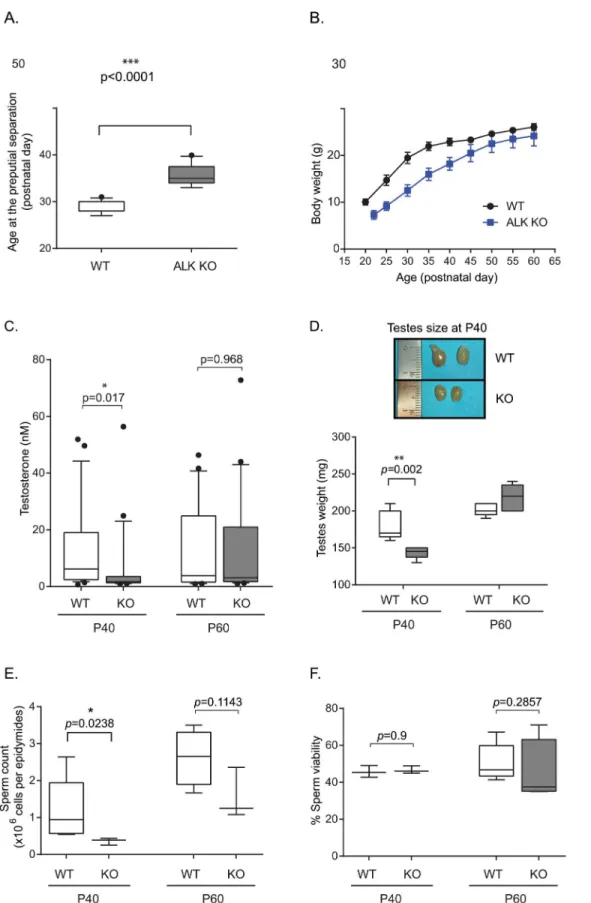
ALK KO male mice exhibit hypogonadotropic hypogonadism
both 40 and 60 days after birth (P40, P60) in blood samples collected from wild type (n = 26 per age) and homozygote ALK mutant male mice (n = 24 per age).
In contrast to wild type mice at P40, ALK kinase KO male mice showed significantly lower levels of serum testosterone (p-value = 0.017). At P60 we were unable to detect significant
dif-ferences between serum samples from wild type and ALK kinase KO males (Fig 2C). This de-crease in serum testosterone levels in ALK mutant males at P40 supports our observation that ALK mutant mice exhibit hypogonadotropic hypogonadism as compared to their wild type lit-termates. Testicular weights and sizes were also determined at P40 and P60, as testicular weight increases at the time of puberty. At P40, the testicular weights of wild type mice were signifi-cantly greater than those of ALK kinase KO mice (180 mg vs 143 mg,P= 0.002;Fig 2D). This
is associated with bigger testes size in wild type mice compared with ALK mutant mice. This difference disappears by P60, when the testicular weights of ALK mutant mice were no longer significantly different from those of wild type mice. Sperm counts, following sperm retrieval from the cauda epididymides, show a significant decline (p-value = 0.0238;Fig 2E) between
WT and ALK kinase KO mice at P40 (1.23x10⁶versus 0.36 x10⁶sperm/epididymides) but not
at P60 (p-value = 0.1143; 2.33x10⁶versus 1.56x10⁶sperm/epididymides). However, sperm
analyses parameters revealed no significant changes in sperm viability (Fig 2F) in ALK kinase KO mice compared to their corresponding controls at either P40 (p-value = 0.9) or P60 (p
-value = 0.2857).
To verify if decreased testosterone levels are associated with any abnormal testicular his-toarchitecture, we performed standard hematoxylin and eosin staining of paraffin sections of testes collected from ALK KO mice and wild type siblings at P40 and P60. As the number of Sertoli cells per seminiferous tubule depends on the stage of the epithelial cycle and the shape of the section, we ensured that the investigated tubule cross-sections were absolutely round and were not close to the turn of the tubule to allow proper quantification. Seminiferous tu-bules of wild type mice consist of well-organized layers of germ cells and Sertoli cells in the pe-ripheral layer. However, germ cells with irregularly arranged nuclei (Fig 3, green arrow) and with fewer Sertoli cells as visualized by GATA4 immunostaining within the seminiferous tu-bules at P40 (Fig 3A, insets) were observed in the ALK KO mouse testes. Reduced nuclear hyperchromasia, especially prominent at the outer cell layers, coupled with disturbed cell po-larity and cell shrinkage were other striking features. Intracytoplasmic vacuoles coupled with apoptotic bodies were major cytological abnormalities. These vacuoles appeared abundantly at P40 and occasionally at P60 within the tubules of the ALK KO mice testis and were rarely ob-served in wild type testes at P40 (Fig 3A, black arrows). The presence of vacuoles and apoptotic bodies may be indicative of degenerative changes and/or disturbed cell maturation. We further performed a GATA4-positive cell count and showed that the number of GATA4-expressing Sertoli cells within the seminiferous tubules in ALK KO mice was significantly lower than that in the wild type animals examined at P40 (P= 0.0002) but not at P60 (P= 0.6547;Fig 3B).
Taken together, these data suggest that ALK KO mice are delayed in their sexual maturation, due in part to decreased testosterone levels and mild testicular disorganization.
P60. Box plots indicate the median (black line), 25th and 75th percentiles (borders of boxes), 10th and 90th percentiles (whiskers) and the outliers (dots) from 20–25 mice per group.*P<0.05 indicates significant difference. (D) Testicular weight (n = 5 per postnatal day and genotype), as well as representative testicular size in wild type mice compared with ALK KO mice is shown. (E) Number of cauda epididymidis-retrieved spermatozoa per epididymis (n = 4 per genotype at P40, n = 6 at P60 for WT animals and n = 4 at P60 for ALK mutant mice). (F) Viability percentage of cauda epididymidis-retrieved spermatozoa (n = 4 per genotype and per age).
Fig 3. Abnormalities in seminiferous tubules from ALK kinase KO testes.(A) Histological morphology of testes stained with standard H & E. Note the irregular arrangement of germ cells at the periphery (green arrow) of the seminiferous tubule and the increased presence of vacuoles (black arrow) in ALK kinase KO mice. Fewer GATA4-expressing cells within the seminiferous tubules are observed at P40 in ALK kinase KO mice as compared with wild type controls. This decrease is less obvious at P60 (insets). (B) Quantification of age-dependent loss of GATA4 immunoreactivity in the Sertoli cells within the seminiferous tubules of wild type and ALK kinase KO mice. N = 10 mice per age and per genotype.
Inhibition of ALK reversibly decreases testosterone level in male mice
These observations are interesting given recent reports of the effects of crizotinib on male NSCLC patients in which a rapid suppression of testosterone levels was observed in men under crizotinib treatment [17]. Our findings in ALK KO mice prompted us to examine whether a similar phenomenon could be observed in wild type mice in response to crizotinib. To test this hypothesis, 26 adult male mice were randomized in two groups: treated and control. Mice aged from 7 to 9 weeks old were treated daily with 20 mg/kg crizotinib or vehicle for two weeks. Three independent blood samples were collected from each group: one prior to treatment, a second directly after the end of treatment and a final sample 5 weeks post-treatment and ana-lyzed for serum testosterone levels (Fig 4). The results show significantly decreased testosterone levels in mice after crizotinib treatment. However, this decrease was reversible, since 5 weeks after crizotinib cessation testosterone levels were once again comparable to normal control lev-els. No significant changes in testosterone levels were observed in control group mice. The ob-served changes in testosterone levels display a rapid onset, returning to normal upontermination of treatment, confirming a direct causal relationship with crizotinib administra-tion. Thus, treatment of mice with crizotinib results in lower levels of serum testosterone, sug-gesting that crizotinib treatment abrogates testosterone production in male mice.
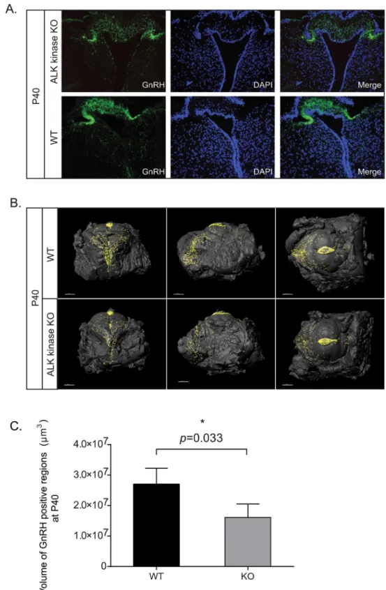
Decreased levels of gonadotropin releasing hormone (GnRH) in ALK KO
mice
It has been postulated that the crizotinib induced reduction of testosterone levels in human males may be due to hypothalamic or pituitary effects in response to treatment with crizotinib [17]. In order to test this, expression of GnRH positive neurons in ALK KO was examined. Lev-els of GnRH in ALK KO hypothalamic slices were lower than those observed in control mice (Fig 5A). To examine this more carefully, hypothalami collected from wild type and ALK KO mice at P40 and P60 was evaluated by optical projection tomography (OPT), allowing direct quantification and three dimensional–spatial assessments of GnRH expressing cells. A clear re-duction in the volume of GnRH positive cells was observed in ALK KO mice at P40 when com-pared with wild type mice; such reduction in GnRH expression was not observed at P60 (Fig 5B, quantified in 5C;S1 Fig, quantified in S1). Taken together, examination of hypothalamic GnRH levels by both convention confocal microscopy and OPT analysis indicates a role for ALK in the modulation of the hypothalamic-pituitary-gonadal axis affecting hypogonadotropic hypogonadism.
Discussion
to be viable but also displayed delayed breeding. This observation was strengthened by the findings that ALK kinase KO male mice exhibit significantly lower serum testosterone levels at P40 when compared with control litter mates. Interestingly, these decreased testosterone levels are accompanied by mild histoarchitectural changes in the testes from these mice, including appearance of vacuolated nuclei in seminiferous tubules, decreased numbers of Sertoli cells and irregularly distributed germ cells. These findings were further strengthened by the finding that ALK kinase KO mice have decreased number of GATA4-expressing cells within the semi-niferous tubules. GATA4 is expressed in Sertoli and Leydig cells and plays an important role in-testes development and also in Sertoli cell function in adult mice [38]. Indeed, adult mice lacking GATA4 in the testes also exhibit vacuoles in the seminiferous tubules [38]. Examina-tion of sperm in ALK kinase KO mice reveals a significant decrease in sperm count, although viability was not affected. Given the reduced testosterone levels and the decreased sperm count in ALK kinase KO mice, it would appear that loss of ALK activity may lead to suboptimal testes function. While we noted clear differences between ALK kinase KO and wild type mice at P40, it would be of interest to examine this further at earlier time points in future studies as the as-sessment of puberty onset by preputial separation indicates that the puberty, while delayed in ALK kinase KO mice, occurs between P35-40.
Fig 4. Crizotinib treatment in mice leads to reduction of serum testosterone levels.Adult male mice were treated with crizotinib daily (at 20 mg/kg). Blood samples were collected prior to crizotinib treatment, one day after the last treatment and five weeks after completion of treatment and analyzed for testosterone. Box plots indicate the median (black line), 25th and 75th percentiles (borders of boxes), 10th and 90th percentiles (whiskers) and the outliers (dots) from 13 mice per group.*P<0.05 indicates significant difference.
Fig 5. ALK knockout mice display reduced GnRH expression.(A) Confocal analysis of GnRH expression in P40 ALK kinase KO. GnRH (green), DAPI (blue). (B) Expression of hypothalamic GnRH at P40 in ALK kinase knockout males. Reconstituted OPT images of representative hypothalami are shown in anterior, lateral and ventral views. GnRH positive regions are presented as (iso-surface rendered) black dots on the iso-surfaces of dissected hypothalami. (C) Quantitation of GnRH positive regions from P40 hypothalami. Values represent the average volumes of GnRH stained regions in hypothalami, 3–4 mice per group.
To further examine this we employed ALK kinase inhibitors, in this case the ATP analogue crizotinib [14], to modulate ALK activity in wild type adult mice. As would be predicted from our findings in the ALK kinase KO animals, treatment with crizotinib resulted in reduction in serum testosterone levels. Some depression of testosterone levels was also observed in control mice after feeding with vehicle, which may be due to use of anesthetic during administration or animal handling[39]. Furthermore, these effects were reversible, with serum testosterone re-turning to normal levels upon cessation of crizotinib treatment. These results are interesting in light of a recent report of rapid-onset hypogonadism secondary to crizotinib treatment in male patients treated for ALK positive non-small cell lung cancer [17]. Thus, while crizotinib is not absolutely specific for ALK among the tyrosine kinase family and targets other than ALK must be considered in these patients, our findings in ALK KO mice suggest that ALK inhibition in patients may result in modulation of testosterone levels.
These findings also have implications for patients being treated with other ALK inhibitors, several of which are now in clinical use [40], including the recently FDA approved ceritinib (LDK378) [41,42]. A young male ALK positive NSCLC patient treated with crizotinib, and subsequently with ceritinib, is of particular interest. This patient displayed reduced plasma tes-tosterone levels which were treated effectively with testes-tosterone supplement. He later became crizotinib resistant and continued ALK inhibitor treatment, in this case with ceritinib, but con-tinues to require testosterone supplement (Helland, Å., personal communication). Since ALK positive lung cancer patients are generally younger and fitter than the majority of lung cancer patients, low levels of testosterone can induce fatigue and reduced libido and impact on quality of life. For patients living with cancer as a chronic disease, quality of life is extremely important, thus clinicians following these patients should be aware of this potential effect and more knowledge is required concerning ALK inhibition in this patient group. Other relevant patient groups are young neuroblastoma patients, as well as other pediatric cancer patients with ALK driven cancers, that may take ALK inhibitors on a long-term basis [43]. Furthermore, informa-tion on testosterone levels in patients taking ALK inhibitors targeting other alterainforma-tions in can-cer, for example MET and ROS, should also be gathered.
While ALK is expressed in testes [11], the decrease of FSH and LH in patients treated with crizotinib suggest that decrease of testosterone may be centrally mediated [17]. LH and FSH are produced by the anterior pituitary in response to GnRH secreted by neurons in the hypo-thalamus into the portal blood system [44]. Our initial examination of GnRH levels in the hy-pothalamus of ALK kinase KO mice identified decreased levels of GnRH in ALK mutant mice. More accurate Optical Projection Tomography analysis allowed quantification and confirmed significantly lower GnRH expression levels in the hypothalamus of ALK kinase KO males at P40 as compared with control littermates. These findings support the hypothesis that downre-gulation of testosterone level either by chemical or genetic depletion of ALK is at least in part initiated upstream in the hypothalamic pituitary gonadal axis. Thus our mouse ALK knockout model displays hypogonadotropic hypogonadism. A number of additionally interesting regula-tors of hypogonadotropic hypogonadism exist, including e.g. the kisspeptins [45]. Kisspeptins bind to the GPR54 receptor on the GnRH-neurons regulating the pulsing of GnRH secretion [46]. While we have not investigated Kisspeptin in this study, it will be interesting to study them further in the context of ALK function.
Supporting Information
S1 Fig. OPT analysis of hypothalamic GnRH at P60.Expression of hypothalamic GnRH at P60 in ALK kinase KO analyzed by OPT. GnRH positive regions are presented as black dots on the iso-surfaces of dissected hypothalami. Quantitation of GnRH positive regions from P60 hypothalami. Values represent the average volumes of GnRH stained regions in hypothalami. P<0.05 indicates significant difference.
(PDF)
Acknowledgments
Work in the author’s laboratories has been supported by grants from the Swedish Cancer Soci-ety (BH 12–0722, RHP 12–0796), the Children’s Cancer Foundation (BH 11/020, RHP 13/ 0049), the Swedish Research Council (RHP 621-2011-5181, BH 521-2012-2831), Lions Cancer Society, Umeå (BH and RHP LP 12–1946), the JC Kempe Foundation (BH and RHP) and FP-People-2011-IAPP (BH).
Author Contributions
Conceived and designed the experiments: BW AE CN UA ME EVL BH RHP. Performed the experiments: BW AE CN ME EVL. Analyzed the data: BW AE CN UA ME EVL OAA BH RHP. Contributed reagents/materials/analysis tools: UA BH RHP. Wrote the paper: BW AE CN UA ME EVL AH OAA BH RHP.
References
1. Morris SW, Kirstein MN, Valentine MB, Dittmer KG, Shapiro DN, Saltman DL, et al. Fusion of a kinase gene, ALK, to a nucleolar protein gene, NPM, in non-Hodgkin's lymphoma. Science. 1994; 263 (5151):1281–4. PMID:8122112.
2. Iwahara T, Fujimoto J, Wen D, Cupples R, Bucay N, Arakawa T, et al. Molecular characterization of ALK, a receptor tyrosine kinase expressed specifically in the nervous system. Oncogene. 1997; 14 (4):439–49. PMID:9053841.
3. Morris SW, Naeve C, Mathew P, James PL, Kirstein MN, Cui X, et al. ALK, the chromosome 2 gene locus altered by the t(2;5) in non-Hodgkin's lymphoma, encodes a novel neural receptor tyrosine kinase that is highly related to leukocyte tyrosine kinase (LTK). Oncogene. 1997; 14(18):2175–88. PMID:
9174053.
4. Hallberg B, Palmer RH. Mechanistic insight into ALK receptor tyrosine kinase in human cancer biology. Nat Rev Cancer. 2013; 13(10):685–700. Epub 2013/09/26. doi:10.1038/nrc3580nrc3580[pii]. PMID:
24060861.
5. Lemmon MA, Schlessinger J. Cell signaling by receptor tyrosine kinases. Cell. 2010; 141(7):1117–34. Epub 2010/07/07. doi:10.1016/j.cell.2010.06.011S0092-8674(10)00665-3[pii]. PMID:20602996; PubMed Central PMCID: PMC2914105.
6. Caren H, Abel F, Kogner P, Martinsson T. High incidence of DNA mutations and gene amplifications of the ALK gene in advanced sporadic neuroblastoma tumours. Biochem J. 2008; 416(2):153–9. PMID:
18990089.
7. Mosse YP, Laudenslager M, Longo L, Cole KA, Wood A, Attiyeh EF, et al. Identification of ALK as a major familial neuroblastoma predisposition gene. Nature. 2008; 455(7215):930–5. PMID:18724359. doi:10.1038/nature07261
8. George RE, Sanda T, Hanna M, Frohling S, Luther W 2nd, Zhang J, et al. Activating mutations in ALK provide a therapeutic target in neuroblastoma. Nature. 2008; 455(7215):975–8. PMID:18923525. doi:
10.1038/nature07397
9. Chen Y, Takita J, Choi YL, Kato M, Ohira M, Sanada M, et al. Oncogenic mutations of ALK kinase in neuroblastoma. Nature. 2008; 455(7215):971–4. PMID:18923524. doi:10.1038/nature07399 10. Janoueix-Lerosey I, Lequin D, Brugieres L, Ribeiro A, de Pontual L, Combaret V, et al. Somatic and
11. Vernersson E, Khoo NK, Henriksson ML, Roos G, Palmer RH, Hallberg B. Characterization of the ex-pression of the ALK receptor tyrosine kinase in mice. Gene Expr Patterns. 2006; 6(5):448–61. PMID:
16458083.
12. Bilsland JG, Wheeldon A, Mead A, Znamenskiy P, Almond S, Waters KA, et al. Behavioral and neuro-chemical alterations in mice deficient in anaplastic lymphoma kinase suggest therapeutic potential for psychiatric indications. Neuropsychopharmacology. 2008; 33(3):685–700. Epub 2007/05/10. doi: 1301446 [pii] doi:10.1038/sj.npp.1301446PMID:17487225.
13. Weiss JB, Xue C, Benice T, Xue L, Morris SW, Raber J. Anaplastic Lymphoma Kinase and Leukocyte Tyrosine Kinase: Functions and genetic interactions in learning, memory and adult neurogenesis. Phar-macol Biochem Behav. 2012; 100(3):566–74. Epub 2011/11/15. doi: S0091-3057(11)00353-4 [pii] doi:
10.1016/j.pbb.2011.10.024PMID:22079349.
14. Christensen JG, Zou HY, Arango ME, Li Q, Lee JH, McDonnell SR, et al. Cytoreductive antitumor activi-ty of PF-2341066, a novel inhibitor of anaplastic lymphoma kinase and c-Met, in experimental models of anaplastic large-cell lymphoma. Molecular cancer therapeutics. 2007; 6(12 Pt 1):3314–22. PMID:
18089725.
15. Kwak EL, Bang YJ, Camidge DR, Shaw AT, Solomon B, Maki RG, et al. Anaplastic lymphoma kinase inhibition in non-small-cell lung cancer. The New England journal of medicine. 2010; 363(18):1693– 703. Epub 2010/10/29. doi:10.1056/NEJMoa1006448PMID:20979469.
16. Ou SH, Bartlett CH, Mino-Kenudson M, Cui J, Iafrate AJ. Crizotinib for the treatment of ALK-rearranged non-small cell lung cancer: a success story to usher in the second decade of molecular targeted thera-py in oncology. Oncologist. 2012; 17(11):1351–75. Epub 2012/09/20. doi:
10.1634/theoncologist.2012-0311theoncologist.2012-0311[pii]. PMID:22989574; PubMed Central PMCID: PMC3500356. 17. Weickhardt AJ, Rothman MS, Salian-Mehta S, Kiseljak-Vassiliades K, Oton AB, Doebele RC, et al.
Rapid-onset hypogonadism secondary to crizotinib use in men with metastatic nonsmall cell lung can-cer. Cancan-cer. 2012; 118(21):5302–9. Epub 2012/04/11. doi:10.1002/cncr.27450PMID:22488744. 18. Tobet SA, Bless EP, Schwarting GA. Developmental aspect of the gonadotropin-releasing hormone
system. Mol Cell Endocrinol. 2001; 185(1–2):173–84. Epub 2001/12/12. doi: S0303720701006165 [pii]. PMID:11738807.
19. Wray S. Development of luteinizing hormone releasing hormone neurones. J Neuroendocrinol. 2001; 13(1):3–11. Epub 2000/12/21. doi: jne609 [pii]. PMID:11123510.
20. Wray S. Molecular mechanisms for migration of placodally derived GnRH neurons. Chem Senses. 2002; 27(6):569–72. Epub 2002/07/27. PMID:12142333.
21. Wray S, Grant P, Gainer H. Evidence that cells expressing luteinizing hormone-releasing hormone mRNA in the mouse are derived from progenitor cells in the olfactory placode. Proc Natl Acad Sci U S A. 1989; 86(20):8132–6. Epub 1989/10/01. PMID:2682637; PubMed Central PMCID: PMC298229. 22. Wray S, Nieburgs A, Elkabes S. Spatiotemporal cell expression of luteinizing hormone-releasing
hor-mone in the prenatal mouse: evidence for an embryonic origin in the olfactory placode. Brain research. 1989; 46(2):309–18. Epub 1989/04/01. PMID:2655994.
23. Charlton H. Neural transplantation in hypogonadal (hpg) mice—physiology and neurobiology. Repro-duction. 2004; 127(1):3–12. Epub 2004/04/02. doi:10.1530/rep.1.00066127/1/3[pii]. PMID:15056765. 24. Korenbrot CC, Huhtaniemi IT, Weiner RI. Preputial separation as an external sign of pubertal
develop-ment in the male rat. Biol Reprod. 1977; 17(2):298–303. PMID:889997.
25. Zaneveld LJ, Polakosk KL. Collection and physical examination of the ejaculate in techniques of human andrology. North Holland Biomedical Press, Amsterdam. 1977;Ed. Hafez ESE.: 147–56. 26. Golde WT, Gollobin P, Rodriguez LL. A rapid, simple, and humane method for submandibular bleeding
of mice using a lancet. Lab Anim (NY). 2005; 34(9):39–43. Epub 2005/10/01. doi: laban1005-39 [pii] doi:10.1038/laban1005-39PMID:16195737.
27. Anjamrooz SH, Movahedin M, Mowla SJ, Bairanvand SP. Assessment of morphological and functional changes in the mouse testis and epididymal sperms following busulfan treatment. Iranian biomedical journal. 2007; 11(1):15–22. PMID:18051700.
28. Cheng Y, Buffone MG, Kouadio M, Goodheart M, Page DC, Gerton GL, et al. Abnormal sperm in mice lacking the Taf7l gene. Mol Cell Biol. 2007; 27(7):2582–9. doi:10.1128/MCB.01722-06PMID:
17242199; PubMed Central PMCID: PMC1899882.
29. Sharpe J, Ahlgren U, Perry P, Hill B, Ross A, Hecksher-Sorensen J, et al. Optical projection tomogra-phy as a tool for 3D microscopy and gene expression studies. Science. 2002; 296(5567):541–5. Epub 2002/04/20. doi:10.1126/science.1068206296/5567/541[pii]. PMID:11964482.
31. Eriksson AU, Svensson C, Hornblad A, Cheddad A, Kostromina E, Eriksson M, et al. Near infrared opti-cal projection tomography for assessments of beta-cell mass distribution in diabetes research. J Vis Exp. 2013;(71: ):e50238. Epub 2013/01/29. doi:10.3791/5023850238[pii]. PMID:23353681; PubMed Central PMCID: PMC3582649.
32. Cheddad A, Svensson C, Sharpe J, Georgsson F, Ahlgren U. Image processing assisted algorithms for optical projection tomography. IEEE Trans Med Imaging. 2012; 31(1):1–15. Epub 2011/07/20. doi:10.
1109/TMI.2011.2161590PMID:21768046.
33. Hornblad A, Cheddad A, Ahlgren U. An improved protocol for optical projection tomography imaging re-veals lobular heterogeneities in pancreatic islet and beta-cell mass distribution. Islets. 2011; 3(4):204– 8. Epub 2011/06/03. doi: 16417 [pii]. PMID:21633198; PubMed Central PMCID: PMC3154448. 34. Frisch RE, Hegsted DM, Yoshinaga K. Body weight and food intake at early estrus of rats on a high-fat
diet. Proc Natl Acad Sci U S A. 1975; 72(10):4172–6. PMID:1060097; PubMed Central PMCID: PMC433162.
35. Huhtaniemi I. Mutations along the pituitary-gonadal axis affecting sexual maturation: novel information from transgenic and knockout mice. Mol Cell Endocrinol. 2006; 254–255:84–90. Epub 2006/05/30. doi: S0303-7207(06)00214-0 [pii] doi:10.1016/j.mce.2006.04.015PMID:16730882.
36. Ebling FJ, Cronin AS. The neurobiology of reproductive development. Neuroreport. 2000; 11(16):R23– 33. Epub 2000/11/30. PMID:11095489.
37. Lasek AW, Lim J, Kliethermes CL, Berger KH, Joslyn G, Brush G, et al. An evolutionary conserved role for anaplastic lymphoma kinase in behavioral responses to ethanol. PLoS One. 2011; 6(7):e22636. Epub 2011/07/30. doi:10.1371/journal.pone.0022636PONE-D-10-06421 [pii]. PMID:21799923; PubMed Central PMCID: PMC3142173.
38. Kyronlahti A, Euler R, Bielinska M, Schoeller EL, Moley KH, Toppari J, et al. GATA4 regulates Sertoli cell function and fertility in adult male mice. Mol Cell Endocrinol. 2011; 333(1):85–95. Epub 2010/12/22. doi:10.1016/j.mce.2010.12.019S0303-7207(10)00598-8[pii]. PMID:21172404; PubMed Central PMCID: PMC3026658.
39. Arantes-Rodrigues R, Henriques A, Pinto-Leite R, Faustino-Rocha A, Pinho-Oliveira J, Teixeira-Guedes C, et al. The effects of repeated oral gavage on the health of male CD-1 mice. Lab Anim (NY). 2012; 41(5):129–34. doi:10.1038/laban0512-129PMID:22517091.
40. Awad MM, Shaw AT. ALK inhibitors in non-small cell lung cancer: crizotinib and beyond. Clin Adv Hematol Oncol. 2014; 12(7):429–39. PMID:25322323; PubMed Central PMCID: PMC4215402. 41. Galkin AV, Melnick JS, Kim S, Hood TL, Li N, Li L, et al. Identification of NVP-TAE684, a potent,
selec-tive, and efficacious inhibitor of NPM-ALK. Proc Natl Acad Sci U S A. 2007; 104(1):270–5. PMID:
17185414.
42. Friboulet L, Li N, Katayama R, Lee CC, Gainor JF, Crystal AS, et al. The ALK inhibitor ceritinib over-comes crizotinib resistance in non-small cell lung cancer. Cancer Discov. 2014; 4(6):662–73. doi:10.
1158/2159-8290.CD-13-0846PMID:24675041; PubMed Central PMCID: PMC4068971.
43. Mosse YP, Lim MS, Voss SD, Wilner K, Ruffner K, Laliberte J, et al. Safety and activity of crizotinib for paediatric patients with refractory solid tumours or anaplastic large-cell lymphoma: a Children's Oncolo-gy Group phase 1 consortium study. Lancet Oncol. 2013; 14(6):472–80. Epub 2013/04/20. doi:10.
1016/S1470-2045(13)70095-0S1470-2045(13)70095-0[pii]. PMID:23598171.
44. Thompson IR, Kaiser UB. GnRH pulse frequency-dependent differential regulation of LH and FSH gene expression. Mol Cell Endocrinol. 2013. Epub 2013/09/24. doi: S0303-7207(13)00374-2 [pii] doi:
10.1016/j.mce.2013.09.012PMID:24056171.
45. de Roux N, Genin E, Carel JC, Matsuda F, Chaussain JL, Milgrom E. Hypogonadotropic hypogonadism due to loss of function of the KiSS1-derived peptide receptor GPR54. Proc Natl Acad Sci U S A. 2003; 100(19):10972–6. Epub 2003/08/29. doi:10.1073/pnas.18343991001834399100[pii]. PMID:
12944565; PubMed Central PMCID: PMC196911.
46. Irwig MS, Fraley GS, Smith JT, Acohido BV, Popa SM, Cunningham MJ, et al. Kisspeptin activation of gonadotropin releasing hormone neurons and regulation of KiSS-1 mRNA in the male rat. Neuroendo-crinology. 2004; 80(4):264–72. Epub 2005/01/25. doi: 83140 [pii] doi:10.1159/000083140PMID: