Identification of Differentially Expressed Proteins from
Leishmania amazonensis
Associated with the Loss of
Virulence of the Parasites
Rubens D. M. Magalha˜es1, Mariana C. Duarte2, Eliciane C. Mattos3, Vivian T. Martins1, Paula S. Lage2,
Miguel A. Cha´vez-Fumagalli2, Daniela P. Lage4, Daniel Menezes-Souza5, Wiliam C. B. Re´gis6,
Maria J. Manso Alves3, Manuel Soto7, Carlos A. P. Tavares1, Ronaldo A. P. Nagen1,
Eduardo A. F. Coelho2,4*
1Departamento de Bioquı´mica e Imunologia, Instituto de Cieˆncias Biolo´gicas, Universidade Federal de Minas Gerais, Belo Horizonte, Minas Gerais, Brazil,2Programa de Po´s-Graduac¸a˜o em Cieˆncias Sau´de: Infectologia e Medicina Tropical, Faculdade de Medicina, Universidade Federal de Minas Gerais, Belo Horizonte, Minas Gerais, Brazil, 3Departamento de Bioquı´mica, Instituto de Quı´mica, Universidade de Sa˜o Paulo, Sa˜o Paulo, Sa˜o Paulo, Brazil,4Departamento de Patologia Clı´nica, COLTEC, Universidade Federal de Minas Gerais, Belo Horizonte, Minas Gerais, Brazil,5Departamento de Parasitologia, Instituto de Cieˆncias Biolo´gicas, Universidade Federal de Minas Gerais, Belo Horizonte, Minas Gerais, Brazil,6Departamento de Bioquı´mica, PUC Minas, Belo Horizonte, Minas Gerais, Brazil,7Centro de Biologı´a Molecular Severo Ochoa (CSIC-UAM), Departamento de Biologı´a Molecular, Universidad Auto´noma de Madrid, Madrid, Spain
Abstract
Background:The present study analyzed whether or not thein vitrocultivation for long periods of time of pre-isolated Leishmania amazonensisfrom lesions of chronically infected BALB/c mice was able to interfere in the parasites’ infectivity usingin vivoand in vitroexperiments. In addition, the proteins that presented a significant decrease or increase in their protein expression content were identified applying a proteomic approach.
Methodology/Principal Findings:Parasites were cultured in vitrofor 150 days. Aliquots were collected on the day 0 of culture (R0), as well as after ten (R10; 50 days of culture), twenty (R20; 100 days of culture), and thirty (R30; 150 days of culture) passages, and were used to analyze the parasites’in vitroandin vivoinfectivity, as well as to perform the proteomic approach. Approximately 837, 967, 935, and 872 spots were found in 2-DE gels prepared from R0, R10, R20, and R30 samples, respectively. A total of 37 spots presented a significant decrease in their intensity of expression, whereas a significant increase in protein content during cultivation could be observed for 19 proteins (both cases.2.0 folds). Some of these identified proteins can be described, such as diagnosis and/or vaccine candidates, while others are involved in the infectivity of Leishmania. It is interesting to note that six proteins, considered hypothetical in Leishmania, showed a significant decrease in their expression and were also identified.
Conclusions/Significance:The present study contributes to the understanding that the cultivation of parasites over long periods of time may well be related to the possible loss of infectivity of L. amazonensis. The identified proteins that presented a significant decrease in their expression during cultivation, including the hypothetical, may also be related to this loss of parasites’ infectivity, and applied in future studies, including vaccine candidates and/or immunotherapeutic targets against leishmaniasis.
Citation:Magalha˜es RDM, Duarte MC, Mattos EC, Martins VT, Lage PS, et al. (2014) Identification of Differentially Expressed Proteins fromLeishmania amazonensis
Associated with the Loss of Virulence of the Parasites. PLoS Negl Trop Dis 8(4): e2764. doi:10.1371/journal.pntd.0002764
Editor:Diane McMahon-Pratt, Yale School of Public Health, United States of America ReceivedSeptember 20, 2013;AcceptedFebruary 16, 2014;PublishedApril 3, 2014
Copyright:ß2014 Magalha˜es et al. This is an open-access article distributed under the terms of the Creative Commons Attribution License, which permits unrestricted use, distribution, and reproduction in any medium, provided the original author and source are credited.
Funding:This work was supported by grants from Pro´-Reitoria de Pesquisa of UFMG (Edital 03/2013), Instituto Nacional de Cieˆncia e Tecnologia em Nano-Biofarmaceˆutica, FAPEMIG (CBB-APQ-00496-11 and CBB-APQ-00819-12), and CNPq (472090/2011-9 and 482976/2012-8). EAFC is a grant recipient of CNPq. MACF is a grant recipient of PNPD/CAPES. This study was also, in part, supported in Spain by grants from Ministerio de Ciencia e Innovacio´n FIS/PI1100095. The funders had no role in study design, data collection and analysis, decision to publish, or preparation of the manuscript.
Competing Interests:The authors have declared that no competing interests exist. * E-mail: [email protected]
Introduction
Leishmaniasis consists of a wide range of diseases present in 98 countries worldwide, where approximately 1.6 million cases occur each year, with an estimated 40,000 deaths [1]. Many geographic regions are endemic for multipleLeishmania species, which is the case in Brazil, where the disease is caused by at least six different species ofLeishmania. Among them,Leishmania amazonensispresents
a particular importance, as it is one of the main species capable of causing human disease with a broad spectrum of clinical manifestations, ranging from cutaneous to visceral leishmaniasis [2,3]. In one study, it was also observed that BALB/c mice experimentally infected withL. amazonensisdeveloped visceraliza-tion of the parasites in different organs, such as the brain, liver, spleen, and bone marrow, characterizing a diagnosis of murine visceral leishmaniasis [4].
It has been postulated that thein vitromaintenance of parasites by cultivation over long periods of time may well diminish their ability to differentiate into amastigote forms [5]. In fact, long-term axenic cultures were one of the first empirical approaches to efficiently identify parasite virulence genes, which later led to the experimental development of attenuated strains [6]. Similarly, the long-termin vitrogrowth of drug-resistant parasites was suggested to mediate the loss of resistance phenotype [7]. It is well-known that parasites can regulate their gene expression, mainly at the post-transcriptional level; however, little is known about the biological mechanisms and the protein expression involved in this process [8]. In this context, the identification of proteins involved either in the infectivity of parasites in the mammal hosts, or in their maintenance in axenic cultures, should be considered relevant.
The proteomic study applied to evaluate the protein expression patterns in Leishmaniaoffers the possibility of assigning potential functions for proteins, including those previously identified by genomics as hypothetical, which should be evaluated, such as vaccine candidates, diagnostic markers, and/or immunotherapeu-tic targets. Several studies have been published evaluating the stage-specific expression and differentiation profiles of proteins in different Leishmania species [9,10,11,12,13,14]. In addition, the discovery of new proteins through proteomics has been recom-mended as one of the main research priorities for further development and improvement of leishmaniasis vaccines [15].
In this context, the identification of proteins involved in parasites’ infectivity should be considered important, given that they could be used in immunological applications to prevent the disease. In the present study, a proteomic approach, based on two-dimensional electrophoresis (2-DE) and mass spectrometry, was carried out to analyze the variation of protein expression profiles in stationary promastigotes of L. amazonensis, which were pre-isolated from lesions of chronically infected BALB/c mice and maintained in axenic cultures over a long period of time. The proteins that presented significant variations in their levels during thein vitrocultivation were identified in an attempt to select new
vaccine candidates and/or immunotherapeutic targets against leishmaniasis. The results showed several known, as well as six hypothetical, L. amazonensis proteins, some of which are well-known proteins involved in the infectivity of Leishmania, while others are described through the metabolic functions of the parasites.
Materials and Methods
Ethics statement
Experiments were performed in compliance with the National Guidelines of the Institutional Animal Care and Use Committee for the Ethical Handling of Research Animals (CEUA) from the Federal University of Minas Gerais (UFMG) (Law number 11.794, 2008), which approved this study on April 25, 2012, under protocol number 092/2012.
Mice and parasites
Female BALB/c mice (8 weeks of age) were obtained from the breeding facilities of the Department of Biochemistry and Immunology, Institute of Biological Sciences, UFMG, and were maintained under specific pathogen-free conditions.L. amazonensis
(IFLA/BR/1967/PH-8) parasites were grown at 24uC in complete Schneider’s medium , supplemented with 20% heat-inactivated fetal bovine serum (FBS), 20 mM L-glutamine, 200 U/ml penicillin, 100mg/ml streptomycin, and 50mg/ml gentamicin, at
pH 7.4. The amastigote-like cells were obtained as described in [16].
In vivoinfection
BALB/c mice (n = 8) were infected subcutaneously in their hind footpad with 16106stationary promastigotes ofL. amazonensis. The course of the disease was monitored at weekly intervals by measuring footpad thickness with a metric caliper, and expressed as the increase in thickness of the infected footpad compared to the non-infected footpad. At week 8 post-infection, animals were sacrificed and their infected footpads, spleen, and liver were harvested for parasite quantification by a limiting-dilution assay [17]. To evaluate thein vivoinfectivity of parasites in the different collected passages, R0 and R30 samples were used to infect BALB/c mice (n = 8, each group). The infection schedule and the parasitological analyses were the same as described above.
Preparation of the parasites for proteomics
Parasites were collected from infected footpads of the animals (8 weeks after infection) and purified to perform the proteomic approach. For this, parasites recovered from lesions were homogenized and immediately washed in Schneider’s medium, which was supplemented with 10% FBS and 1% penicillin G/ streptomycin sulfate solution, and subsequently cultured in complete Schneider’s medium. Passages ofin vitro cultures were performed every five days, until the thirtieth passage (150 days after). Aliquots were collected on day 0 of culture (R0, first passage), as well as 50 (R10), 100 (R20) and 150 (R30) days after the beginning of the cultures, and quantified for the experiments.
Evaluation ofin vitroinfectivity
Aliquots containing parasites of R0, R10, R20, and R30 passages were centrifuged for 10 min and 5,0006g, at 4uC. The
supernatant was removed, and the pellet containing the parasites was washed 3 times with sterile PBS. Murine macrophages collected from BALB/c mice were plated on round glass coverslips within the wells of a 24-well culture plate, at a concentration of 56105 cells per coverslip in RPMI 1640 medium, which was
Author Summary
Leishmania amazonensiscan induce a diversity of clinical manifestations in mammal hosts, including tegumentary and visceral leishmaniasis. The present study evaluated the variation of infectivity of L. amazonensis, which was pre-isolated from lesions of chronically infected mice and in vitrocultured for 150 days, in turn connecting these results with the profile of parasite protein expression using a proteomic approach. Parasites were recovered after the first passage, as well as after 50, 100, and 150 days of axenic cultures, and were subsequently evaluated. A total of 37 proteins presented a significant decrease, whereas 19 proteins presented a significant increase in their protein expression content in the assays (both cases .2.0 fold). Some of the identified proteins have been reported in prior literature, including diagnosis and/or vaccine candi-dates for leishmaniasis, while others proved to be involved in the infectivity ofLeishmania. It is interesting to note that proteins related to the parasites’ metabolism were also the majority of the proteins identified in the old cultures ofL. amazonensis, suggesting a possible relation between the metabolic state of parasites and their possible loss of infectivity. In conclusion, the proteins identified in this study represent a contribution to the discovery of new vaccine candidates and/or immunotherapeutic targets against leishmaniasis.
Proteomic Approach Applied toLeishmania amazonensis
supplemented with 20% FBS, 2 mM L-glutamine, 200 U/mL penicillin G, and 100mg/mL streptomycin sulfate, at pH 7.4. After 2 h of incubation at 37uC in 5% CO2, stationary promastigotes ofL. amazonensiswere quantified and added to the wells (16106and 56106, for a ratio of 1:2 or 1:10 macrophage per parasites, respectively). The cultures were incubated for 24 h at 37uC in 5% CO2. Next, the cells were washed and stained to determine the percentages of infected macrophages and the number of intra-macrophage amastigotes by counting 200 cells in triplicate [18]. An optical microscopy was also used to check the stationary profile of allin vitrocultures, and a prior titration curve was performed to determine the best time of infection for the macrophages (data not shown).
Preparation of total extract ofLeishmania
The total extraction of proteins ofL. amazonensiswas performed following a technical protocol [19]. Briefly, 26108 stationary promastigotes were dissolved in a DeStreak rehydratation solution, containing phosphatases (5 mM NaF, 2 mM Na3VO4, and 50 mM Nab-glycerophosphate) and proteases (Protease Inhibitor Cocktail; plus 1 mM PMSF) inhibitors. After homogenization, samples were disrupted by sonication in an ice bath for 15 min by applying a continuous pulse and centrifuged at 20,0006g for
7 min, at 4uC. The supernatant was collected, and the protein concentration was estimated using the Bradford method [20]. Aliquots were immediately frozen at280uC, until use. For each passage, cellular material was extracted from the parasites harvested from two different animals, and two independent culture bottles of each animal were grown separately, totaling four individual samples.
Isoelectric focusing
The isoelectric focalization (IEF) was performed using the Ettan IPGphor3 system. For the first-dimension electrophoresis, 650mg of total extracts were added to a volume of 250mL with a rehydration solution containing a DeStreak rehydratation solution in 1% immobilized pH gradient buffer (IPG-buffer, pH 4–7). Next, samples were applied to IPG strips (13 cm, pH 4–7; GE Healthcare) for passive rehydration for 18 h at room temperature. After gel rehydration, IEF was performed at 1,000 V for 800 V/h; 8,000 V for 11,500 V/h; holding at 8,000 V for 7,500 V/h.
SDS-PAGE
After IEF, each strip was incubated for 15 min with 1% dithiothreitol (DTT) in the equilibrium buffer [75 mM Tris-HCl buffer, pH 8.8; 6 M urea, 39% (v/v) glycerol, and 2% (w/v) SDS], followed by a second incubation step for 15 min in 2.5% iodoacetamide diluted in equilibrium buffer. IPG strips were washed with milli-Q water, transferred to a 12% polyacrilamide, and sealed with an agarose solution (0.5% agarose in running buffer, containing 25 mM Tris, 192 mM glycine, and 0.1% SDS, pH 8.3). The protein standard was purchased from BioRad (pre-stained SDS-PAGE broad range). Electrophoresis was performed using a SE 600 ruby standard dual cooled vertical unit system connected to a MultiTemp III cooling bath. Proteins were separated at 30 mA/gel.
Protein digestion, peptide extraction, and spot handling The 2-DE gels were stained with colloidal Coomassie Brilliant Blue G-250, following a defined technical procedure [21]. For image analysis, 16 stained gels were scanned using an ImageS-canner III. Analyses were carried out using ImageMaster 2D Platinum 7.0 software. This software identifies spots on a gel image
(300 dpi) by comparing the number of pixels in the background image to the number of pixels that make up the image of the spot itself. The spots present in the images are differentiated from other gels by determining the spot’s position through the manual insertion of image markers. The parameters used for spot detection included: minimal area of 5 pixels, with a smooth factor of 4 and a saliency of 80. The reference gel (higher number of spots) was used to match corresponding protein spots within different gels. The intensity volume of individual spots was normalized by the total intensity volume (value of the intensity volumes obtained from all spots in the same 2-DE gel) so as to remain relatively independent of variations due to protein loading and staining, performed by considering the total volume of all spots in the images. All of the spots selected by software were checked manually. The statistical test of analysis of variance (One-way ANOVA) was performed at a 1% statistical significance level (P,0.01) to determine the mean values of spot intensity for each passage (R0, R10, R20, and R30) in an attempt to determine the significant changes among the passages. Additionally, this study applied a cut-off of at least 2-fold of the core value of intensity of all spots selected by the program, which were the same in each passage. The obtained fold value was the number obtained by the ratio between the higher and lower core values of each spot’s passage. Spots that presented significant variations within the passages were manually excised and destained with a solution containing 50% methanol and 2.5% acetic acid. The proteins were reduced in 10 mM DTT and alkylated using 50 mM iodoacetamide. Limited protein enzymatic digestion was per-formed with 0.4 or 0.8mg of trypsin for larger spots. Excess protease was removed and replaced by 25 mM ammonium bicarbonate. Digestion was performed at 37uC for 18 h. Peptide extraction was performed twice for 15 min, using 30mL of a solution containing 50% acetonitrile and 5% formic acid. The digested samples were dried using a speed-vac.
Protein identification and database search
The identification of proteins was performed at the Mass Spectrometry Laboratory of the Brazilian Biosciences National Laboratory (LNBio, CNPEM/ABTLuS, Campinas, Sa˜o Paulo, Brazil). This procedure was conducted using an ESI-Quad-TOF apparatus attached to a UPLC system. The mass spectra were processed by the Protein Lynx V 2.1 program and analyzed by the MASCOT MS/MS Ion Search program (http://www. matrixscience.com). The following parameters were used for this analysis: enzyme, trypsin; allowing of up to 1 missed cleavage; fixed modification, carbamidomethyl (C); variable modification, oxidation (M); peptide tolerance, 60.1 Da; MS/MS tolerance, 60.1 Da; and a peptide charge of 1+, 2+, and 3+. The database
Leishmania(dated June 2012) was used for protein identification, the records of which can be found in the NCBI concerning
Leishmania spp.(49,496 sequences; 30,861,888 residues). All data regarding the proteins evaluated in the present study were harvested from NCBI, UniProt, and Gene Ontology databases.
Immunoblotting 2-DE analysis
To validate the proteins identified in this study, such as the significant decrease or increase in their expression content after cultivation, Western blot experiments and 2-DE gel quantitation were performed. Whole cell extracts of stationary promastigotes and amastigotes-like forms of L. amazonensis were separated electrophoretically from R0 and R30 passages and transferred onto cellulose membranes (Schleicher & Schull, Dassel, Germany) by semi-dry blotting for 2 h at 400 mA. Membranes were blocked in 5% (w/v) low-fat dried milk diluted in TBS plus 0.05% Tween Proteomic Approach Applied toLeishmania amazonensis
20 for 16 h at 4uC. Next, the membranes were washed 3 times with a solution containing TBS and 0.05% Tween 20 (TBS-T, 10 min each) and were pre-incubated with anti-a-tubulin (1:1,000 dilution), anti-HSP83 (1:1,000 dilution), anti-GRP78 (1:2,000 dilution), or anti-paraflagellar rod protein 1D (1:2,000 dilution) antibodies for 2 h at room temperature. After, membranes were washed 6 times with TBS-T (10 min each) and incubated with a peroxidase-conjugated anti-rabbit IgG secondary antibody (1:40,000 dilution) for 2 h at room temperature. After having been washed 7 times with TBS-T (10 min each), the reaction was processed using ECLTMWestern Blotting Detection Reagent and ImageQuant LAS4000 equipment. The Ponceau S staining of each membrane was used as a loading control (data not shown). The band intensity of each protein was quantified by Image J software. The normalized values were obtained in the comparison between R0 and R30 of each parasite stage. The experiments were performed in triplicate, and the Student’s t-test (P,0.05) was employed in the statistical analyses.
Statistical analysis
The statistical analysis of the in vitro and in vivo infectivity experiments was performed using the GraphPad Prism software (version 5.0 for Windows). The differences were evaluated by one-way ANOVA analysis, followed by the Bonferroni’ test. Differ-ences were considered significant when P,0.05. Statistical analyses evaluating the intensity and variation of the protein expression profile in the 2-DE gels and immunoblotting were also performed, as described above. The data are representative of three independent experiments, performed in triplicate, which presented similar results.
Results
Evaluation ofin vivo andin vitroinfectivity
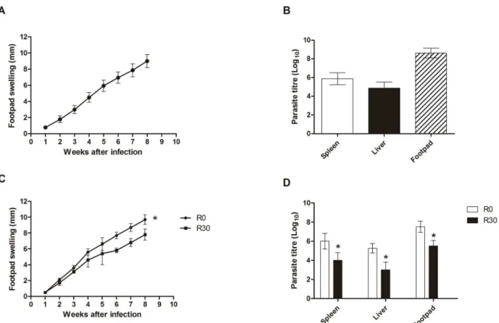
BALB/c mice (n = 8) subcutaneously infected withL. amazonensis
were monitored for 8 weeks by measuring the footpad thickness, given that the footpad swelling was similar in all evaluated animals (Figure 1A). The number of parasites recovered in the infected footpads, spleen, and liver of the infected animals was evaluated, and the results showed values of 8.660.5, 5.960.6, and 4.960.6 log, respectively (Figure 1B). In this context and due to the high homogeneity of infections in the mice, represented by similar values of footpad swelling and parasite loads, the parasites were recovered from lesions and used in the axenic cultures to perform the proteomic analyses of this study.
To evaluate the variation of thein vivoinfectivity between the different passages of L. amazonensis, stationary promastigotes obtained from R0 and R30 samples were used to infect BALB/c mice (n = 8 per group, with 16106 stationary promastigotes injected in each mouse). Animals infected with R30, as compared to the animals infected with R0, presented a significantly lower edema in the infected footpads at 8 weeks after infection (Figure 1C). The lower lesion size observed in the R30 group, when compared to the values obtained in the R0 group, was related to the lower parasite load observed when evaluating the infected footpads, spleen, and liver of these animals (Figure 1D).
For the evaluation of the in vitro infectivity, stationary promastigotes recovered in all passages (R0, R10, R20, and R30) were quantified and employed in the experiments. It could be observed that by using 2 parasites to infect 1 macrophage, parasites obtained from the R0 passage presented an infection average of 65.161.5% and a number of amastigotes per macrophage of 2.260.1. By contrast, using the R30 sample, the infection average was 14.962.3% and the number of amastigotes
per macrophage was 0.560.1. When 10 parasites were used to infect 1 macrophage, the infection average of the R0 group was 96.962.6% and the number of amastigotes per macrophage was 7.460.4. On the other hand, using parasites from the R30 group, the infection average was 59.562.2% and the number of amastigotes per macrophage was 3.860.4 (Table 1).
Analyses of protein expression inLeishmania amazonensis
Electrofocusing was performed using 13 cm pH 4–7 IPG strips after having investigated the best strip to isolate the total extracts. Strips of 13 cm were chosen because they provide a better range of separation of proteins by their pI without the gels becoming difficult to handling. Two ranges of pH were evaluated: 3–10 and 4–7. This study opted for a narrower pH range, given that most of the identified spots were located in this region. Although some spots located outside the pH 4–7 have been missed, the most spots were obtained within of this range due a better separation. After 2-DE gels had been applied, approximately 837 spots were found in the R0 sample, while 967, 935, and 872 spots were identified in the R10, R20, and R30 samples, respectively. Figure 2 is representative of the gels obtained in each condition. The 2-DE profiles and the number of observed spots in the different passages were reproducible in terms of both the total number of protein spots and their relative positions and intensities in four 2-DE gels performed for each passage (data not shown). After 2-DE analysis, 315 spots, which presented a significant variation in their intensities, were selected for identification by mass spectrometry. From all these spots, 258 were identified as proteins, and 164 unique proteins were identified. Of these, 58 proteins showed that the intensity of their corresponding spots either increased (19 spots) or decreased (37 spots) during the passages from R0 to R30, always maintaining a 2-fold minimal variation. It is also important to report that, upon performing thein vitroinfection experiments, a stabilization of the infectivity could be observed between the R20 and R30 samples (Table 1).
Identification of proteins of interest
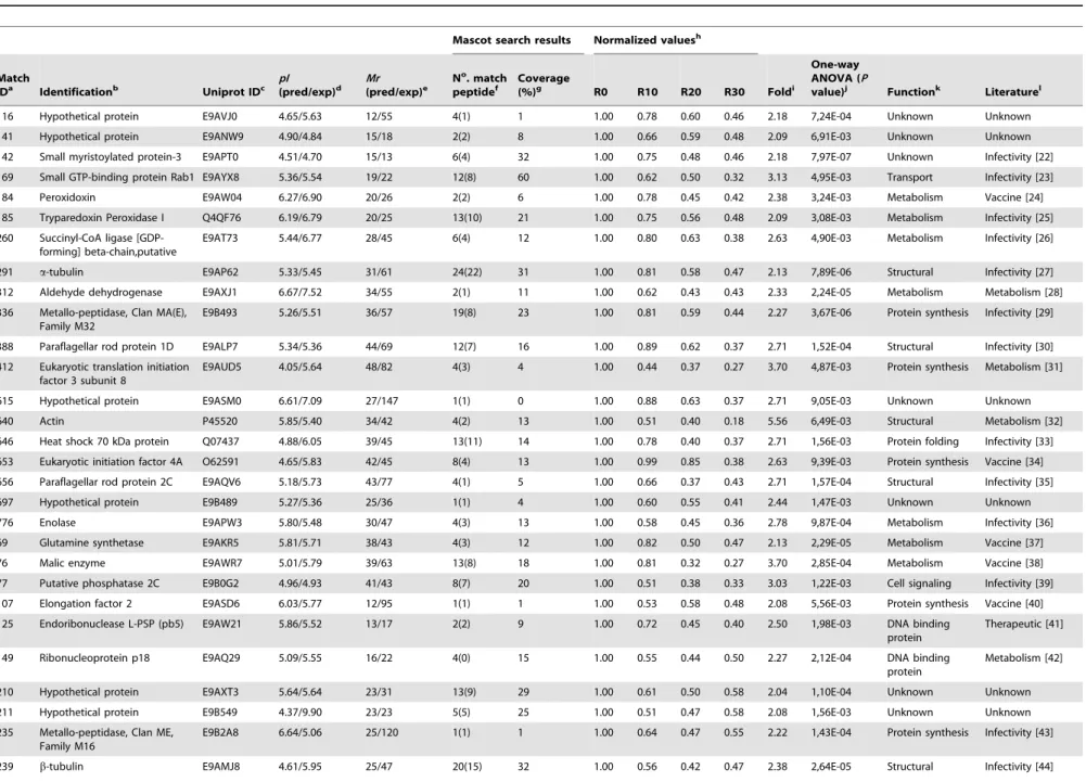
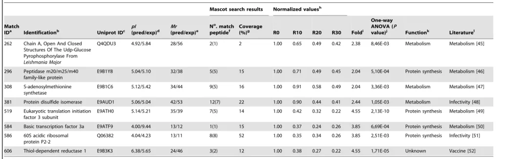
Among the 37 proteins that presented a significant decrease in their content during the axenic passages, six were hypothetical, while another 31 were known proteins, like described in Table 2 [22–52]. Some of these proteins present biological functions described in prior literature, such as tryparedoxin peroxidase [25], metallo-peptidases [29], heat shock protein HSP70 [33], and protein disulfide isomerase [48], all of which are involved with the parasites’ infectivity. Possible targets for therapeutic interventions, such as S-adenosylmethionine synthetase [47]; proteins identified as diagnosis candidates, such as acidic ribosomal protein P2; and vaccine candidates, such as the eukaryotic initiation factor 4A [34] and thiol-dependent reduc-tase 1 [52], were also identified. Proteins involved in the flagellum motility inLeishmania, such as a small myristoylated protein [22], and others related to metabolic functions, such as aldehyde dehydrogenase [28], were also identified. Evaluating the proteins that presented a significant increase in their content, including one hypothetical and 18 known proteins, could be identified. Data are showed in Table 3 [53–70]. In relation to known proteins, the majority are commonly involved in the parasites’ metabolism, such as nucleosome assembly proteins [57], 6-phosphogluconolactonase [62], and rieske iron sulfur proteins [66], while others, such as mannose-1-phosphate guanyltransfer-ase [56] and short chain dehydrogenguanyltransfer-ase [65], have been employed as candidates for immunotherapeutic targets.
Proteomic Approach Applied toLeishmania amazonensis
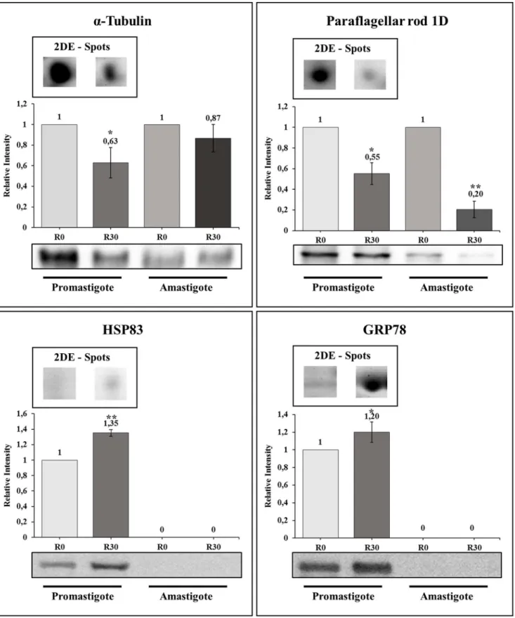
Immunoblotting validation
Some of the identified proteins that presented a significant increase or decrease in their contents in the axenic cultures were used to validate the results found in this study. In this context, two
of them presenting a significant decrease in their expression content, namelya-tubulin and paraflagellar rod protein 1D, and two of them, which presented an increase in their expression, namely HSP83 and GRP78, were used in the Western blot Figure 1. Infection of BALB/c mice.Mice (n = 8) were infected subcutaneously with 16106stationary promastigotes ofLeishmania amazonensis.
Lesion development in the infected footpads was monitored weekly, up to 8 weeks after infection. Mean6standard deviation (SD) are shown in (A). Parasite load in the infected footpads, spleen, and liver was analyzed in all animals (B). Other mice (n = 8, per group) were subcutaneously infected with 16106stationary promastigotes ofL. amazonensisobtained from R0 or R30 passages, and the lesion development was monitored up to 8 weeks
after infection. Mean6SD of the groups are shown (C). The parasite load in the infected footpads, spleen, and liver was also evaluated in these groups (D). The experiments were repeated three times, and presented similar results. *Significant difference between the R0 and R30 groups (P,
0.05).
doi:10.1371/journal.pntd.0002764.g001
Table 1.Evaluation ofin vitroinfection.
Ratio Percentage of infected macrophages
R0 R10 R20 R30
1:2 65.161.5 37.863.2 19.263.6 14.962.3
1:10 96.962.6 82.861.4 64.162.5 59.562.2
Ratio Number of amastigotes per macrophage
R0 R10 R20 R30
1:2 2.260.1 1.660.1 0.760.2 0.560.1
1:10 7.460.4 5.260.8 4.360.2 3.860.4
Murine macrophages (56105cells) were infected with stationary promastigotes ofL. amazonensis(16106and 56106, by a ratio of 1:2 or 1:10 macrophage per parasites,
respectively) and the cultures were incubated for 24 h at 37uC, 5% CO2. Next, free parasites were removed and the percentage of infected cells and the number of
amastigotes per macrophage in each passage (R0, R10, R20, and R30) were analyzed by counting 200 cells in triplicate. Mean6SD is shown. Data shown are representative of three separate experiments, performed in triplicate, which presented similar results.
doi:10.1371/journal.pntd.0002764.t001
Proteomic Approach Applied toLeishmania amazonensis
experiments (Figure 3). When promastigote extracts were employed, the selected proteins showed a variation that runs in line with the results obtained in the 2-DE gels. In addition, the decrease in the level ofa-tubulin and paraflagellar rod protein 1D detected in the promastigote forms are also maintained when the R30 forms are axenically derived into the amastigote stage of the parasite.
Discussion
Leishmania amazonensis is a member of the Leishmania mexicana
complex, and it is the etiological agent for a broad spectrum of disease in South American countries [5]. The mechanisms ofin vivo
persistence are of particular interest to this parasite species, given that several lines indicate thatL. amazonensis, when compared to otherLeishmaniaspecies, is particularly adept at surviving attacks from intracellular killing mechanisms [5,14]. Taking this into account, the present study applied a proteomic approach to analyze the variation of the protein expression profile from L. amazonensis, which was pre-isolated from lesions of chronically infected BALB/c mice and maintained inin vitro cultures over a long period of time. The purpose of this study was to verify whether or not thein vitrocultivation, performed over a 150-day period, could in fact decrease the parasites’ infectivity, as well as to identify proteins that could present a relation with a possible loss of infectivity inL. amazonensis.
Studies have shown that the maintenance ofLeishmaniain axenic cultures over long periods of time constitutes a relevant factor in the reduction of infectivity inL. infantum[71] andL. major[72]. In one study, the loss of infectivity inL. infantumwas related to the maintenance of the parasites after 105 days of successivein vitro
passages [73]. Proteomic analyses have been employed successfully to identify proteins expressed in both promastigote and amastigote stages of Leishmania spp., as well as to evaluate the stage-specific proteins and protein expression profile in the parasites
[8,11,74,75,76,77]. In the present study, proteins that presented a significant variation in their content, observed using 2-DE gels and analyzed by bioinformatics programs, were identified in an attempt to select possible targets for future immunological interventions in leishmaniasis. For this, stationary promastigotes were used in the same concentration in all passages so as to perform the experiments properly. In general, an increase of
Leishmania promastigote infectivity can also be observed when parasites pass from the logarithmic phase (days 1–3) to the stationary phase (days 4–6) of their growth cycle inin vitrocultures [78,79,80,81]. In the present study, it could be observed that the percentage of the stationary promastigotes found in all cultures was homogeneous, suggesting that the changes found in the protein expression profile and in the infectivity values of the parasites submitted to axenic cultures, could not only be associated with or depend on the reduction in the number of infective promastigotes present in thein vitrocultures.
Another important aspect here was the reduction in thein vitro
andin vivoinfectivity observed from R0 to R30 samples. In thein vitro experiments performed using murine macrophages, in addition to a significant decrease found in the percentage of infected macrophages, a marked reduction in the number of intracellular amastigotes could be observed. Evaluating in vitro
cultures performed up to 300 days after infection (R60), as compared to R30, no significant difference was found in the percentage of infected macrophages, and in the number of intra-macrophage amastigotes (data not shown). In addition, when R0 and R30 cultures were used to infect BALB/c mice, it could be observed that animals infected with R0 developed a more progressive disease than did those infected with the R30 sample, confirming the results obtained fromin vitroexperiments, though no significant difference could be observed between R20 and R30 in the infectivity experiments. Furthermore, the present study’s data are in accordance with Moreira et al. (2012), which showed that L. infantum promastigotes present a significant loss of their Figure 2. Two-dimensional profiles of cultures fromLeishmania amazonensis.The 2-DE gels were obtained after the separation of stationary promastigotes extracts (R0, R10, R20, and R30 passages; 650mg of each extract) by 2-DE (first dimension: IEF pH range 4–7; second dimension: 12% SDS-PAGE) and staining with colloidal Coomassie Brilliant Blue G-250. The gel fragments in the lower portion of the figures represent evaluated amplifications (see within the dotted lines). 2-DE gels of each passage were derived from four independent protein preparations of each passage. One representative preparation of each sample is showed in this study.
doi:10.1371/journal.pntd.0002764.g002
Proteomic Approach Applied toLeishmania amazonensis
Table 2.Identification of proteins that presented a significant decrease in their expression content.
Mascot search results Normalized valuesh
Match
IDa Identificationb Uniprot IDc pI (pred/exp)d
Mr (pred/exp)e
No. match
peptidef
Coverage
(%)g R0 R10 R20 R30 Foldi
One-way ANOVA (P
value)j Functionk Literaturel
116 Hypothetical protein E9AVJ0 4.65/5.63 12/55 4(1) 1 1.00 0.78 0.60 0.46 2.18 7,24E-04 Unknown Unknown
141 Hypothetical protein E9ANW9 4.90/4.84 15/18 2(2) 8 1.00 0.66 0.59 0.48 2.09 6,91E-03 Unknown Unknown
142 Small myristoylated protein-3 E9APT0 4.51/4.70 15/13 6(4) 32 1.00 0.75 0.48 0.46 2.18 7,97E-07 Unknown Infectivity [22]
169 Small GTP-binding protein Rab1 E9AYX8 5.36/5.54 19/22 12(8) 60 1.00 0.62 0.50 0.32 3.13 4,95E-03 Transport Infectivity [23]
184 Peroxidoxin E9AW04 6.27/6.90 20/26 2(2) 6 1.00 0.78 0.45 0.42 2.38 3,24E-03 Metabolism Vaccine [24]
185 Tryparedoxin Peroxidase I Q4QF76 6.19/6.79 20/25 13(10) 21 1.00 0.75 0.56 0.48 2.09 3,08E-03 Metabolism Infectivity [25]
260 Succinyl-CoA ligase [GDP-forming] beta-chain,putative
E9AT73 5.44/6.77 28/45 6(4) 12 1.00 0.80 0.63 0.38 2.63 4,90E-03 Metabolism Infectivity [26]
291 a-tubulin E9AP62 5.33/5.45 31/61 24(22) 31 1.00 0.81 0.58 0.47 2.13 7,89E-06 Structural Infectivity [27]
312 Aldehyde dehydrogenase E9AXJ1 6.67/7.52 34/55 2(1) 11 1.00 0.62 0.43 0.43 2.33 2,24E-05 Metabolism Metabolism [28]
336 Metallo-peptidase, Clan MA(E), Family M32
E9B493 5.26/5.51 36/57 19(8) 23 1.00 0.81 0.59 0.44 2.27 3,67E-06 Protein synthesis Infectivity [29]
388 Paraflagellar rod protein 1D E9ALP7 5.34/5.36 44/69 12(7) 16 1.00 0.89 0.62 0.37 2.71 1,52E-04 Structural Infectivity [30]
412 Eukaryotic translation initiation factor 3 subunit 8
E9AUD5 4.05/5.64 48/82 4(3) 4 1.00 0.44 0.37 0.27 3.70 4,87E-03 Protein synthesis Metabolism [31]
615 Hypothetical protein E9ASM0 6.61/7.09 27/147 1(1) 0 1.00 0.88 0.63 0.37 2.71 9,05E-03 Unknown Unknown
640 Actin P45520 5.85/5.40 34/42 4(2) 13 1.00 0.51 0.40 0.18 5.56 6,49E-03 Structural Metabolism [32]
646 Heat shock 70 kDa protein Q07437 4.88/6.05 39/45 13(11) 14 1.00 0.78 0.40 0.37 2.71 1,56E-03 Protein folding Infectivity [33]
653 Eukaryotic initiation factor 4A O62591 4.65/5.83 42/45 8(4) 13 1.00 0.99 0.85 0.38 2.63 9,39E-03 Protein synthesis Vaccine [34]
656 Paraflagellar rod protein 2C E9AQV6 5.18/5.73 43/77 4(1) 5 1.00 0.66 0.37 0.43 2.71 1,57E-04 Structural Infectivity [35]
697 Hypothetical protein E9B489 5.27/5.36 25/36 1(1) 4 1.00 0.60 0.55 0.41 2.44 1,47E-03 Unknown Unknown
776 Enolase E9APW3 5.80/5.48 30/47 4(3) 13 1.00 0.58 0.45 0.36 2.78 9,87E-04 Metabolism Infectivity [36]
69 Glutamine synthetase E9AKR5 5.81/5.71 38/43 4(3) 12 1.00 0.82 0.50 0.47 2.13 2,29E-05 Metabolism Vaccine [37]
76 Malic enzyme E9AWR7 5.01/5.79 39/63 13(8) 18 1.00 0.81 0.32 0.27 3.70 2,85E-04 Metabolism Vaccine [38]
77 Putative phosphatase 2C E9B0G2 4.96/4.93 41/43 8(7) 20 1.00 0.51 0.38 0.33 3.03 1,22E-03 Cell signaling Infectivity [39]
107 Elongation factor 2 E9ASD6 6.03/5.77 12/95 1(1) 1 1.00 0.53 0.58 0.48 2.08 5,56E-03 Protein synthesis Vaccine [40]
125 Endoribonuclease L-PSP (pb5) E9AW21 5.86/5.52 13/17 2(2) 9 1.00 0.72 0.45 0.40 2.50 1,98E-03 DNA binding protein
Therapeutic [41]
149 Ribonucleoprotein p18 E9AQ29 5.09/5.55 16/22 4(0) 15 1.00 0.55 0.44 0.50 2.27 2,12E-04 DNA binding protein
Metabolism [42]
210 Hypothetical protein E9AXT3 5.64/5.64 23/31 13(9) 29 1.00 0.61 0.50 0.58 2.04 1,10E-04 Unknown Unknown
211 Hypothetical protein E9B549 4.37/9.90 23/23 5(5) 25 1.00 0.51 0.47 0.58 2.08 1,56E-03 Unknown Unknown
235 Metallo-peptidase, Clan ME, Family M16
E9B2A8 6.64/5.06 25/120 1(1) 1 1.00 0.64 0.47 0.55 2.22 1,43E-04 Protein synthesis Infectivity [43]
239 b-tubulin E9AMJ8 4.61/5.95 25/47 20(15) 32 1.00 0.56 0.42 0.47 2.38 2,64E-05 Structural Infectivity [44]
Proteomic
Approach
Applied
to
Leishman
ia
amazonensis
PLOS
Neglected
Tropical
Diseases
|
www.plosntds
.org
7
April
2014
|
Volume
8
|
Issue
4
|
Table 2.Cont.
Mascot search results Normalized valuesh
Match
IDa Identificationb Uniprot IDc pI (pred/exp)d
Mr (pred/exp)e
No. match
peptidef
Coverage
(%)g R0 R10 R20 R30 Foldi
One-way ANOVA (P
value)j Functionk Literaturel
262 Chain A, Open And Closed Structures Of The Udp-Glucose Pyrophosphorylase From Leishmania Major
Q4QDU3 4.92/5.84 28/56 2(1) 2 1.00 0.65 0.49 0.42 2.38 8,46E-03 Metabolism Metabolism [45]
296 Peptidase m20/m25/m40 family-like protein
E9B1Y8 5.04/5.10 32/38 5(5) 15 1.00 0.71 0.49 0.45 2.04 5,10E-04 Protein synthesis Metabolism [46]
308 S-adenosylmethionine synthetase
E9B1C6 5.12/5.42 34/44 9(5) 16 1.00 0.91 0.58 0.49 2.04 3,36E-03 Metabolism Metabolism [47]
381 Protein disulfide isomerase E9AUD1 5.06/5.04 42/53 12(7) 22 1.00 0.90 0.44 0.41 2.44 1,05E-03 Metabolism Infectivity [48]
519 Eukaryotic translation initiation factor 3 subunit
E9ATH0 5.14/5.21 35/39 7(5) 14 1.00 0.42 0.32 0.22 4.55 2,13E-10 Protein synthesis Metabolism [49]
584 Basic transcription factor 3a E9ATF9 4.00/9.44 13/12 1(1) 15 1.00 0.37 0.24 0.26 3.85 6,69E-04 Protein synthesis Metabolism [50]
586 60S acidic ribosomal protein P2-2
Q06382 4.04/4.23 13/11 8(8) 52 1.00 0.35 0.34 0.26 3.85 2,51E-03 Protein synthesis Infectivity [51]
606 Thiol-dependent reductase 1 E9B3K3 6.38/5.65 24/46 3(2) 12 1.00 0.38 0.27 0.22 4.55 1,71E-05 Unknown Vaccine [52]
a)Spots match ID number obtained from ImageMaster Platinum; b)Name of the identified protein;
c)Uniprot identification code;
d)Experimentally predicted and expected isoelectric point (pI); e)Experimentally predicted and expected molecular weight (
Mr, in kDa); f)Number of identified peptides by MS;
g)Percentage of the protein sequence covered by identified peptides;
h)Normalized data from R0 represented by mean values of each condition divided by R30 value; i)Fold represents the maximum spot intensity mean value of the conditions divided by the smallest value; j)One-way ANOVA (P,0.01) obtained from spot analysis;
k)Biological functions according to NCBI, UniProt, and Gene Ontology databases;
l)Biological activity and/or immunological application described in other studies: [22] Tull et al., 2010; [23] Oliveira et al., 2006; [24] Daifalla et al., 2011; [25] Iyer et al., 2008; [26] Hunger-Glaser et al., 1999; [27] Werbovetz et al., 1999;
[28] Feng et al., 2011; [29] Niemirowicz et al., 2007; [30] Hunger-Glaser et al., 1997; [31] Alcolea et al., 2009; [32] Bhaskar et al., 2012; [33] Khanra et al., 2012; [34] Berberich et al., 2003; [35] Moore et al., 1996; [36] Swenerton et al., 2011; [37] Hummadi et al., 2006; [38] Martins et al., 2006; [39] Burns et al., 1993; [40] Kushawaha et al., 2011; [41] Misra et al., 2005; [42] Bringaud et al., 1995; [43] Eggleson et al., 1999; [44] Mureev et al., 2007; [45] Steiner et al., 2007; [46] Martı´nez-Rodrı´guez et al., 2012; [47] Drummelsmith et al., 2004; [48] Achour et al., 2002; [49] Buda et al., 2013; [50] Alcolea et al., 2011; [51] Martı´n et al., 2009; [52] Silva et al., 2012. The proteins were identified through the data included in the NCBI database (dated June 2012) forLeishmania spp.
doi:10.1371/journal.pntd.0002764.t002
Proteomic
Approach
Applied
to
Leishman
ia
amazonensis
PLOS
Neglected
Tropical
Diseases
|
www.plosntds
.org
8
April
2014
|
Volume
8
|
Issue
4
|
Table 3.Identification of proteins that presented a significant increase in their expression content.
Mascot search results Normalized valuesh
Match
IDa Identificationb Uniprot IDc pI(pred/exp)d Mr(pred/exp)e
No. match
peptidef
Coverage
(%)g R0 R10 R20 R30 Foldi
One-way ANOVA
(Pvalue)j Functionk Literaturel
8 Calreticulin E9B259 4.52/4.51 50/45 3(3) 5 1.00 3.26 4.40 4.49 4.49 6.96E-03 Protein folding
Metabolism [53]
12 Isocitrate dehydrogenase
E9B494 5.44/5.51 40/47 16(6) 28 1.00 1.71 1.76 2.51 2.51 2.41E-03 Metabolism Metabolism [54]
303 60S acidic ribosomal subunit protein
E8NHJ8 5.07/5.00 33/35 26(21) 45 1.00 3.15 4.72 4.75 4.75 9.76E-05 Protein synthesis
Diagnosis [55]
326 Mannose-1-phosphate guanyltransferase
E9AW11 5.67/5.29 36/42 10(7) 23 1.00 2.07 2.25 3.24 3.24 1.72E-04 Metabolism Metabolism [56]
392 Nucleosome assembly protein
E9ARZ6 4.64/4.64 45/40 17(9) 25 1.00 2.17 2.49 2.61 2.61 1.78E-04 DNA binding protein
Metabolism [57]
420 ATPase beta subunit E9AXJ6 5.02/5.14 49/56 60(51) 49 1.00 1.74 1.89 2.02 2.02 9.55E-04 Metabolism Metabolism [58]
432 T-complex protein 1, theta subunit
E9AUC7 5.27/5.24 54/59 27(18) 49 1.00 1.54 2.05 3.35 3.35 3.65E-04 Protein folding Metabolism [59]
458 Chain A, Protein Structure Of Usp FromL. Majorin Apo-Form
D3G6S4 5.36/5.34 63/69 4(4) 3 1.00 1.82 3.11 3.24 3.24 2.57E-03 Metabolism Metabolism [60]
739 Hs1vu complex proteolytic subunit-like,hs1vu complex proteolytic subunit-like, threonine peptidase, Clan T(1), family T1B
E9ATI1 5.24/6.09 22/25 4(1) 9 1.00 1.65 1.87 2.05 2.05 3.70E-04 Protein synthesis
Metabolism [61]
767 6-phosphogluconolactonase E9AYQ1 5.50/5.22 26/29 2(2) 8 1.00 1.39 1.71 2.27 2.27 1.63E-03 Metabolism Metabolism [62]
40 Heat shock protein 83; HSP 83
P27741 6.27/5.00 31/81 1(1) 1 1.00 1.88 2.92 2.93 2.93 3.76E-03 Protein folding Diagnosis [63]
62 2-hydroxy-3-oxopropionate reductase
E9B0E2 5.77/5.40 26/31 6(5) 25 1.00 2.39 3.13 3.42 3.42 6.53E-05 Metabolism Metabolism [64]
230 Short chain dehydrogenase E9B602 6.57/6.31 25/28 3(1) 9 1.00 2.16 2.61 2.62 2.62 8.48E-04 Metabolism Therapeutic [65]
279 Reiske iron-sulfur protein precursor
E9B632 5.57/6.02 29/34 9(7) 43 1.00 1.83 2.43 2.89 2.89 6.34E-06 Metabolism Metabolism [66]
327 Vacuolar ATPase subunit-like protein
E9AKM1 4.93/4.85 36/42 13(5) 25 1.00 1.75 2.39 2.49 2.49 3.74E-03 Metabolism Metabolism [67]
510 Cyclin 1 E9AMR1 5.99/5.67 31/36 3(1) 13 1.00 2.26 2.28 2.78 2.78 8.78E-03 Protein synthesis
Metabolism [68]
529 Protein transport protein Sec13
E9B2C5 5.69/5.51 34/37 2(1) 9 1.00 2.45 2.78 3.00 3.00 2.79E-03 Unknown Metabolism [69]
676 Hypothetical protein
E9ATK7 4.98/4.91 98/119 33(22) 27 1.00 1.75 2.75 2.80 2.80 3.84E-03 Unknown Unknown
Proteomic
Approach
Applied
to
Leishman
ia
amazonensis
PLOS
Neglected
Tropical
Diseases
|
www.plosntds
.org
9
April
2014
|
Volume
8
|
Issue
4
|
infectivity after 100 days ofin vitrocultures, suggesting that this condition may well be related to specific modifications in the protein differentiation content of parasites [73].
In relation to the identified proteins that presented a decreased expression from R0 to R30, several had already been described in other published studies, such as proteins involved in the infectivity of Leishmania or in other parasite species. For example, peroxidoxin is a protein expressed in the endoplasmic reticulum of Trypanosomatides and is involved in cellular resistance to reactive oxygen species [82], been also a virulence factor described inTrypanosoma cruzi[83]. The malic enzyme is involved in the virulence of Xanthomonas campestris
[84], while aldehyde dehydrogenase acts in the protection of mammal cells against damage evoked by osmotic and saline stress [85]. S-adenosylmethionine synthetase in L. panamensis
[86] andL. major[87] is related to drug resistance. Enolase is a membrane protein that plays a role in the infectivity of
Leishmania, as it is involved in the interaction between the parasites and host cells [88]. The carboxypeptidase family (M32) has also been identified as a virulence factor inT. cruzi
[89] and operates in the catabolism of peptides, favoring the growth and multiplication of parasites [90]. Phosphatase 2C is considered a virulence factor inToxoplasma gondii [91], while tryparedoxin peroxidase in L. donovani is involved in drug resistance [92].
Evaluating the databases of proteins that presented an increased expression from R0 to R30, most present metabolic functions described in prior literature, such as those related to cellular stress, recovery of improperly folded proteins, and the restoration of core functions. In this context, phosphatase 1 guanyltransferase mannose is involved in oxidative stress in yeast [93], while isocitrate dehydrogenase is involved in cellular stress inCryptococcus neoformans [94]. The glucose regulated protein 78 kDa is a membrane protein that is up-regulated in conditions of cellular stress and that can lead to cell cycle arrest [95]. The protein complex Hs1VU-like proteolytic subunit is a peptidase that is over-expressed and correlated to the accumulation of improperly folded proteins within the cells [96]. Calreticulin is involved in cellular processes related to protein folding, calcium homeostasis, apop-tosis, and cell differentiation [97].
Western blot assays with four identified proteins were performed to validate 2-DE gel quantification results. When promastigote samples were analyzed, a significant correlation could be observed when comparing the two techniques used for proteins with a decreased or increased expression in aged cultures (Figure 3). When axenic amastigote extracts were employed for Western blots, a decrease in the level ofa-tubulin and paraflagellar rod protein 1D observed in the 2-DE was also detected. Unfortunately, the lack of signs when antibodies against HSP83 and GRP78 were employed made it impossible to confirm whether or not the increase in protein expression associated with the loss of infectivity is maintained in the amastigote forms.
In conclusion, the data presented in the present study could contribute to a better understanding of the biological processes involved in a possible loss of infectivity of L. amazonensis when submitted to in vitro cultures over a long period of time, as described for otherLeishmaniaspecies. Furthermore, the identified proteins presenting a significant decrease in their protein content during cultivation, including the hypothetical, should be evaluated in future studies, including vaccine candidates and/or immuno-therapeutic targets against leishmaniasis. Additional studies are warranted in an attempt to address the major concern that identified proteins are indeed involved in the possible loss of virulence in the parasites cultured over long periods of time.
Table 3. Cont. Mascot search results Normalized values h Match ID a Identification b Uniprot ID c pI (pred/exp) d Mr (pred/exp) e N o. match peptide f Coverage (%) g R0 R10 R20 R30 Fold i
One-way ANOVA (P
value) j Function k Literature l 735 G lucose-regulate d p rotein 78; GRP78 E9AZT9 5.15/5.18 67/72 27(22) 28 1.00 4.69 4.84 4.87 4.87 9.57E-05 Protein folding V accine [70] a) Spots m atch ID number obtained from ImageMaster Platinum; b) Name of the identified protein; c) Uniprot identification code; d) Experimentally predicted and expected isoelectric point ( pI ); e) Experimentally predicted and expected molecular weight ( Mr , in kDa); f) Number of identified peptides by MS; g) Percentage o f the protein sequence covered by identified peptides; h) Normalized data from R 0 represented by mean values of each condition d ivided by R30 value; i)Fold represents the maximum spot intensity mean value o f the conditions divided by the smallest value; j) One-way ANOVA ( P , 0.01) obtained from spot analysis; k) Biological functions according to NCBI, UniProt, and Gene Ontology d atabases; l)Biological activity and/or immunological application d escribed in other studies: [53] Joshi et al., 1996; [54] Tielens et al., 2010; [55] Soto et al. ,1996; [56] Lackovic e t al., 2010; [57] Scher e t al., 2012; [58] Sa ´nchez-Can ˜ete et al., 2009; [59] Peris et al., 1994; [60] Steiner et al., 2007; [61] Jaramillo et al., 2011; [62] Duclert-Savatier et al., 2009; [63] Celeste et al., 2004; [64] Liu e t al., 2011; [65] Leblanc et al., 1998; [66] P riest et al., 1996; [67] B akker-Grunwald, 1992; [68] Banerjee et al., 2006; [69] Casanova et al., 2008; [70] Jensen et al., 2001. The p roteins w ere identified through the data included in the NCBI database (dated June 2012) for Leishmania spp. doi:10.1371/journal.pntd .0002764.t003
Proteomic Approach Applied toLeishmania amazonensis
Figure 3. Immunoblotting validation of some proteins inLeishmania amazonensis.Representative immunoblotting of some proteins that presented a significant decrease or increase in their expression content between R0 and R30 passages, using promastigote and amastigotes-like forms ofL. amazonensis, are shown here. For each protein [a-tubulin, in A; paraflagellar rod protein 1D, in B; glucose-regulated protein 78 (GRP78) in C, and heat shock protein 83 (HSP83), in D], this image presents one example of correspondent 2-DE spot of promastigote form obtained from R0 or R30 passages. The antibodies used to validate each spot are described in the material and methods section. Asterisks represent the comparison between the expression of the protein in the R0 condition in relation to the R30 sample in each parasite stage, applying the Student’s t-test (P,0.05), and the numbers represent the relative variation of each protein in comparison to R0 of each parasite stage. All experiments were performed in triplicate.
doi:10.1371/journal.pntd.0002764.g003
Proteomic Approach Applied toLeishmania amazonensis
Acknowledgments
The authors would like to thank to the Mass Spectrometry Laboratory of the Brazilian Biosciences National Laboratory to the Mass Spectrometry Laboratory (LNBio, CNPEM/ABTLuS, Campinas, Brazil) for its support in the mass spectrometry experiments, as well as to Dra Daniela Castanheira Bartholomeu (Department of Parasitology, UFMG), for providing the antibodies employed in the immunoblotting experiments.
Author Contributions
Conceived and designed the experiments: EAFC CAPT MS RDMM MACF RAPN. Performed the experiments: RDMM MCD ECM VTM PSL DPL DMS. Analyzed the data: EAFC CAPT MS RDMM MACF. Contributed reagents/materials/analysis tools: MJMA WCBR. Wrote the paper: EAFC CAPT RDMM MS.
References
1. World Health Organization (2010) Control of the Leishmaniasis: report of a meeting of the WHO Expert Committee on the Control of Leishmaniases, Geneva, 22–26 March 2010. Available: http://whqlibdoc.who.int/trs/WHO_ TRS_949_eng.pdf. 2010 Accessed 15 June 2013.
2. Garcez LM, Goto H, Ramos PK, Brigido MC, Gomes PAF, et al. (2002) Leishmania (Leishmania) amazonensis-induced cutaneous leishmaniasis in the primateCebus apella: a model for vaccine trials. Int J Parasitol 32: 1755–1764. 3. Barral A, Pedral-Sampaio D, Momen H, Mc Mahon-Pratt D, Jesus AR, et al.
(1991). Leishmaniasis in Bahia, Brazil: evidence thatLeishmania amazonensis produces a wide spectrum of clinical disease. Am J Trop Med Hyg 44: 536–546. 4. Abreu-Silva AL, Calabrese KS, Cupolilo SMN, Cardoso FO, Souza CSF, et al. (2004) Histopathological studies of visceralizedLeishmania (Leishmania) amazonensis in mice experimentally infected. Vet Parasitol 121: 179–187.
5. Grimaldi JrG, Tesh RB (1993) Leishmaniasis of the New World: current concepts and implications for future research. Clin Microbiol Rev 6: 230–250. 6. Mitchell GF, Handman E, Spithill TW (1984) Vaccination against cutaneous leishmaniasis in mice using nonpathogenic cloned promastigotes ofLeishmania majorand importance of route of injection. Aust J Exp Biol Med Sci 62: 145–153. 7. Hadighi R, Mohebali M, Boucher P, Hajjaran H, Khamesipour A, et al. (2006) Unresponsiveness to Glucantime treatment in Iranian cutaneous leishmaniasis due to drug-resistantLeishmania tropicaparasites. PLoS Med 3: e162. 8. Coelho VTS, Oliveira JS, Valadares DG, Duarte MC, Cha´vez-Fumagalli MA,
et al. (2012) Identification of proteins in promastigote and amastigote-like Leishmaniausing an immunoproteomic approach. PLoS Negl Trop Dis 6: e1430. 9. Drummelsmith J, Brochu V, Girard I, Messier N, Ouellette M (2003) Proteome mapping of the protozoan parasiteLeishmaniaand application to the study of drug targets and resistance mechanisms. Mol Cell Proteomics 2: 146–155. 10. Chenik M, Lakhal S, Ben Khalef N, Zribi L, Louzir H, et al. (2006) Approaches
for the identification of potential excreted/secreted proteins ofLeishmania major parasites. Parasitology 132: 493–509.
11. Leifso K, Cohen-Freue G, Dogra N, Murray A, Mc Master WR (2007) Genomic and proteomic expression analysis ofLeishmaniapromastigote and amastigote life stages: theLeishmaniagenome is constitutively expressed. Mol Biochem Parasitol 152: 35–46.
12. Morales MA, Watanabe R, Laurent C, Lenormand P, Rousselle JC, et al. (2008) Phosphoproteomic analysis ofLeishmania donovanipro- and amastigote stages. Proteomics 8: 350–363.
13. Rosenzweig D, Smith D, Myler PJ, Olafson RW, Zilberstein D (2008) Post-translational modification of cellular proteins during Leishmania donovani differentiation. Proteomics 8: 1843–1850.
14. Paape D, Barrios-Lerena ME, Le Bihan T, Mackay L, Aebischer T (2010) Gel free analysis of the proteome of intracellularLeishmania mexicana. Mol Biochem Parasitol 169: 108–114.
15. Costa CHN, Peters NC, Maruyama SR, Brito JrEC, Santos IKFM (2011) Vaccines for the leishmaniases: Proposals for a research agenda. The working group on research priorities for development of leishmaniasis vaccines. PLoS Negl Trop Dis 5: e943.
16. Doyle PS, Engel JC, Pimenta PFP, Silva PP, Dwyer DM (1991)Leishmania donovani: long-term culture of axenic amastigotes at 37uC. Exp Parasitol 73: 326– 334.
17. Coelho EA, Tavares CA, Carvalho FA, Chaves KF, Teixeira KN, et al. (2003) Immune responses induced by theLeishmania (Leishmania) donovaniA2 antigen, but not by the LACK antigen, are protective against experimental Leishmania (Leishmania) amazonensisinfection. Infect Immun 71: 3988–3994.
18. Valadares DG, Duarte MC, Oliveira JS, Martins VT, Costa LE, et al. (2011) Leishmanicidal activity of theAgaricus blazeiMurill in differentLeishmaniaspecies. Parasitol Int 60: 357–363.
19. Lewis TS, Hunt JB, Aveline LD, Jonscher KR, Louie DF, et al. (2000) Identification of novel MAP kinase pathway signalling targets by functional proteomics and mass spectrometry. Mol Cell 6: 1343–1354.
20. Bradford MM (1976) A rapid and sensitive method for the quantification of microgram quantities of protein utilizing the principle of protein-dye binding. Anal Biochem 72: 248–254.
21. Neuhoff V, Arold N, Taube D, Ehrhardt W (1988) Improved staining of proteins in polyacrilamide gels including isoeletric focusing gels with clear background at nanogram sensitivity using Coomassie Brilliant Blue G-250 and R-250. Electrophoresis 9: 255–262.
22. Tull D, Naderer T, Spurck T, Mertens HD, Heng J, et al. (2010) Membrane protein SMP-1 is required for normal flagellum function inLeishmania. J Cell Sci 123: 544–554.
23. Oliveira AH, Ruiz JC, Cruz AK, Greene LJ, Rosa JC, et al. (2006) Subproteomic analysis of soluble proteins of the microsomal fraction from two Leishmaniaspecies. Comp Biochem Physiol Part D Genomics Proteomics 1: 300– 308.
24. Daifalla NS, Bayih AG, Gedamu L (2011) Immunogenicity ofLeishmania donovani iron superoxide dismutase B1 and peroxidoxin 4 in BALB/c mice: the contribution of toll-like receptor agonists as adjuvant. Exp Parasitol 129: 292– 298.
25. Iyer J P, Kaprakkaden A, Choudhary ML, Shaha C (2008) Crucial role of cytosolic tryparedoxin peroxidase inLeishmania donovanisurvival, drug response and virulence. Mol Microbiol 68: 372–391.
26. Hunger-Glaser I, Brun R, Linder M Seebeck T (1999) Inhibition of succinyl CoA synthetase histidine-phosphorylation inTrypanosoma bruceiby an inhibitor of bacterial two-component systems. Mol Biochem Parasitol 100: 53–59. 27. Werbovetz KA, Brendle JJ, Sackett DL (1999) Purification, characterization, and
drug susceptibility of tubulin fromLeishmania.Mol Biochem Parasitol 98: 53–65. 28. Feng X, Feistel T, Buffalo C, Mc Cormack A, Kruvand E, et al. (2011) Remodeling of protein and mRNA expression inLeishmania mexicanainduced by deletion of glucose transporter genes. Mol Biochem Parasitol 175: 39–48. 29. Niemirowicz G, Parussini F, Agu¨ero F, Cazzulo JJ (2007) Two metallo
carboxypeptidases from the protozoan Trypanosoma cruzibelong to the M32 family, found so far only in prokaryotes. Biochem J 401: 399–410.
30. Hunger-Glaser I, Seebeck T (1997) Deletion of the genes for the paraflagellar rod protein PFR-A in Trypanosoma brucei is probably lethal. Mol Biochem Parasitol 90: 347–351.
31. Alcolea PJ, Alonso A, Sa´nchez-Gorostiaga A, Moreno-Paz M, Go´mez MJ, et al. (2009) Genome-wide analysis reveals increased levels of transcripts related with infectivity in peanut lectin non-agglutinated promastigotes ofLeishmania infantum. Genomics 93: 551–564.
32. Bhaskar, Kumari N, Goyal N (2012) Cloning, characterization and sub-cellular localization of gamma subunit of T-complex protein-1 (chaperonin) from Leishmania donovani. Biochem Biophys Res Commun 429: 70–74.
33. Khanra S, Datta S, Mondal D, Saha P, Bandopadhyay SK, et al. (2012) RFLPs of ITS, ITS1 and hsp70 amplicons and sequencing of ITS1 of recent clinical isolates of Kala-azar from India and Bangladesh confirms the association of Leishmania tropicawith the disease. Acta Trop 124: 229–234.
34. Berberich C, Ramı´rez-Pineda JR, Hambrecht C, Alber G, Skeiky YA, et al. (2003) Dendritic cell (DC)-based protection against an intracellular pathogen is dependent upon DC-derived IL-12 and can be induced by molecularly defined antigens. J Immunol 170: 3171–3179.
35. Moore LL, Santrich C, LeBowitz JH (1996) Stage-specific expression of the Leishmania mexicanaparaflagellar rod protein PFR-2. Mol Biochem Parasitol 80: 125–135.
36. Swenerton RK, Zhang S, Sajid M, Medzihradszky KF, Craik CS, et al. (2011) The oligopeptidase B of Leishmania regulates parasite enolase and immune evasion. J Biol Chem 286: 429–440.
37. Hummadi YM, Al-Bashir NM, Najim RA (2006)Leishmania majorandLeishmania tropica: II. Effect of an immunomodulator, S(2) complex on the enzymes of the parasites. Exp Parasitol 112: 85–91.
38. Martins DRA, Jeronimo SMB, Donelson JE, Wilson ME (2006)Leishmania chagasiT-cell antigens identified through a double library screen. Infect Immun 74: 6940–6948.
39. Burns JMJr, Parsons M, Rosman DE, Reed SG (1993) Molecular cloning and characterization of a 42 kDa protein phosphatase ofLeishmania chagasi. J Biol Chem 268: 17155–17161.
40. Kushawaha PK, Gupta R, Sundar S, Sahasrabuddhe AA, Dube A (2011) Elongation factor-2, a Th1 stimulatory protein ofLeishmania donovani, generates strong IFN-cand IL-12 response in curedLeishmania-infected patients/hamsters and protects hamsters againstLeishmaniachallenge. J Immunol 187: 6417–6427. 41. Misra S, Bennett J, Friew YN, Abdulghani J, Irvin-Wilson CV, et al. (2005) A type II ribonuclease H fromLeishmaniamitochondria: an enzyme essential for the growth of the parasite. Mol and Biochem Parasitol 143: 135–145.
42. Bringaud F, Peris M, Zen KH, Simpson L (1995) Characterization of two nuclear-encoded protein components of mitochondrial ribonucleoprotein complexes fromLeishmania tarentolae. Mol Biochem Parasitol 71: 65–79. 43. Eggleson KK, Duffin KL, Goldberg DE (1999) Identification and
characteriza-tion of falcilysin, a metallopeptidase involved in hemoglobin catabolism within the malaria parasitePlasmodium falciparum. J Biol Chem 274: 32411–32417. 44. Mureev S, Kushnir S, Kolesnikov AA, Breitling R, Alexandrov K (2007)
Construction and analysis of Leishmania tarentolae transgenic strains free of selection markers. Mol Biochem Parasitol 155: 71–83.
Proteomic Approach Applied toLeishmania amazonensis
45. Steiner T, Lamerz AC, Hess P, Breithaupt C, Krapp S, et al. (2007) Open and closed structures of the UDP-glucose pyrophosphorylase fromLeishmania major. J Biol Chem 282: 13003–13010.
46. Martı´nez-Rodrı´guez S, Garcı´a-Pino A, Heras-Va´zquez FJ, Clemente-Jime´nez JM, Rodrı´guez-Vico F, et al. (2012) Mutational and structural analysis of L-N-carbamoylase reveals new insights into a peptidase M20/M25/M40 family member. J Bacteriol 194: 5759–5768.
47. Drummelsmith J, Girard I, Trudel N, Ouellette M (2004) Differential protein expression analysis of Leishmania major reveals novel roles for methionine adenosyltransferase and S-adenosylmethionine in methotrexate resistance. J Biol Chem 279: 33273–33280.
48. Achour YB, Chenik M, Louzir H, Dellagi K (2002) Identification of a disulfide isomerase protein ofLeishmania majoras a putative virulence factor. Infect Immun 70: 3576–3585.
49. Buda P, Reinbothe T, Nagaraj V, Mahdi T, Luan C, et al. (2013) Eukaryotic translation initiation factor 3 subunit e controls intracellular calcium homeostasis by regulation of cav1.2 surface expression. PLoS One 8: e64462.
50. Alcolea PJ, Alonso A, Larraga V (2011) Genome-wide gene expression profile induced by exposure to cadmium acetate inLeishmania infantumpromastigotes. Int Microbiol 14: 1–11.
51. Martı´n OA, Villegas ME, Aguilar CF (2009) Three-dimensional studies of pathogenic peptides from the c-terminal of Trypanosoma cruzi ribosomal P proteins and their interaction with a monoclonal antibody structural model. PMC Biophys 2: 4.
52. Silva AM, Tavares J, Silvestre R, Ouaissi A, Coombs GH, et al. (2012) Characterization of Leishmania infantum thiol-dependent reductase 1 and evaluation of its potential to induce immune protection. Parasite Immunol 34: 345–350.
53. Joshi M, Pogue GP, Duncan RC, Lee NS, Singh NK, et al. (1996) Isolation and characterization ofLeishmania donovanicalreticulin gene and its conservation of the RNA binding activity. Mol Biochem Parasitol 81: 53–64.
54. Tielens AG, Van Grinsven KW, Henze K, Van Hellemond JJ, Martin W (2010) Acetate formation in the energy metabolism of parasitic helminths and protists. Int J Parasitol 40: 387–397.
55. Soto M, Requena JM, Quijada L, Alonso C (1996) Specific serodiagnosis of human leishmaniasis with recombinantLeishmaniaP2 acidic ribosomal proteins. Clin Diagn Lab Immunol 3: 387–391.
56. Lackovic K, Parisot JP, Sleebs N, Baell JB, Debien L, et al. (2010) Inhibitors of Leishmania GDP-mannose pyrophosphorylase identified by high-throughput screening of small-molecule chemical library. Antimicrob Agents Chemother 54: 1712–1719.
57. Scher R, Garcia JB, Pascoalino B, Schenkman S, Cruz AK (2012) Character-ization of anti-silencing factor 1 inLeishmania major. Mem Inst Oswaldo Cruz 107: 377–386.
58. Sa´nchez-Can˜ete MP, Carvalho L, Pe´rez-Victoria FJ, Gamarro F, Castanys S (2009) Low plasma membrane expression of the miltefosine transport complex renders Leishmania braziliensis refractory to the drug. Antimicrob Agents Chemother 53: 1305–1313.
59. Peris M, Frech GC, Simpson AM, Bringaud F, Byrne E, et al. (1994) Characterization of two classes of ribonucleoprotein complexes possibly involved in RNA editing fromLeishmania tarentolaemitochondria. EMBO J l 13: 1664– 1672.
60. Steiner T, Lamerz AC, Hess P, Breithaupt C, Krapp S, et al. (2007) Open and Closed Structures of the UDP-glucose Pyrophosphorylase fromLeishmania major. J Biol Chem 282: 13003–13010.
61. Jaramillo M, Gomez MA, Larsson O, Shio MT, Topisirovic I, et al. (2011) Leishmaniarepression of host translation through mTOR cleavage is required for parasite survival and infection. Cell Host Microbe 9: 331–341.
62. Duclert-Savatier N, Poggi L, Miclet E, Lopes P, Ouazzani J, et al. (2009) Insights into the enzymatic mechanism of 6-phosphogluconolactonase fromTrypanosoma bruceiusing structural data and molecular dynamics simulation. J Mol Biol 388: 1009–1021.
63. Celeste BJ, Angel SO, Castro LGM, Gidlund M, Goto H (2004)Leishmania infantum heat shock protein 83 for the serodiagnosis of tegumentary leishmaniasis. Braz J Med Biol Res 37: 1591–1593.
64. Liu Y, Koh CMJ, Sun L, Ji L (2011) Tartronate semialdehyde reductase defines a novel rate-limiting step in assimilation and bioconversion of glycerol inUstilago maydis. PLoS One 6: e16438.
65. Leblanc E, Papadopoulou B, Bernatchez C, Ouellette M (1998) Residues involved in co-factor and substrate binding of the short-chain dehydrogenase/ reductase PTR1 producing methotrexate resistance inLeishmania. Eur J Biochem 251: 768–774.
66. Priest JW, Hajduk SL (1996)In vitroimport of the rieske iron-sulfur protein by trypanosome mitocondria. J Biol Chem 271: 20060–20069.
67. Bakker-Grunwald T (1992) Ion transport in parasitic protozoa. J Exp Biol 172: 311–322.
68. Banerjee S, Sen A, Das P, Saha P (2006)Leishmania donovanicyclin 1 (LdCyc1) forms a complex with cell cycle kinase subunit CRK3 (LdCRK3) and is possibly involved in S-phase-related activities. FEMS Microbiol Lett 256: 75–82. 69. Casanova M, Portale`s P, Blaineau C, Crobu L, Bastien P, et al. (2008) Inhibition
of active nuclear transport is an intrinsic trigger of programmed cell death in trypanosomatids. Cell Death Differ 15: 1910–1920.
70. Jensen AT, Curtis J, Montgomery J, Handman E, Theander TG (2001) Molecular and immunological characterisation of the glucose regulated protein 78 ofLeishmania donovani. Biochim Biophys Acta 1549: 73–87.
71. Grimm F, Brun R, Jenni L (1991) Promastigote infectivity inLeishmania infantum. Parasitol Res 77: 185–191.
72. Segovia M, Artero JM, Mellado E, Chance ML (1992) Effects of long-term in vitro cultivation on the virulence of cloned lines ofLeishmania majorpromastigotes. Ann Trop Med Parasitol 86: 347–354.
73. Moreira D, Santare´m N, Loureiro I, Tavares J, Silva AM, et al. (2012) Impact of continuous axenic cultivation inLeishmania infantumvirulence. PLoS Negl Trop Dis 6: e1469.
74. Pawar H, Sahasrabuddhe NA, Renuse S, Keerthikumar S, Sharma J, et al. (2012) A proteogenomic approach to map the proteome of an unsequenced pathogen -Leishmania donovani. Proteomics 12: 832–844.
75. Bente M, Harder S, Wiesgigl M, Heukeshoven J, Gelhaus C, et al. (2003) Developmentally induced changes of the proteome in the protozoan parasite Leishmania donovani. Proteomics 3: 1811–1829.
76. Nugent PG, Karsani SA, Wait R, Tempero J, Smith DF (2004) Proteomic analysis ofLeishmania mexicanadifferentiation. Mol Biochem Parasitol 136: 51–62. 77. Mc Nicoll F, Drummelsmith J, Muller M, Madore E, Boilard N, et al. (2006) A combined proteomic and transcriptomic approach to the study of stage differentiation inLeishmania infantum. Proteomics 6: 3567–3581.
78. Walker J, Vasquez JJ, Gomez MA, Drummelsmith J, Burchmore R, et al. (2006) Identification of developmentally-regulated proteins inLeishmania panamensisby proteome profiling of promastigote and axenic amastigotes. Mol Biochem Parasitol 147: 64–73.
79. Sacks DL, Perkins PV (1984) Identification of an infective stage ofLeishmania promastigotes. Science 223: 1417–1419.
80. Da Silva R, Sacks DL (1987) Metacyclogenesis is a major determinant of Leishmania promastigote virulence and attenuation. Infect Immun 55: 2802– 2806.
81. Bates PA (1994) Complete developmental cycle ofLeishmania mexicanain axenic culture. Parasitology 8: 1–9.
82. Demasi APD, Martinez EF, Napimoga MH, Freitas LL, Vassallo J, et al. (2013) Expression of peroxiredoxins I and IV in multiple myeloma: association with immunoglobulin accumulation. Virchows Arch 463: 47–55.
83. Piacenza L, Peluffo G, Alvarez MN, Martı´nez A, Radi R (2012)Trypanosoma cruzi antioxidant enzymes as virulence factors in Chagas’ disease. Antioxid Redox Signal 19: 723–734.
84. Tang D-J, He Y-Q, Feng J-X, He B-R, Jiang B-L, et al. (2005)Xanthomonas campestrispossesses a single gluconeogenic pathway that is required for virulence. J Bacteriol 187: 6231–6237.
85. Brocker C, Lassen N, Estey T, Pappa A, Cantore M, et al. (2010) Aldehyde dehydrogenase 7A1 (ALDH7A1) is a novel enzyme involved in cellular defense against hyperosmotic stress. J Biol Chem 285: 18452–18463.
86. Walker J, Gongora R, Vasquez J-J, Drummelsmith J, Burchmore R, et al. (2012) Discovery of factors linked to antimony resistance in Leishmania panamensis through differential proteome analysis. Mol Biochem Parasitol 183: 166–176. 87. Drummelsmith J, Girard I, Trudel N, Ouellette M (2004) Differential protein
expression analysis of Leishmania major reveals novel roles for methionine adenosyltransferase and S-adenosylmethionine in methotrexate resistance. J Biol Chem 279: 33273–33280.
88. Ghosh AK, Jacobs-Lorena M (2011) Surface-expressed enolases ofPlasmodium and other pathogens. Mem Inst Oswaldo Cruz 106: 85–90.
89. Alvarez VE, Niemirowicz GT, Cazzulo JJ (2012) The peptidases ofTrypanosoma cruzi: digestive enzymes, virulence factors, and mediators of autophagy and programmed cell death. Biochim Biophys Acta 1824: 195–206.
90. Isaza CE, Zhong X, Rosas LE, White JD, Chen RP-Y, et al. (2008) A proposed role for Leishmania major carboxypeptidase in peptide catabolism. Biochem Biophys Res Commun 373: 25–29.
91. Jan G, Delorme V, Saksouk N, Abrivard M, Gonzalez V, et al. (2009) A Toxoplasmatype 2C serine-threonine phosphatase is involved in parasite growth in the mammalian host cell. Microbes Infect 11: 935–945.
92. Iyer JP, Kaprakkaden A, Choudhary ML, Shaha C (2008) Crucial role of cytosolic tryparedoxin peroxidase inLeishmania donovanisurvival, drug response and virulence. Mol Microbiol 68: 372–391.
93. Suslu KG, Palabiyik B, Temizkan G (2011) Genes involved in glucose repression and oxidative stress response in the fission yeastSchizosaccharomyces pombe. Genet Mol Res 10: 4041–4047.
94. Brown SM, Upadhya R, Shoemaker JD, Lodge JK (2010) Isocitrate dehydrogenase is important for nitrosative stress resistance in Cryptococcus neoformans, but oxidative stress resistance is not dependent on glucose-6-phosphate dehydrogenase. Eukaryot Cell 9: 971–980.
95. Poblete-Castro I, Binger D, Rodrigues A, Becker J, Martins-dos-Santos VAP, et al. (2013) In-silico-driven metabolic engineering of Pseudomonas putida for enhanced production of poly-hydroxyalkanoates. Metab Eng 15: 113–123. 96. Yoo SJ, Seol JH, Kang MS, Chung CH (1996) Poly-L-lysine activates both
peptide and ATP hydrolysis by the ATP-dependent HslVU protease in Escherichia coli. Biochem Biophys Res Commun 229: 531–535.
97. Ramı´rez G, Valck C, Aguilar L, Kemmerling U, Lo´pez-Mun˜oz R, et al. (2012) Roles ofTrypanosoma cruzicalreticulin in parasite-host interactions and in tumor growth. Mol Immunol 52: 133–140.
Proteomic Approach Applied toLeishmania amazonensis