Proteomic Analysis of Duodenal Tissue from
Escherichia coli
F18-Resistant and
-Susceptible Weaned Piglets
Zhengchang Wu1, Riwei Xia1, Xuemei Yin1, Yongjiu Huo1, Guoqiang Zhu2, Shenglong Wu1, Wenbin Bao1*
1Key Laboratory for Animal Genetics, Breeding, Reproduction and Molecular Design of Jiangsu Province, College of Animal Science and Technology, Yangzhou University, Yangzhou, 225009, P. R. China,
2College of Veterinary Medicine, Yangzhou University, Yangzhou, Jiangsu, P. R. China
Abstract
Diarrhea and edema disease in weaned piglets due to infection byEscherichia coliF18 is a leading cause of economic loss in the pig industry. Resistance toE.coliF18 depends on ex-pression of receptors on intestinal epithelial cells, and individual immunity. This study was conducted in Sutai pigE.coliF18-resistant and -susceptible full sib-pair individuals, identified on the basis of resource populations and verification of adhesion assays. The molecular mechanism underlyingE.coliF18 resistance was investigated through analysis of the ex-pression ofE.coliF18 receptor associated and innate immunity proteins, using proteomics and bioinformatics techniques. Two-dimensional electrophoresis analysis revealed a total of 20 differentially expressed proteins inE.coliF18-resistant and -susceptible groups (10 upre-gulated and 10 downreupre-gulated). A total of 16 differentially expressed proteins were identified by MALDI TOF/TOF mass spectral analysis. According to gene ontology and pathway analy-sis, differentially expressed proteins were mainly involved in cell adhesion, immune response and other biologically relevant functions. Network analysis of interactions between differen-tially expressed proteins indicated a likelihood of their involvement inE.coliF18 infection. The expression levels of several important proteins including actin beta (ACTB), vinculin (VCL), heat stress proteins (HSPs) and transferrin (TF) inE.coliF18-resistant and -suscepti-ble individuals were verified by Western blotting, supporting the identification of ACTB, VCL, HSPs and TF as promising candidate proteins for association withE.coliF18 susceptibility.
Introduction
Escherichia colistrain F18 is the main pathogenic bacterium responsible for post-weaning diar-rhea (PWD) and edema disease (ED) in piglets. The pathogenicity ofE.coliF18 depends on ex-pression of receptors for theE.coliF18 pilus on the brush border of piglet alvine epithelial cells. Piglets lacking this receptor exhibit resistance toE.coliF18 [1]. However, specificE.coli
F18 binding receptors in intestinal tracts of piglets have not yet been identified.
a11111
OPEN ACCESS
Citation:Wu Z, Xia R, Yin X, Huo Y, Zhu G, Wu S, et al. (2015) Proteomic Analysis of Duodenal Tissue fromEscherichia coliF18-Resistant and -Susceptible Weaned Piglets. PLoS ONE 10(6): e0127164. doi:10.1371/journal.pone.0127164
Academic Editor:Qin Zhang, China Agricultrual University, CHINA
Received:November 14, 2014
Accepted:April 13, 2015
Published:June 8, 2015
Copyright:© 2015 Wu et al. This is an open access article distributed under the terms of theCreative Commons Attribution License, which permits unrestricted use, distribution, and reproduction in any medium, provided the original author and source are credited.
Data Availability Statement:All relevant data are within the paper and its Supporting Information files.
Funding:National Natural Science Funds (31372285, 31172183); Genetically Modified Organisms Technology Major Project (2014ZX08006-001B); Science and Technology Supporting Project of Jiangsu Province (BE2012330, BY2012157, BE2013345); Priority Academic Program
Development of Jiangsu Higher Education Institutions (PAPD).
Many studies have focused on the molecular mechanism controlling the adhesion ofE.coli
F18 to its receptor. Using the candidate gene approach and linkage analysis, Vogeli et al. sug-gested that the alpha (1,2)-fucosyltransferase (FUT1) gene on chromosome 6qll is a candidate gene for controlling adhesion to theE.coliF18 receptor. Furthermore, it was observed that G to A mutation of the M307 site in theFUT1gene was related to receptor formation and could be used as a genetic marker for screening. Specifically, the AA genotype was associated withE.
coliF18 resistance, while the AG and GG genotypes were associated with susceptibility [2]. However, analysis ofFUT1gene polymorphism in dozens of local pig breeds in China revealed that the AA genotype associated withE.coliF18 resistance is not represented [3], which sug-gested that it is not feasible to select for theFUT1resistance genotype (AA genotype) directly from Chinese native pig breeds. It is well known that Chinese local pig breeds generally exhibit characteristic strong resistance and tolerance to disease, suggesting that indigenous Chinese pigs and foreign pig breeds have different molecular mechanisms and physiological functions related to the formation and structure of receptor molecules or innate and adaptive immunity againstE.coliF18 infection. Therefore, it is necessary to seek effective molecular markers forE.
coliF18 resistance in Chinese pig breeds.
A comprehensive understanding of the regulation and range of gene expression is very im-portant to elucidate the biological role of the encoded protein. Changes in gene expression con-figuration can also provide evidence concerning regulatory mechanisms, cellular functions and biochemical pathways [4]. Accordingly, researcher interest has focused on the identification and characterization of the functional protein products encoded by the genome, whether inde-pendent or organized as a tissue [5]. Protein-based methods are important because they char-acterize translational regulation and post-translational modifications. Among these methods, two-dimensional electrophoresis (2DE) and matrix-assisted laser desorption/ionization-time of flight mass spectrometry (MALDI-TOF MS) monitoring of proteome levels are considered to be very useful tools.
In order to elucidate the genetic basis ofE.coliF18-resistance in Chinese local pig breeds, Sutai pigs were selected as the experimental animal. Sutai pig is the hybridization product of Duroc and Taihu pigs after 15 years of cross-breeding. In 1999, it was approved by National Committee of Livestock and Poultry Species as a new breed. Using a small number individuals of AG genotype (9.2%) inFUT1gene detected from Sutai pigs, our group conducted proper se-lection and assortative mating. After five years of continuous molecular sese-lection and breeding, two resource populations of Sutai pigs were established, one beingE.coliF18-resistant (AA type) and the otherE.coliF18-sensitive (AG or GG type).Escherichia coliare a group of Gram-negative flagellated bacteria that normally reside and multiply in the intestinal tract of animals. Veterinary pathology experiments have demonstrated that the duodenum and jejunum are main sites ofE.coliF18 strain colonization and replication. In previous studies, duodena were collected forE.coliF18 adhesion and high-throughput sequencing studies [6,7]. The present study used proteomics and bioinformatics techniques to compare differential protein expres-sion in duodenal tissues of weaned Sutai piglets in a complete sib-pair group showing sensitivi-ty and resistance toEscherichia coliF18, respectively. Identification and analysis of all
differentially expressed proteins was performed by mass spectrometry (MALDI/TOF/
proteins could be further validated in Chinese local pig breeds in order to improveE.coliF18 resistance breeding in such Chinese local pig breeds.
Materials and Methods
Ethics statement
The animal study proposal was approved by the Institutional Animal Care and Use Committee (IACUC) of the Yangzhou University Animal Experiments Ethics Committee with permit number SYXK(Su) IACUC 2012–0029. All piglet experimental procedures were performed in accordance with the Regulations for the Administration of Affairs Concerning Experimental Animals approved by the State Council of the People’s Republic of China.
Experimental materials
Forty full-sib healthy individuals from eight families were selected from theE.coliF18 disease-resistant basic breeding colony of Sutai pigs in the Suzhou Sutai Pig Breeding Center, bred fol-lowing complete screening ofE.coliF18-susceptible and -resistant individuals. Further analysis and verification ofE.coliF18 resistance/susceptibility was conducted using the adhesion test for intestinal epithelial cells: the F18ab fimbriae standard strain 107/86 (O139: K12: H1) was provided as a gift by the veterinary laboratory at the Institute of Microbiology, University of Pennsylvania. Expression of recombinantE.coliF18 fimbriae was from the vector pET22b car-rying the F18 operon. Expression of F18ac fimbriae that contained thefedoperon of recombi-nantE.colirE.coli1534 was optionally induced by IPTG, and the surface expression of the F18ab fimbriae FedF subunit ofE.colipnirBMisL-fedF was constructed and stored [8,9]. An-aerobic bacteria were washed with phosphate buffer saline (PBS), and the bacterial concentra-tion was adjusted to approximately 1 × 10⁹CFU/mL. After adding 1% mannose (w/v) to 0.5 mL of wild-type or recombinant bacteria, the suspensions were incubated at 37°C for 30 min, mixed with 0.5 mL of small intestinal cells, incubated at 37°C for 30 min, and finally centri-fuged at 1,000 rpm (201 ×g) for 5 min. At this time, 50μL of the preparation was extracted
after resuspension in PBR (0.24 g/L KH2PO4, 1.44 g/L Na2HPO4, 0.42 g/L KCl, 9 g/L NaCl,
0.25 g/L CaCl2). The mixture was deposited on a glass slide, air dried, heat-fixed, and stained
with methylene blue for 3–5 min. Then, bacterial association with the intestinal cells was visu-alized microscopically [10]. Fifteen centimeters of duodenal tissue were obtained according to the approach that Alwan et al. used for the isolation and preparation of intestinal epithelial cells [11]. The adhesion of bacteria was evaluated quantitatively by counting the mean number of bacteria adhering along a 50μm villous brush border at 20 randomly selected sites for each
was placed into 1.5 mL nuclease-free Eppendorf tubes, frozen in liquid nitrogen and stored at -80°C until further use.
Protein extraction and quantification
Samples were washed with normal saline, homogenized in liquid nitrogen and 100μL lysis
buffer (9.5 M urea, 4% CHAPS, 65 mM DTT and ampholyte with 2% carrier (pH 3-10NL, GE healthcare, Piscataway, NJ, USA)), and an enzyme inhibitor cocktail (Roche, Basel, Switzer-land) was added. A small sample of the homogenized tissue was subjected to ultrasonication (eight periods of 6 s at 80 W at 10 s intervals). The homogenate was incubated on ice for 1 h and centrifuged for 20 min at 21,480 ×g(Eppendorf Centrifuge 5810R, Baltimore, MD, USA). The supernatant was collected for analysis.“Bio-Rad Protein assay”reagent (Bio-Rad Labora-tories, CA, USA) was used for protein quantification.
Two-dimensional gel electrophoresis and data analysis
Experimental 2-DE was performed according to the IPGphor method [13]. Protein samples (100μL) were subjected to isoelectric focusing (IEF) using hydrated gel strips (13 cm, pH 3–10)
(Electrophoresis conditions were 30 V for 12 h, 500 V for 1 h, 1000 V for 1 h, 8000 V for 6 h and 500 V for 4 h). IPG gel strips were immediately placed into 2 mL balance buffer A (6 M urea, 0.05 M Tris-HCl (pH 8.8), 2% SDS, 30% (w/v) glycerol and trace bromophenol blue were mixed and stored at -20°C. 1% DTT was added before use) and 2 mL balance buffer B (6 M urea, 0.05M Tris-HCl (pH 8.8), 2% SDS and 30% (w/v) glycerol) were added. Samples were stored at -20°C. Samples were equilibrated for 15 min and 1.25% iodoacetamide was added be-fore use. Equilibrated IPG gel strips were used for the second-dimension SDS-PAGE electro-phoresis for 30 min at 15 mA/gel. The current was then increased to 30 mA/gel. SDS
polyacrylamide gels were stained with silver nitrate [14] and images were acquired and analyzed using a GS-710 optical density scanner with Imagemaster software (GE Healthcare, Piscataway, NJ, USA). Three replicate 2-DE spectra were analyzed to identify statistically significant differ-ences in protein expression (quantitative ratio between two groups of samples>1.5).
Enzymolysis and mass spectrum analysis of differentially expressed
proteins
Spots of differentially expressed proteins were cut from the gels and decolorized using 30 mM K3Fe(CN)6and 100 mM Na2S2O3(1:1 v/v), freeze-dried and incubated for approximately 20 h
at 37°C in sequencing-grade trypsin solution (Promega, Madison, WI, USA). The enzymolysis
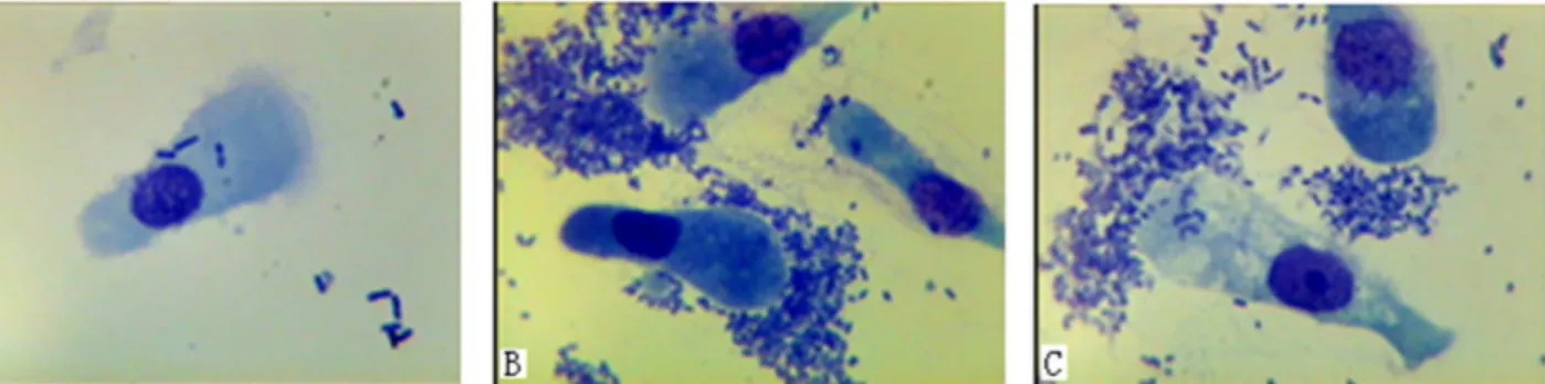
Fig 1. Adhesion test of intestinal epithelial cells fromE.coliF18-resistant and -sensitive piglets.(A) representsE.coliF18-resistant piglets; (B) and
(C) representE.coliF18-sensitive piglets. Images were recorded using an oil immersion lens at 1000 × magnification.
liquid was removed into a separate tube and 100μL 60% ACN/0.1% TFA were added into the
original tubes which were subjected to ultrasonic processing for 15 min. The resultant solutions were combined and freeze-dried. Samples were desalted using Ziptips (Millipore, Bedford, MA, USA) according to the manufacturer’s instructions.
Samples were mixed with 5 mg/mL HCCA matrix at a ratio of 1:1 for second-stage mass spectral analysis (MS/MS) using a 4800 Plus MALDI TOF/TOF Analyzer (Applied Biosystems, USA) with the following conditions: acceleration voltage 2 kV; PMF mass scanning range 800–
4,000 Da; parent ions with signal-noise ratio>50. Peptide mass fingerprint spectra were
ana-lyzed with Data Explore Software to generate corresponding protein peptide sequences. For protein identification, the MS/MS spectra were searched using Mascot software (Matrix Sci-ence, London, United Kingdom;http://www.matrixscience.com) using the genome data ofSus scrofafrom NCBInr (http://www.ncbi.nih.gov).
Bioinformatics analysis of differentially expressed proteins
Differentially expressed proteins corresponding to porcine genes were defined as differential genes. Gene Ontology (GO) Analysis (http://www.geneontology.org/) and Pathway Analysis (http://www.genome.jp/kegg/) of differential genes were carried out to identify all gene-related functions. Using R software (http://www.r-project.org/), Fisher’s exact testing and multiple hy-pothesis testing were carried out to determine the statistical significance (P-value) and false dis-covery rate (FDR) of each differential gene-involved GO Pathway, based on the method of Benjamini and Hochberg [15]. Differential expressed genes within a significant GO Pathway were screened; significant screening standards were defined asP<0.05 and FDR<0.05. The
interaction between differential genes and gene products was identified using the KEGG database.
Real-time PCR analysis
Total RNA (500 ng) was reverse transcribed in a final reaction volume of 10μL using a
Pri-merScript RT reagent kit (TaKaRa Biotechnology Dalian Co., Ltd). The reactions contained 5μL 5 × PrimerScript Buffer, 0.5μL PrimerScript RT Enzyme Mix I, 0.5μL Oligo (dT), 0.5μL
random hexamers and RNase-free H2O. The cDNA was synthesized at 37°C for 15 min
fol-lowed by a termination step at 85°C for 5 s and then stored at−20°C. Real-time PCR
amplifi-cation was performed in 20μL reaction mixtures containing 1μL cDNA, 0.4μL 50 × ROX
Reference Dye II, 10μL 2 × SYBR Green Real-time PCR Master Mix, 7.8μL dd H2O, 0.4μL
(10μM) of each pair of specific primers orGAPDHprimers. All primer sequences are shown
inS1 Table. Real-time PCR was performed on an ABI 7500 system (Applied Biosystems, Fos-ter City, CA, USA). PCR cycling parameFos-ters were: 95°C for 15 s; and then 95°C for 5 s fol-lowed by 62°C for 30 s for 40 cycles. Dissociation curve analysis was performed at the end of the 40 cycles to verify PCR product identity. Each sample was tested three times to obtain average data.
Western blot analysis
To confirm the differential expression of proteins identified by 2-DE inE.coliF18-resistent and -susceptible individuals, total proteins were extracted using a NE-PER kit (Nuclear and Cytoplasmic Extraction Reagents, Thermo Fisher Scientific Inc.) according to the manufac-turer’s protocol. Protein levels were normalized using a BCA kit (Thermo Fisher Scientific Inc.). SDS-PAGE conditions were, 10μL protein loaded to a 10% gel run at 120 V for 90 min.
HSP27 (0.47 mg/mL, 22.9 KDa) or VCL (0.23 mg/mL, 116.9 KDa) (Abmart, Inc.). The sec-ondary antibody was HRP conjugated goat anti-rabbit IgG (Beijing ComWin Biotech Co., Ltd., 1:2500).
Results
Electrophoretic analysis of duodenal proteins in
E
.
coli
F18-resistent and
-susceptible individuals
Duodenal proteins fromE.coliF18-resistent and -susceptible individuals were analyzed by 2-DE. Gel images were obtained with improved reproducibility and higher resolution following silver nitrate staining (S1 Fig). The apparent molecular weights of the majority of protein spots ranged from 14 kDa to 97 kDa, with isoelectric points distributed between 4 and 10. Very few proteins were identified in extremely acidic or alkaline regions.
Differential display of duodenal proteins in
E
.
coli
F18 -resistant and
-susceptible individuals
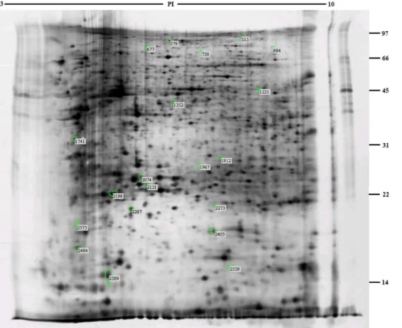
A total of 20 differential protein spots showed a significant change in expression on analysis with Imagemaster software (Fig 2). The group IDs of these protein spots were 515, 579, 677, 684, 730, 1221, 1352, 1741, 1912, 1967, 2074, 2131, 2198, 2273, 2287, 2373, 2403, 2484, 2558 and 2589, respectively. Among these, 10 spots were highly expressed in theE.coliF18-resistant
Fig 2. Analysis of two-dimensional electrophoresis highlighting differentially expressed proteins in duodenal tissues ofE.coliF18-resistant and -susceptible individuals.
group and 10 different spots were highly expressed in theE.coliF18-susceptible group (S2 Table).
Mass spectral analysis of differentially expressed duodenal proteins in
E
.
coli
F18-resistant and -susceptible individuals
MALDI-MS/MS analysis was undertaken for the 20 differentially expressed proteins identified by comparison of the two groups. Peptide fingerprint spectra were successfully obtained for 16 protein spots. Target proteins were identified by searching for homologous proteins and pep-tides in the NCBInr database (http://www.ncbi.nih.gov).
A total of 12 proteins were identified among the 16 differentially expressed protein spots (Table 1). Among these, two groups of spots (2287, 2589, 2484 and 2074, 1967, 1912) repre-sented actin alpha 2 (ACTA2) and albumin (ALB) respectively. Other differentially expressed proteins included four different upregulated proteins in theE.coliF18-resistant group: trans-ferrin (TF); similar to collapsin response mediator protein-2A (LOC100151886); ribosomal protein SA (RPSA); and similar to AGAP005293-PA (LOC100153507). There were also six upregulated proteins in theE.coliF18-susceptible group: vinculin (VCL); aconitase 2 (ACO2); actin, alpha cardiac muscle 1 (ACTC1); actin beta (ACTB); heat shock protein 27 kDa (HSP27); and smooth muscle protein 22-alpha (SM22A).
Gene ontology and pathway analyses of differentially expressed
proteins
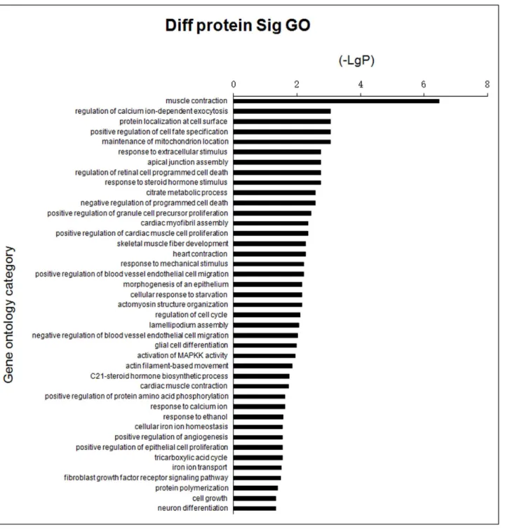
GO analysis was used to identify genes with known functions, including muscle contraction, cell surface protein localization, response to extracellular stimuli and activation of MAPKK (Fig 3,S3 Table). Pathways of differential genes corresponding to the differentially expressed proteins identified in the KEGG database included regulation of the actin cytoskeleton, adhe-rens junction formation, leukocyte transendothelial migration and focal adhesion (Fig 4,S4 Table). In general, each GO or pathway included several of the differentially expressed genes, although no differentially expressed gene was limited to one GO or pathway.
Network diagram analysis of interactions between differentially
expressed proteins
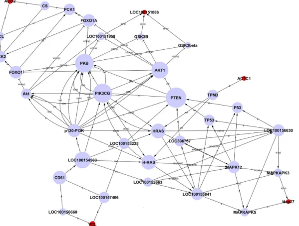
Network diagrams of interactions between differentially expressed proteins were constructed based on the KEGG database information by combining proteins in all significant pathways in order to identify any correlation of target proteins (Fig 5). Due to the small number of differen-tially expressed proteins identified in this study, and limitations on the information in the data-base, the network diagram included only five differential proteins, ACO2, LOC100151886, ACTC1, VCL and HSP27 (indicated by red spots inFig 5), plus connecting proteins. These five proteins were located upstream or downstream of the network. According to the analysis meth-od of graph theory, the nmeth-odes most upstream or downstream of a network are of particular im-portance, which implies that the five differentially expressed proteins considered in the analysis are likely to be related toE.coliF18 infection to some extent (S5 Table).
Verification of differentially expressed proteins related to
E
.
coli
F18
infection
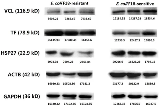
(VCL), actin beta (ACTB) and heat shock protein 27 kDa (HSP27) from theE.coli F18-suscep-tible group. In real-time PCR analysis (Fig 6), it was obvious that expression of TF mRNA in tissue from theE.coliF18-resistant group is significantly higher than in tissue from the suscep-tible group (P<0.05), while the expression levels of VCL, ACTB and HSP27 were significantly
higher in theE.coliF18- susceptible group (P<0.05). Western blot analysis (Fig 7) was
consis-tent with these qPCR results. These data confirm the accuracy of the 2-DE.
Discussion
This study investigated theE.coliF18-susceptiblity status of complete sib-pair healthy individ-uals with extreme phenotypes from a Sutai pig resource colony, using an alvine epithelial cell
Table 1. Differential proteins in duodenal tissues of individuals between theE.coliF18-resistant group and the susceptible group. Spot Protein gi Protein
MW Protein PI Protein score Protein Score C.I% RNA_nucleotide accession
Gene ID Symbol Description Regulation
2373 194037939 56732.2 4.69 116 100 XM_001924781.1 100153507 LOC100153507 Similar to AGAP005293-PA
2.373
1741 122069666 33021.5 4.8 191 100 —— 641351 RPSA Ribosomal protein
SA/37-kDa laminin Receptor precursor/ (LRP)/ 67-kDa laminin receptor (LR) 2.066
730 194041527 62681.8 5.95 113 100 XM_001927796.1 100151886 LOC100151886 Similar to collapsin Response mediator protein-2A
1.881
579 833800 78954.1 6.73 83 99.988 X12386.1 396996 TF Transferrin 1.776
2484 254771936 42381 5.23 79 99.97 FJ547477.1 42381 ACTA2 Actin, alpha 2,
smooth muscle, aorta
1.646
1967 833798 71361.6 5.92 417 100 X12422.1 396960 ALB Albumin 1.603
2287 257470979 42381 5.23 183 100 NM_001164650.1 733615 ACTA2 Actin, alpha 2, smooth muscle, aorta
-2.856
677 17979613 117373.7 5.89 139 100 AF165172.1 396974 VCL Vinculin -2.395
515 113159 86448.5 8.24 77 99.955 —— 396999 ACO2 Aconitase 2,
mitochondrial
-2.334
1912 833798 71361.6 5.92 252 100 X12422.1 396960 ALB Albumin -2.109
2131 50916342 14268.2 5.94 146 100 AY574049.1 493184 HSP 27 Heat shock protein
27kDa
-1.592
2589 254771936 42381 5.23 300 100 FJ547477.1 42381 ACTA2 Actin, alpha 2,
smooth muscle, aorta
-1.589
2403 2984713 10156.2 4.93 152 100 AF053629.1 397021 SM22A Smooth muscle
protein 22-alpha
-1.584
1352 187692583 27588.8 4.98 132 100 EU655628.1 100158242 ACTC1 Actin, alpha, cardiac muscle 1
-1.570
2198 45269029 45162.4 5.55 363 100 AY550069.1 414396 ACTB Actin, beta -1.559
2074 833798 71361.6 5.92 358 100 X12422.1 396960 ALB Albumin -1.558
“Spot”represents the serial number of a protein in two-dimensional electrophoresis;“Protein MW”represents protein molecular weight;“Protein PI” represents protein isoelectric point;“Protein Score C.I%>95”represents successful identification; The positive value of“Regulation”represents up-regulated proteins in theE.coliF18-resistant group compared with the susceptible group, the negative value represents down-regulated proteins.
adhesion assay system. Sib pairs were matched for factors including body weight, hair color, growth performance and genetic background as far as possible, and consistency of the feeding environment was maintained in order to minimize the number of differentially expressed pro-teins. Screening by 2-DE revealed only 20 differentially expressed proteins; sixteen were identi-fied by mass spectral analysis. Excluding redundant proteins, only 12 significant proteins remained, including ACTB, VCL, HSP27 and TF, which were validated by Western blotting.
Fig 3. Bar chart showing GO analysis of differentially expressed proteins in groups ofE.coliF18-resistant and -susceptible individuals.Y-axis represents the name of GO; X-axis represents the minus logarithm of p-value (-LgP).
On functional analysis, these 12 significant proteins were found to be involved mainly in cell adhesion and immune responses. Weaned piglet morbidity depends critically onE.coli
F18-specific receptor expression in the intestinal epithelial cell mucosa. Mouricout et al. pro-posed that the biochemical structures of theE.coliF18 receptor comprised glycoprotein or glu-cosamines [16]. The results of this present study indicated that cell adhesion function-related proteins were represented among the differentially expressed proteins, with upregulated ex-pression of ribosomal protein SA (RPSA) in theE.coliF18-resistant group and upregulated ex-pression of ACTB and vinculin (VCL) in the susceptible group.
RPSA, also known as the 37 kDa laminin receptor precursor (37LRP), the 67 kDa laminin receptor (LR) and ribophorin P40 (P40), is a multifunctional protein encoded by the 37LRP full-length gene [17]. LR is a transmembrane protein containing a variety of monosaccharides and is involved in intercellular mutual recognition and intracellular and extracellular informa-tion transfer. This protein also funcinforma-tions as the host cell surface receptor for laminin, prion protein and many viruses. LR upregulation is closely related to the occurrence of several tissue cancers [18–20]. However, previous studies showed that bacteria and bacterial toxins predomi-nantly combine with glycolipids, with few interactions between the microorganisms and glyco-proteins. It can be speculated that the significance of glycolipids lies in the membrane surface location of the polysaccharides of glycolipids, while glycoprotein glycans are located more dis-tally. The majority of adhesion molecules bind to oligosaccharides containing Galβ4Glc. Occa-sionally, adhesion molecules combine with Galβ4Glc at the end of lactose ceramide, although core oligosaccharides are usually covered by other monosaccharides (such as blood group anti-gens) [21]. Coddens et al. showed that F18-fimbriatedE.coliselectively interact with glyco-sphingolipids having blood group ABH determinants on the type 1 core and blood group A type 4 heptaglycosylceramide [22]. We therefore concluded that LR was not associated with
Fig 4. Bar chart showing pathway analysis of differentially expressed proteins in groups ofE.coliF18-resistant and -susceptible individuals. Y-axis represents the name of pathway; X-Y-axis represents the minus logarithm of p-value (-LgP).
Fig 5. Network of interactions of differentially expressed proteins.Note: Each circle represents a protein (red, differential protein; blue, linking protein). The area of each circle represents the value of degree, i.e. the extent of interactions between one protein and other proteins.
doi:10.1371/journal.pone.0127164.g005
Fig 6. Differential expression ofVCL,TF,HSP27andACTBgenes in duodenal tissue betweenE.coliF18-resistant and -susceptible groups, analyzed by Real-time PCR.
formation of theE.coliF18 specific receptor; upregulation of the laminin receptor in theE.coli
F18-resistant pig group in the present work was possibly caused by the high complexity of pro-teins expressed within the host cells.
The cytoskeletal proteins ACTB and vinculin (VCL) were upregulated in theE.coli F18-sus-ceptible group in our analyses. Studies have shown thatShigellawith the type III secretion sys-tem secretes a series of effector molecules after contact with host cells and interacts with the cell membrane to activate signal transduction pathways associated with cytoskeleton formation and arrangement. This results in rearrangement of the cytoskeleton and further invasion of host cells [23]. Susceptibility toE.coliF18 infections in pigs has been shown to be dependent on the presence of the F18 receptor (F18R) on the porcine intestinal epithelial cells surface [24,25]. However, further research is required to establish whether tight junctions preventing entry of pathogenic bacteria into intestinal epithelial cells of lower tissues can resistE.coli
F18 infection.
Differential protein analysis in this study also revealed upregulation of transferrin (TF) in theE.coliF18-resistant group, and of heat stress proteins (HSPs) in the susceptible group. These two protein types are mainly involved in immune responses. The complete sib pairs in this study were not infected withE.coliF18 and did not have diarrhea or ED symptoms. The immune system is essentially inactive under these conditions and therefore, our findings indi-cated that TF and HSPs might have an effect on innate immune responses toE.coliF18 infec-tion. Heat stress proteins, also known as heat shock proteins, are a group of highly conserved, soluble, intracellular proteins, which are synthesized rapidly under conditions of stress. How-ever, these proteins are also expressed under normal conditions and account for 5–10% of the total cell protein content [26]. David et al. detected expression of HSPs, predominantly HSP27, in heart, liver, lung and other tissues of newborn piglets [27]. Similarly, HSP27 was detected in duodenal tissues of healthy weaned piglets in the present study. HSPs play an important
Fig 7. Differential expression of VCL, TF, HSP27 and ACTB proteins in duodenal tissue betweenE.
coliF18-resistant and -susceptible groups, analyzed by western blot.Each image shows three samples from individual animals.
physiological role in cell growth, development and differentiation, gene transcription, and in protein synthesis, folding and decomposition. HSPs are also critically involved in anti-infective immunity and autoimmunity. Studies have shown that HSPs interact with pathogens invading host cells to form antigenic peptides which are taken up by macrophages and presented by MHC class I molecules via the endogenous pathway, thus stimulating a CD8+T cell response [28,29]. Furthermore, as a Gram-negative bacterium,E.coliF18 releases endotoxin (LPS) after death, which strongly activates the MAPK signaling pathway that regulates inflammatory reac-tions through a multi-stage kinase cascade [30]. For example, LPS activates the p38 pathway, which stimulates intracellular protein kinases such as MAPKAPK2/3 (MAPK activated protein kinase 2/3) and PRAK (p38 regulated/activated protein kinase). These members of the serine/ threonine kinase family are phosphorylated and activated, resulting in HSP27 activation, which mediates cytoskeleton reconstruction and participates in cellular stress responses [31].
Transferrin (TF) is a single-chain glycoprotein. Grange et al. hypothesized that the complex of transferrin with cellular transferrin receptor acts as a specific receptor for theE.coliK88ab pilus [32]. Furthermore, many studies have indicated that the transferrin gene is related toE.
coliK88ab and K88ac resistance [33–35]. Apart from its role in regulation of cell proliferation and the immune system, transferrin is also involved in iron transport and metabolism and has the ability to chelate iron. Iron is an important growth factor for many bacteria [36]. Therefore, transferrin exerts antibacterial and disease-resistance functions as a result of competition with bacteria for iron resources. We speculate that transferrin plays additional roles in the resistance of the piglet intestinal tract toE.coliF18.
Conclusions
Differentially expressed proteins related to adhesion and immunity were identified in screens ofE.coliF18-resistant and -susceptible groups of piglets. 2-DE and Western blot analysis iden-tified four promising candidate proteins, VCL, ACTB, TF and HSP27, which may be related to
E.coliF18 infection. Based on the physiological functions of these proteins, we proposed that transferrin (TF) was the most plausible candidate for theE.coliF18 receptor. In further work, we will study the function of these proteins by reconstructing the TALEN/CRISPR-Cas9 vector and establishing knock-out cell lines in porcine intestinal epithelial cells. Meanwhile further validation and analysis was performed by using the adhesion test for intestinal epithelial cells. Such analysis is required to elucidate the molecular mechanism of porcineE.coliF18 resistance for disease-resistant breeding.
Supporting Information
S1 Fig. Two-dimensional electrophoresis images of duodenal proteins fromE.coli F18-re-sistant and -susceptible individuals.
(DOC)
S1 Table. Primer sequence for differentially expressed genes.The selected genes were identi-fied by real-time PCR. The housekeeping gene,GAPDHwas used as the internal control. The data were analyzed by cycle threshold (C(t)) method.
(DOC)
S2 Table. Information list of differential points of individual duodenal tissues with quanti-tative ratio>1.5 betweenE.coliF18-resistant and -susceptible groups.Note:“-”denotes
down-regulation; values in Group ID column used values of corresponding points in original gel images.
S3 Table. Analysis of significant function of differential protein-corresponding differential genes (41 items).
(DOC)
S4 Table. Differential protein-corresponding differential genes-involved in significant sig-nal transduction pathways (six items).
(DOC)
S5 Table. Interactions of differential proteins with upstream and downstream proteins in interaction network of differential proteins.Note: Degree refers to the extent of the interac-tion between any protein and other proteins. Among these, Indegree represents the number of upstream proteins regulating a protein (arrow points to the protein), Outdegree represents the number of downstream proteins regulated by a protein (arrow points from this protein to the other proteins); Degree is equal to the sum of Indegree and Outdegree, and the line segment be-tween two proteins represents their interaction.
(DOC)
Acknowledgments
We thank Ph.D. Pengpeng Xia, Mingxu Zhou for experimental help. We thank Prof. JinYu Wang for comments on the manuscript. We thank Shanghai Biotechnology Co., Ltd. Shanghai, China for technical assistance.
Author Contributions
Conceived and designed the experiments: WBB SLW. Performed the experiments: ZCW RWX XMY. Analyzed the data: YJH ZCW. Contributed reagents/materials/analysis tools: GQZ YJH. Wrote the paper: ZCW.
References
1. Benin AM, Ducher-Suchaux MF. Relationship between virulence and adherence of various enterotoxi-genic Escherichia coli: strains to isolated intestinal epithelial cells from Chinese Meishan and European large white pigs. Am J Vet Res. 1991; 52: 45–49. PMID:2021252
2. Vogeli P, Meijerink E, Fries R, Stricker C, Bertschinger HU. A molecular test for the detection of E. coli F18 receptors: a breakthrough in the struggle against ED and post-weaning diarrhea in Swine. Schweiz Arch Tierheilkd. 1997; 139: 479–484. PMID:9480539
3. Bao WB, Wu SL, Musa HH, Zhu GQ, Chen GH. Genetic variation at the alpha (1, 2) fucosyltransferase (FUT1) gene in Asian wild boar and Chinese and Western commercial pig breeds. J Anim Breed Genet. 2008; 125: 427–430. doi:10.1111/j.1439-0388.2008.00722.xPMID:19134079
4. Junghans P, Kaehne T, Beyer M, Metges CC, Schwerin M. Dietary protein-related changes in hepatic transcription correspond to modifications in hepatic protein expression in growing pigs. J Nutr. 2004; 134: 43–47. PMID:14704291
5. Yi SS, Oh SJ, Kim IY, Yeom HJ, Yeom SC, Hwang SY, et al. Proteomic analysis of liver in miniature pigs according to developmental stages using two-dimensional electrophoresis and matrix-assisted laser desorption/ionization-time of flight mass spectrometry. Lab Anim Res. 2013; 29: 162–167. doi: 10.5625/lar.2013.29.3.162PMID:24106511
6. Bao WB, Ye L, Zhu J, Pan ZY, Du ZD, Zhu GQ, et al. Microarray analysis of differential gene expression in sensitive and resistant pig toEscherichia coliF18. Anim Genet. 2012; 43: 525–534. doi:10.1111/j. 1365-2052.2011.02287.xPMID:22497274
7. Ye L, Su XM, Wu ZC, Zheng XR, Wang J, Zi C, et al. Analysis of differential miRNA expression in the duodenum ofEscherichia coliF18-sensitive and-resistant weaned piglets. PLoS ONE. 2012; 7:
e43741. doi:10.1371/journal.pone.0043741PMID:22937089
9. Zhang JJ, Zhu GQ. Cloning and expression of F18 fimbrial operon gene clusters from ecterotoxigenic Escherichia coli and their bioactivity. Acta Microbiol Sin. 2007; 47: 790–794. PMID:18062250
10. Wu SL, Yuan ZW, Ju HP, Zhu GQ, Wang JY, Song CY, et al. Study on genetically susceptible piglets of small intestinal epithelium receptors to pathogenic F18 fimbrialEscherichia coliadhesion in vitro. Chin
J Vet Sci. 2006; 26: 622–625.
11. Alwan A, Designan T, Sullivan MO, Kelly J, Farrelly CO. Quantitative assay of Salmonella adherence to intestinal epithelial cells: a new method for assessing novel intervention products. J Microbiol Meth. 1998; 33: 163–170. doi:10.1016/S0167-7012(98)00052-9
12. Coddens A, Verdonck F, Tiels P, Rasschaert K, Goddeeris BM, Cox E. The age-dependent expression of the F18+E.colireceptor on porcine gut epithelial cells is positively correlated with the presence of
histo-blood group antigens. Vet Microbiol. 2007; 122: 332–341. doi:10.1016/j.vetmic.2007.02.007 PMID:17353102
13. Görg A, Weiss W, Dunn MJ. Current two-dimensional electrophoresis technology for proteomics. Prote-omics. 2004; 4: 3665–3685. doi:10.1002/pmic.200401031PMID:15543535
14. Heukeshoven J, Dernick R. Simplified method for silver staining of proteins inpolyacrylamide gels and the mechanism of silver staining. Electrophoresis. 1985; 6: 103–116. doi:10.1002/elps.1150060302
15. Benjamini Y, Hochberg Y. Controlling the false discovery rate: a practical and powerful approach to multiple testing. J R Statist Soc B; 1995, 57: 289–300.
16. Mouricout M, Vedrine B, Grange PH. Interactions between the enteric pathogen and the host. An as-sortment of bacterial lectins and a set of glycoconjugate receptors. Adv Exp Med Biol. 1997; 412: 109–123. PMID:9192003
17. Ardini E, Pesole G, Tagliabue E, Magnifico A, Castronovo V, Sobel ME, et al. The 67-kDa LR originated from a ribosomal protein that acquired a dual function during evolution. Mol Biol Evol. 1998; 15: 1017– 1025. PMID:9718729
18. Gauczynski S, Peyrin JM, Haik S, Leucht C, Hundt C, Rieger R et al. The 37-kDa/67-kDa laminin recep-tor acts as the cell-surface receprecep-tor for the cellular prion protein. EMBO J. 2001; 20: 5863–5875. doi: 10.1093/emboj/20.21.5863PMID:11689427
19. Jamieson KV, Hubbard SR, Meruelo D. Structure-Guided Identification of a Laminin Binding Site on the Laminin Receptor Precursor. J Mol Biol. 2011; 405: 24–32. doi:10.1016/j.jmb.2010.10.028PMID: 21040730
20. Akache B, Grimm D, Pandey K, Yant SR, Xu H, Kay MA. The 37/67-kilodalton laminin receptor is a re-ceptor for adeno-associated virus serotypes 8, 2, 3, and 9. J Virol. 2006; 80: 9831–9836. doi:10.1128/ JVI.00878-06PMID:16973587
21. Varki A, Cummings R, Esko J, Freeze H, Hart G, Marth J. Essentials of Glycobiology. Cold Spring Har-bor LaHar-boratory Press, Cold Spring HarHar-bor, New York, 1999.
22. Coddens A, Diswall M,Ångstrom J, Breimer ME, Goddeeris B, Cox E, et al. Recognition of Blood
Group ABH Type 1 Determinants by the FedF Adhesin of F18-fimbriatedEscherichia coli. J Biol Chem.
2009; 284: 9713–9726. doi:10.1074/jbc.M807866200PMID:19208633
23. Zhu L, Wang H. Type Three Secretion System and Pathogenesis of Shigella. Acta Microbiologica Sinica. 2010; 50: 1446–1451. PMID:21268888
24. Bertschinger HU, Bachmann M, Mettler C, Pospischi A, Schraner EM, Stamm M, et al. Adhesive fimbri-ae produced in vivo byEscherichia coli O139: K12 (B): H1 associated with enterotoxaemia in pigs. Vet
Microbiol. 1990; 25: 267–281. PMID:1980757
25. Frydendahl K, Kåre Jensen T, Strodl Andersen J, Fredholm M, Evans G. Association between the por-cineEscherichia coliF18 receptor genotype and phenotype and susceptibility to colonisation and
post-weaning diarrhoea caused byE.coliO138: F18. Vet Microbiol. 2003; 93: 39–51. PMID:12591205 26. Lindquist S, Craig EA. The heat-shock proteins. Annu Rev Genet. 1988; 22: 631–677. doi:10.1146/
annurev.ge.22.120188.003215PMID:2853609
27. David JC, Landry J, Grongnet JF. Perinatal expression of heat-shock protein 27 in brain regions and non neural tissues of the piglet. J M Neurosci. 2000; 5: 109–120. doi:10.1385/JMN:15:2:109PMID: 11220784
28. Srivastava PK. Interaction of heat shock proteins with peptides and antigen presenting cells: chaperon-ing of the innate and adaptive immune responses. Annu Rev Immunol. 2002; 20: 395–425. PMID: 11861608
30. Dziareke R, Jin YP, Gupta D. Differential activation of extracellular signal-regulated kinase (EBX) 1, ERK2, P38 and c-Jun N-terminal kinase mitogen-activated protein kinases bybacterial peptidoglycan. J Infect Dis. 1996; 174: 777–785. doi:10.1093/infdis/174.4.777PMID:8843216
31. New L, Jiang Y, Zhao M, Liu K, Zhu W, Flood LJ, et al. PRAK, a novel proteinkinase regulated by the P38 MAP kinase. EMBO J. 1998; 17: 3372–3384. doi:10.1093/emboj/17.12.3372PMID:9628874
32. Grange PA, Mouricout MA. Transferrin associated with the porcine intestinal mucosa is a receptor spe-cific for K88ab fimbriae ofEscherichia coli. Infect Immun. 1996; 64: 606–610. PMID:8550214 33. Bilsma IG, Bouw J. Inheritance of K88-mediated adhesion forEscherichia colijejunal brush borders in
pigs: a genetic analysis. Vet Res Commun. 1987; 11: 509–518. doi:10.1007/BF00396368PMID: 2897739
34. Edfors-Lilia I, Gustafsson U, Duval-Iflash Y, Ellergren H, Johansson M, Juneja RK, et al. The porcine in-testinal receptor forEscherichia coliK88ab,K88ac: regional location on chromosome 13 and influence of IgG response to the K88 antigen. Anim Genet. 1995; 26: 237–242. doi:10.1111/j.1365-2052.1995. tb03250.xPMID:7661395
35. Mouricout M, Vedrine B, Grange P. Approach to the molecular basis and genetics of susceptibility/re-sistance to colebacillosis in porcine species. Proceeding of international conference on animal biotech-nology. Beijing: International Acadamic Pubishers. 1997: 47–50.