*e-mail: [email protected]
Article presented at the IV Congresso Latino Americano de Órgãos Artificiais e Biomateriais (COLAOB 2006), August 8 and 11, 2006, Caxambu, MG, Brazil
Effect of Sterilization on the Properties of CDHA-OCP-β-TCP Biomaterial
Loreley Morejón-Alonsoa*, Raúl García Carrodeguasa, José Ángel Delgado García-Menocala,
José Antonio Alonso Pérezb, Salvador Martínez Manentc
a
Centro de Biomateriales, Universidad de La Habana, La Habana, Cuba, 10600 A. P. 6130
bCentro de Investigaciones para la Industria Minero Metalúrgica,
Ministerio de la Industria Básica, Carretera Varona Km 1½, Boyeros, La Habana, Cuba
cDept. of Crystallography and Mineralogy, Faculty of Geology, Universitat de Barcelona,
C/Marti i Franqués s/n, ES-08028 Barcelona, Spain
Received: August 4, 2006; Revised: January 8, 2007
The effect of the method of sterilization on the physical, chemical and mechanical properties of a new bone repairing material was studied. The material was obtained by thermal hydrolysis of β-tricalcium phosphate/ orthophosphoric acid cement and was composed of calcium deficient hydroxyapatite, octacalcium phosphate (OCP), and β-tricalcium phosphate. Partial decomposition of the OCP was observed after sterilization for the three methods. Decomposition increased to the following sequence of sterilization methods: ethylene oxide; autoclaving; dry oven. On the other hand, mechanical strength decreased with regard to non sterilized material in the sterilization sequence: ethylene oxide; dry oven; autoclaving. The compressive strength was 8.5 ± 1.0; 9.0 ± 1.2; 8.2 ± 0.8 and 6.5 ± 1.0 MPa, whereas diametral tensile strength was 2.1 ± 0.3; 2.5 ± 0.1; 1.9 ± 0.9 and 1.6 ± 0.3 for the material sterilized by ethylene oxide, dry oven, and autoclaving, respectively. Several compositional and microstuctural changes were detected after dry heat and autoclave sterilization. Ethylene oxide sterilization had lesser effect on the chemical composition and strength than dry heat and autoclaving.
Keywords: octacalcium phosphate, calcium-deficient hydroxiapatite, tricalcium phosphate, sterilization
1. Introduction
Calcium phosphate bioceramics have been successfully employed as bone repairing and substituting materials in many dental and orthopedic applications due to their excellent biocompatibility and osteoconductivity1-6. Commercial hydroxyapatite (HA), β-tricalcium
phosphate (β-TCP), and biphasic calcium phosphate (BCP) ceramics are currently available in the form of granules and blocks manufac-tured by sintering of powders with the adequate stoichiometry. How-ever, the customary manufacturing process of these ceramics brings about limitations in terms of the size and shape of the implants8-11.
To overcome this handicap it has been recently proposed a new calcium phosphate biomaterial composed by β-TCP, calcium defi-cient hydroxyapatite (CDHA), and octacalcium phosphate (OCP), which can be obtained in practically any desired shape and size12.
The new biomaterial is obtained by hydrothermal processing of conventional dicalcium phosphate dihydrate (DCPD) cement13 and
has been proposed to manufacture tailored bone implants for cranio-maxillo-facial surgery12.
On the other hand, materials implanted into the body must be sterile to avoid subsequent infection that can lead to serious illness or death14. Several sterilization methods have been used for implants, i.e.
gamma and laser irradiation, plasma cleaning, steam sterilization, dry heat or dry oven, chemical treatment with ethylene oxide, etc.15-17.
An effective sterilization method must guarantee the required sterility assurance level with a minimum deleterious effect on the chemical, physical and biological properties of the implant. For ex-ample, it has been reported that steam sterilization causes dehydration of DCPD, hydration of CaO present in some BCP, and has no effect on CDHA18. However, published results on the effect of sterilization
by steam, hydrogen peroxide plasma, ethylene oxide and gamma rays have shown no significant effect on the surface chemistry and in vitro bioactivity of pseudowollastonite bioceramics19.
Among the methods employed to sterilize biomaterials, steam sterilization or autoclaving (AC), dry oven (DO) and ethylene oxide (EtO) are the simplest, cheapest and more commonly available. All of them involve processing above room temperature which may induce decomposition of the hydrated phases present in the new calcium phosphate biomaterial studied in this work, composed by CDHA, OCP, and β-TCP.
Thus, it was considered worthy to study the influence of the most common sterilization methods on the physical-chemical and mechani-cal properties of the CDHA-OCP-β-TCP composite biomaterial.
2. Materials and Methods
2.1. Materials
β-TCP: “Tricalcium phosphate”-labeled reagent (Dried, Extra Pure, Merck 2143) (300 g) with a Ca/P atom ratio of 1.55, and CaHPO4.2H2O (Extra Pure, Riedel-de-Häen 04231) (33.96 g) were ball milled in water for 4 hours. The obtained slurry was dried at 120 ±5 °C. The dry mixture, with a Ca/P atom ratio of 1.50 was heated at 1100 °C during 6 hours. The final product was milled and sieved trough 125 µm mesh20. The powder obtained was characterized
Hydrolysis solution: Saturated solution of Na2HPO4 (Panreac, Prs-Codex) at 27 °C, approximately 1 M.
“Pure” OCP: Was prepared by hydrolysis of DCPD in Na2HPO4/ NaH2PO4 buffer (pH = 6.5; total PO4 = 1 mol/L) at 60 °C for 72 hours. After washing with distilled water and drying to constant weight in air at room temperature, its phase purity was checked by x ray diffraction.
“Pure” CDHA: Was prepared and characterized in the same way than “pure” OCP but using a buffer pH of 8.9.
2.2. Preparation of CDHA-OCP-
β
-TCP composite samples
β-TCP powder and liquid were mixed in a liquid/powder ratio of 0.8 mL/g. The resulting slurry was cast into the holes of silicone moulds and let set. The mould containing the cast material was stored at 100% relative humidity for 24 hours at 27 ± 1 °C. Moulds with cavities of 6 mm diameter x 12 mm height, and 12 mm diameter x 6 mm height were employed.The cylinders or disks were removed and placed into glass flasks with hydrolysis solution in a volume/weight ratio of 5 g/mL. The flasks were sealed and placed in a thermostatic bath at 60 ±1 °C during 72 hours. After this time, the samples were removed from the flasks, washed with distilled water, and dried in air at room temperature.
2.3. Sterilization procedures
Steam sterilization (AC). The standard procedure used for surgical materials21 was employed. Samples were wrapped in porous paper
and sterilized at 121 °C during 20 minutes in a SAKURA FOA 3053 steam sterilizer.
Dry heat sterilization (DO). Dry heat sterilization was carried at 190 °C for 2 hours in a TERRUZI TCA 110P air oven on paper wrapped samples.
Ethylene Oxide (EtO). Samples were placed in sealed LDPE bags and exposed to 80/20 ethylene oxide/carbon dioxide atmosphere (1000 mg/L) during 6 hours at 60 °C. After sterilization, the samples were detoxified at same temperature during 8 hours and aerated for 15 days to remove residual EtO and stored22,23. The sterilization
was carried out using an adapted sterilization chamber SAKURA EO 100.
2.4. Characterization techniques
The qualitative mineralogical composition of samples was studied by XRD in a diffractometer D5000 with a Kristalloflex goniometer (Siemens) and Cu-target. Diffractograms were recorded employing Ni-filtered radiation (λ= 1.5406 Å), and anodic voltage and current of 50 kV and 30 mA, respectively. The step size was 0.05° and the time/step ratio was 1.5 seconds.
The microstructure of samples (surface and fracture surface) was examined by Scanning Electron Microscopy (SEM) using a Scan-ning Electron Microscope JEOL JSM 6300 (Japan). Samples were previously coated with a thin layer of gold.
Compressive (CS) and Diametral Tensile (DTS) strengths were measured in a Universal Testing Machine (Microtest) with a load measuring cell of 5 kN. For CS, cylindrical samples were employed and disc-shaped ones for DTS measurements. Samples were placed between two stainless steel loading plates using carton pads to dis-tribute load. Loading rate was 1 mm/min. The number of replica (n) was six, and Student Multiple Comparison Test was used to compare mean values.
Differential Thermal and Thermogravimetric Analysis were carried out in a Q-1500D Derivatograph in alumina crucibles. The experimental conditions were: heating rate 10 °C/min, maximum temperature 1000 °C, TG sensitivity 100 mg, DTG and DTA
sensi-tivities 500 µV, and α-Al2O3 was the reference.
The thermograms of “pure” OCP and CDHA were obtained and employed to calculate the amounts of these phases present in the experimental materials. The amount of OCP in the materials was calculated from the weight loss between room temperature and 550 °C and the loss experimented by “pure” OCP in the same temperature interval. In a similar way the amount of CDHA was estimated from the weight losses between 550 and 1000 °C of the materials and “pure” CDHA. The amount of β-TCP was estimated as the rest to complete 100 wt. (%).
3. Results and Discussion
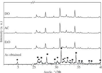
The x ray diffraction pattern of the as obtained
CDHA-OCP-β-TCP biomaterial is shown in the Figure 1. As expected, only three crystalline phases could be identified: OCP (Ca8H2(PO4)6.5H2O, JCPDS 26-1056), CDHA (Ca9HPO4(PO4)5OH, JCPDS 46-905), and
β-TCP (β-Ca3(PO4)2, JCPDS 9-169).
The same phases were present after EtO, AC, and DO steriliza-tion (Figure 1). The intensity of the characteristic diffracsteriliza-tion peaks of OCP clearly decreased with sterilization. This effect increased in the order EtO, AC, DO. On the other hand, the intensity of the characteristic peaks of CDHA and β-TCP seemed not be affected by sterilization.
During the sterilization by the procedures employed in this work, the material is heated to temperatures of 60 °C for EtO, 121 °C for AC, and 190 °C for DO, for several periods of time. It is has been reported that OCP decomposes by heating and the resulting products depend on the temperature range and the duration of heating. By heating at 180 °C the OCP crystalline network collapses and poorly crystallized hydroxyapatite and anhydrous calcium hydrogen phos-phate are formed according to the Equation 124.
2Ca8H2(PO4)6.5H2O → Ca10(PO4)6(OH)2 + (1) 6CaHPO4 + 8H20
However, the reaction products are very hard to detect by powder diffraction techniques and their presence can only be clearly estab-lished by the more sensitive single crystal diffraction technique. On the other hand, CDHA and β-TCP are thermally stable at the temperatures existing during the sterilization of the material by the procedures employed in this work24.
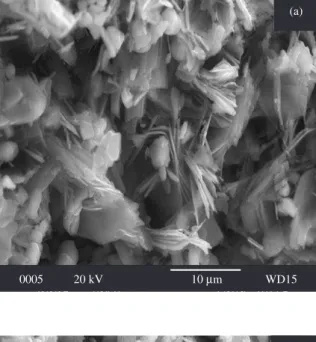
The micrographs of fracture and external surfaces of the
biomate-5 25 30 35 40
Angle, °(2Q)
As obtained
Intensity (a. u.) EtO
AC DO
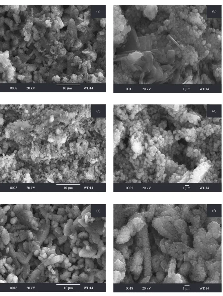
rial are shown in Figures 2 and 3.
Examination by SEM of the fracture surface of the sterilized samples (Figure 2) revealed the presence of large rombohedral particles of β-TCP remaining from the setting reaction of the ce-ment, and aggregates of plate- and ribbon-like crystals of OCP (Figure 2a). Rounded agglomerates of small crystals of CDHA originated from the partial decomposition of OCP were also observed in samples sterilized by AC and DO (Figures 2b and 2c). These morphologies corresponded to those described in the literature for the corresponding pure phases25. As expected, surface micrographs
of the EtO-sterilized sample (Figure 3a and b, up) showed typical globular CDHA and scarce plates of OCP25; however, a considerably
decrease in the porosity of the surface layer of CDHA was found in the AC-sterilized sample, probably due to additional hydrolysis of the inner OCP into CDHA at the high H2O partial pressure and temperature used during sterilization (Figures 3a and b, center). During hydrolysis to CDHA, the OCP crystals existing in the inner core of the material first dissolve in the water provided by the AC sterilizing atmosphere. Part of the CDHA precipitates in the inner porosity of the material, but the saturated solution ascend by capillary forces through the interconnected pore channels to the openings of pores at the material surface, where additional CDHA precipitates as water is removed from the saturated solution by evaporation at the end of the sterilization process. As result the open pores at the surface become clogged by the precipitated CDHA rendering a denser surface layer, even when the inner core of the material has experienced an increase in porosity.
After DO sterilization the surface resembled that of the EtO-steri-lized sample, only globular like CDHA could be identified, crystals of OCP were not observed (Figure 3a and b, down).
The TG, DTG, and DTA plots of the as obtained
CDHA-OCP-β-TCP biomaterial are shown in Figure 4. The main weight loss that starts at 50-60 °C and finishes over 200 °C could be attributed to the OCP decomposition according to Equation 1. The peaks on DTG and DTA plots in the 30-200 °C range evidenced that the process represented in Equation 1 did not proceed in one step but consisted of several partial dehydration processes as previously described;26.
The second step in the TG plot, starting at 250 °C and finishing around 550 °C, can be related to the decomposition of the CaHPO4 produced in Equation 1, according to Equation 224.
2CaHPO4→β – Ca2P2O7 + H2O (2) The final weight loss is attributed to the decomposition of the CDHA into HA and β-TCP as represented in Equation 324.
Ca9HPO4(PO4)5OH →β – 3Ca3(PO4)2 + H2O (3) The residues of the thermal analysis of the as obtained and steri-lized materials were characterized by x ray diffraction as a mixture of β-TCP and β-Ca2P2O7 (JCPDS 9-346).
The TG-DTG-DTA plots for the sterilized materials were very similar to that shown in Figure 4, being the magnitude of the weight losses the unique difference.
The results of the semiquatitative phase composition estimated from the thermogravimetric analysis of samples before and after sterilization by the different methods are listed in Table 1.
The higher the temperature involved in the sterilization, the lower the OCP content in the sterilized material due to the increment of extension of the reaction showed in Equation 1. For the material sterilized with EtO, the content of OCP was abnormally high, even greater than in the as obtained material. An explanation was not found to this result, although it could indicate the presence of adsorbed EtO in the pores of the material, not completely removed during the detoxification process, or the formation of volatile addition products 0005 20 kV 10Mm WD15
(a)
0022 20 kV 10Mm WD14 (b)
0012 20 kV 10Mm WD13 (c)
Figure 2. SEM micrographs of the fracture surface after sterilization. a) EtO;
0008 20 kV 10Mm WD14 (a)
0011 20 kV 1Mm WD14 (b)
0023 20 kV 10Mm WD14 (c)
0025 20 kV 1Mm WD14 (d)
0016 20 kV 10Mm WD14 (e)
0018 20 kV 1Mm WD14 (f)
Figure 3. SEM micrographs of the external surface after sterilization. a) EtO; low magnification; b) EtO, high magnification; c) AC, low magnification; d) AC,
Table 1. Semi-quantitative phase composition of the studied material before and after the treatment by the different sterilization methods, as estimated from thermo gravimetric analysis data.
Sample Maximum T reached during sterilization (°C)
OCP (%) CDHA (%) β -TCP (%)
As obtained R.T. 39 14 47
EtO 60 43 22 35
AC 121 33 18 49
DO 190 28 17 55
R.T.: Room temperature; EtO: ethylene oxide; AC: autoclaving; DO: dry oven.
100 200 300 400 500 600 700 800
W
eight loss (%)
Temperature (°C) Eq. 3 Eq. 1 V oltage ( M V) TG DTG DTA Eq. 2 0 -10 -20 -30 -40 -50 450 400 350 300 250 200 150
Figure 4. DTA, TG and DTG of CDHA-OCP-β-TCP biomaterial before
sterilization.
as glycol or its derivatives during the sterilization process from the reaction of EtO with adsorbed or hydration water present in the mate-rial21. Further research is needed to clarify this effect.
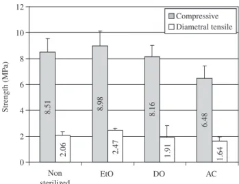
The results of strength measurements are displayed in Figure 5. There was no significant difference among compressive strength values of the as obtained, EtO sterilized, and DO sterilized materi-als. The value of compressive strength of AC sterilized material was significantly lower than the others. There was no significant difference among the values of diametral tensile strength of the as obtained and sterilized materials.
4. Conclusions
The effect of sterilization by autoclaving, dry oven, and ethylene oxide treatment on a new biomaterial composed of octacalcium phos-phate, calcium deficient hydroxiapatite, and β-tricalcium phosphate was investigated. The sterilization methods employed were selected on the basis of their availability, simplicity and cost.
Dry oven sterilization produced the decomposition of over a quarter of the OCP originally present; however, it did not affect the strength of the material. On the other hand, AC had little effect on the composition but significantly decreased the compressive strength of the material. Sterilization by EtO neither affected the phase composition nor the strength of the material. According to these results, EtO sterilization is the best choice among the sterilization methods studied from the compositional and mechanical points of view. However care must be taken to remove the residual EtO or its by-products after sterilization.
Tests need to be conducted to establish the sterility level reached in the studied material by every of the sterilization methods employed in this work.
Acknowledgments
To CYTED Network VIII.J “Biomateriales para la Salud” and Project CICYT MAT2003-08331-C02-01, ICV, CSIC, Spain.
References
1. Hulbert SF, Bokros JC, Hench LL, Wilson J, Heimke G. Ceramics in Clini-cal Applications: Past, Present and Future. Vincenzini P. ed. Amsterdam: Elsevier; 1987.
2. de Groot K. Bioceramics of Calcium Phosphate. Boca Raton: CRC Press; 1983.
Figure 5. Compressive and diametral tensile strength of as obtained and
sterilized material.
2.06 2.47 1.91 1.64
Non sterilized
EtO DO AC
Strength (MP a) Compressive Diametral tensile 12 10 8 6 4 2 0 8.16 6.48 8.98 8.51
3. Ducheyne P, Lemmons J. Bioceramics: Materials Characteristics Versus in Vivo Behaviour. New York Academy of Sciences. 1988; vol. 523. 4. Williams DF. The Biocompatibility and Clinical Uses of Calcium
Phos-phate Ceramics. In Biocompatibility of Tissue and Analogs. Williams D.F. ed. Boca Raton: CRC Press; 1985.
5. Kolberg Ocular Products. Inc. Kolberg-Bioeye Hydroxyapatite Implant. 1998.
6. LeGeros RZ, Lin S, Rohanizadeh R, Mijares D, Legeros JP. Biphasic calcium phosphate bioceramics: preparation, properties and applica-tions. Journal of Material Science: Materials in Medicine. 2003; 14(3):201-209.
7. LeGeros RZ. Crystallographic studies of the carbonate substitution in the apatite structure [Ph. D. Thesis]. New York: New York University; 1967.
8. LeGeros RZ, LeGeros JP. An introduction to bioceramics. Dense hy-droxyapatite. In: Advanced Series in Ceramics. Hench LL, Wilson J, editor. Boca Raton: World Scientific; 1993.
9. Li J, Hermansson L. Mechanical Evaluation of Hot Isostatically Pressed Hydroxylapatite. Interceramic 2, 1990. p. 13-15.
10. Hubbard W. Phisiological Calcium Phosphate as orthopedic implant material [Ph. D. Thesis]. Wisconsin: Marquette University; 1974. 11. Lehr JR, Brown EH, Frazler AW, Smith JP, Thrasher RD. Crystallographic
properties of fertilizer compounds: TVA Chem. Engr. Bull. Alabama: National Fertilizer Development Center; 1967.
13. Bohner M, Lemaitre L. Hydraulic properties of tricalcium phosphate-phosphoric acid-water mixtures. Third euroceramic; 1993 Sept. 12-17; Castellón de La Plana: Faenza Editrice Ibérica SL; 1993. p. 95-100. 14. Kowalsky JB, Morrisey RF. Implants and devices. Sterilization of
Im-plants. In: Biomaterials Science. An Introduction to Materials in Medicine. editor: Ratner B.D. San Diego: Academic Press; 1996.
15. Goldman M, Pruitt L. Comparison of the effects of gamma radiation and low temperature hydrogen peroxide gas plasma sterilization on the molecular structure, fatigue resistance, and wear behavior of UHMWPE. J Biomed Mater Res. 1998; 40(3):378-384.
16. Noah EM, Chen J, Jiao X, Heschel I, Pallua N. Impact of sterilization on the porous design and cell behavior in collagen sponges prepared for tissue engineering. Biomaterials. 2002; 23(14):2855-2861.
17. Takechi M, Miyamoto Y, Momota Y, Yuasa T, Tatehara S, Nagayama M, Ishikawa K. Effects of various sterilization methods on the setting and mechanical properties of apatite cement. J Biomed Mater Res B Appl Biomater. 2004; 69(1):58-63.
18. Dorozhkin SV, Schmitt M, Bouler JM, Daculsi G. Chemical trans-formation of some biologically relevant calcium phosphates in aque-ous media during a steam sterilization. J Mat Sci: Mater Med. 2000; 11(12):779-786.
19. De Aza PN, De Aza AH, Herrera A, López-Prats FA, Pena P. The influ-ence of sterilization techniques on the in vitro bioactivity of wollastonite. J Am Cer Soc. 2006; 89(8):2619-2624.
20. Morejón-Alonso L. Preparation of calcium phosphate monoliths by an hydrothermal route [Thesis for Materials Science M.Sc.]. Havana: University of Havana; 2005.
21. Castro Torres M. Manual de Procedimientos de Enfermería. La Habana: Editorial Ciencias Médicas; 2002.
22. Hidalgo R, Castellanos VM, Chiroles S, Villavicencio O. Dispositivos médicos de uso único procesados por esterilización química mediante óxido de etileno. Rev Cub Hig Epidemiol. 2002; 40(2):89-94. 23. AENOR UNE-EN ISO 10993-7. Evaluación biológica de productos
sanitarios. Parte 7. Residuos de la esterilización por óxido de etileno. In. Madrid; 1996.
24. Elliot JC. Structure and chemistry of the apatites and other calcium or-tophosphates. In: Studies in Inorganic Chemistry. Amsterdam: Elsevier; 1994.
25. LeGeros RZ. Calcium phosphates in oral biology and medicine. San Francisco California: KARGER; 1991.