João Frederico Lourenço dos Santos Barata
Morgado
Licenciado em Bioquímica
Preparation of biopolymer-drug
formulations for cancer drug delivery
Dissertação para obtenção do Grau de Mestre em
Bioquímica
Orientador: Doutora Maria Filomena Andrade de Freitas
Investigadora sénior, FCT-UNL
Co-orientador: Maria Alexandra Núncio de Carvalho
Ramos Fernandes,Professora Doutora, FCT/UNL
Júri:
Presidente: Prof. Doutor Pedro António de Brito Tavares
Arguente: Prof. Doutor Vítor Manuel Delgado Alves
Vogal: Prof. Doutora Maria Filomena Andrade de Freitas
Setembro de 2018
v
Copyright
“Preparation of biopolymer-drug formulations for cancer drug delivery”
Copyright © João Frederico Lourenço dos Santos Barata Morgado, Faculdade de Ciências e
Tecnologia, Universidade Nova de Lisboa
A Faculdade de Ciências e Tecnologia e a Universidade Nova de Lisboa têm o direito, perpétuo e sem limites geográficos, de arquivar e publicar esta dissertação através de exemplares impressos reproduzidos em papel ou de forma digital, ou por qualquer outro meio conhecido ou que venha a ser inventado, e de a divulgar através de repositórios científicos e de admitir a sua cópia e distribuição com objetivos educacionais ou de investigação, não comerciais, desde que seja dado crédito ao autor e editor.
vii First of all, I would like to thank my thesis advisor, Dr. Filomena Freitas, for all the knowledge and know-how shared over the course of this thesis, for all the patience and availability demonstrated to me, for always cheering me up whenever things didn’t work out, and for always promptly devising new ways to keep this thesis moving forward towards proper completion, despite all the bad results obtained.
I would also like the thank my thesis co-advisor, Dr. Alexandra Fernandes, for always pushing me to think on my own rather than depending solely on my advisors, which made me grow as a person and a scientist, for the patience to always answer to my questions whenever I couldn’t get there alone, no matter how dull they were, and for all the new ideas devised when confronted with my bad results, which kept me moving forward.
To both my advisors, thank you very much.
A special thanks goes to Dr. Pedro Viana Batista, for helping me think outside the box and showing me that a bad result is just a matter of perspective.
I would like to leave a thank you to all the people in the two research groups I have been a part of during this year, BioEng and Human Genetics and Cancer Therapeutics, especially to João Pereira, Diana Araújo, Sílvia Baptista, Inês Farinha, Patrícia Reis, Patrícia Freitas, Cristiana Torres (of the BioEng group), Catarina Roma-Rodrigues, Luis Raposo, Andreia Carvalho and Catarina Brás (of the Human Genetics and Cancer Therapeutics) for integrating me in their respective groups, for all the brainstorming, discussions and advices given, as well as for the support provided to me in completing this thesis.
A thank you to my fellow master students in both research groups, Joana Almeida (Human Genetics and Cancer Therapeutics group), Bruno Guerreiro, Patrícia Serrano and Ana Teresa (BioEng) for all the companionship shown to me over this year.
A word of appreciation also to Ana Teresa, Mariana Matos and Liane Meneses for supplying me with the mixed cultures biomass for PHAs extraction, as well as some already extracted polymer.
To my family, which has supported me all my life, inciting me to pursue my dreams, a very heartfelt thank you. For all their invaluable support over my academic course, both during and before college, and for always being there for me, thank you.
Finally, to my girlfriend, Magda Ferreira, for all her love and support, for all the hours spent hearing me rambling and thinking out loud, for all the ideas and brainstorms, for all the nights spent awake on my expense, for all the troubles she went through, for helping me in every way she could, thank you very much.
ix Mannans are highly water-soluble mannose heteropolymers produced by a number of organisms, including yeasts. Polyhydroxyalkanoates (PHAs) are aliphatic polyesters produced by numerous bacteria as a carbon and energy source, with interesting thermal and mechanical properties.
The main objective of this thesis was to prepare different polymeric structures base on mannans and PHAs for use in the pharmaceutical and biomedical areas. Mannans were produced by Komagataella
pastoris using glycerol as carbon source, extracted with a heat-alkali treatment and purified using
dialysis. PHAs were produced by mixed cultures using fermented fruit pulp waste, extracted using chloroform or hypochlorite and purified in ethanol.
A successful deproteinization and an unsuccessful phosphorylation procedure was performed in mannans. The results show a decrease in protein content of 69.49 ± 0.44 % and a decrease in phosphate content of 60.45 ± 1.23 %, respectively.
Mannans were tested in normal fibroblasts, HCT116 and A2780 cell lines for their cytotoxicity, by MTS assay, and no cytotoxicity was discovered. They were then used to prepare gel structures, and gelled using di- and tri-valent cations of iron and copper at low temperature (4 ⁰C) and alkaline pH. Gel particles were obtained in the above conditions and tested for their stability in water. Particles made using tri-valent iron were found the most stable.
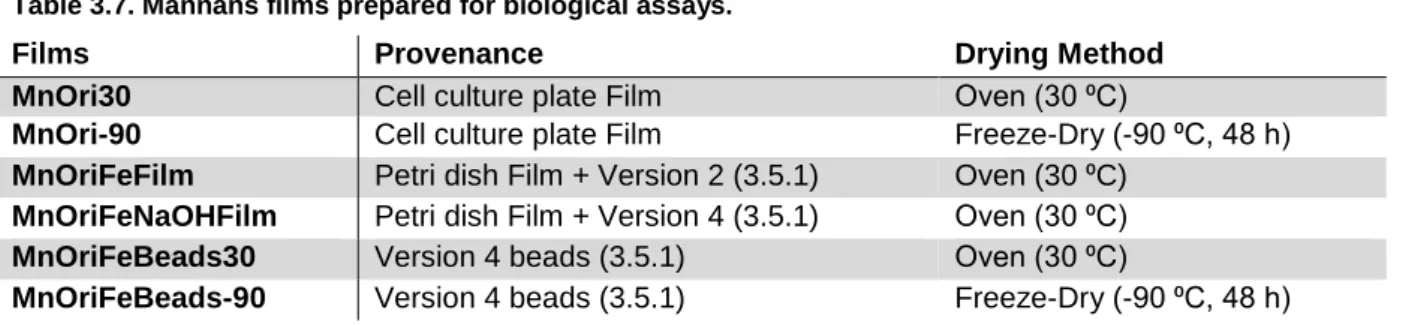
Mannans were also used to produce films. Films were obtained from i) mannans in water dried at 30 ⁰C or freeze dried, ii) from the previously produced films (30 ⁰C) and then coated with iron, at neutral pH or followed by immersion in an alkaline solution and, iii) from gel beads dried at 30 ⁰C or freeze dried. Films were tested for cell adhesion in vitro using normal fibroblasts, but no positive results were found.
PHAs with different HV ratios were used to produce films, pure and blended with mannans, using chloroform as solvent by a solvent casting method. The produced films were tested for cell adhesion in
vitro, using fibroblasts and MCF7-GFP. Pure PHA films were deemed good matrices for this
application with cells adherent to their surface, whereas blend matrices failed in this regard. Some pure PHA matrices were then tested for their cytotoxicity using MCF7-GFP, and with the exception of co-polymer PHBHV with HV content of 18 % (extracted with hypochlorite), they were found to be non-cytotoxic, rendering them useful for biomedical applications such as wound dressing or drug delivery.
Keywords: Mannans; Polyhydroxyalkanoates; Mannans gel beads; Polymeric Films; Cytotoxicity
xi Mananos são heteropolímeros de manose altamente solúveis em água produzidos por vários organismos, incluindo leveduras. Polihidroxialcanoatos (PHAs) são poliésteres alifáticos produzidos por diversas bactérias como fonte de carbono e energia, com interessantes propriedades térmicas e mecânicas.
O principal objetivo desta tese foi preparar diferentes estruturas poliméricas baseadas em mananos e PHAs para uso nas áreas farmacêutica e biomédica. Os mananos foram produzidos por
Komagataella pastoris usando glicerol como fonte de carbono, extraídos com um tratamento térmico
e alcalino, e purificados por diálise. Os PHA foram produzidos por culturas mistas utilizando resíduos de polpa de fruta fermentada, extraídos usando clorofórmio ou hipoclorito e purificados em etanol. Foi realizada em mananos uma desproteinização com sucesso e um procedimento de fosforilação sem sucesso. Os resultados mostram uma diminuição no conteúdo em proteína de 69,49 ± 0,44 % e uma diminuição no conteúdo em fosfato de 60,45 ± 1,23 %, respectivamente.
Os mananos foram testados em fibroblastos normais e nas linhas celulares HCT116 e A2780 para avaliar a sua citotoxicidade, através de um ensaio MTS, e não foi descoberta nenhuma citotoxicidade. Estes foram então usados para preparar estruturas de gel, e gelificaram usando catiões di- e trivalentes de ferro e cobre a baixa temperatura (4 ⁰C) e pH alcalino. As partículas de gel foram obtidas nas condições acima e testadas quanto à sua estabilidade em água. As partículas feitas com ferro trivalente foram consideradas as mais estáveis.
Este polímero foi ainda utilizado para produzir filmes. Os filmes foram obtidos de i) mananos em água, secos a 30 ⁰C ou liofilizados, ii) filmes produzidos anteriormente (30 ⁰C) e revestidos com ferro, a pH neutro ou seguidos de imersão em solução alcalina e, iii) esferas de gel secas a 30 ⁰C ou liofilizadas. Estes filmes foram testados quanto à adesão celular in vitro utilizando fibroblastos normais, mas não foram encontrados resultados positivos.
Foram utilizados PHAs com diferentes rácios de HV para produzir filmes, puros e misturados com mananos, utilizando clorofórmio como solvente. Os filmes produzidos foram testados quanto à adesão celular in vitro, utilizando fibroblastos e MCF7-GFP. Os filmes de PHA puro foram considerados boas matrizes para esta aplicação, com células aderentes à sua superfície, enquanto que nas matrizes de mistura se verificou pouca adesão celular. Algumas matrizes de PHA puro foram então testadas quanto à sua citotoxicidade usando MCF7-GFP e, com exceção do co-polímero PHBHV com teor de HV de 18 % (extraído com hipoclorito), verificou-se não serem citotóxicas, tornando-se úteis para aplicações biomédicas tais como revestimento de feridas e libertação controlada de fármacos.
Palavras-chave: Mananos; Polihidroxialcanoatos; Partículas de gel de mananos; Filmes poliméricos;
xiii Acknowledgements ... vii Abstract ... ix Resumo ... xi Index ... xiii Index of figures ... xv
Index of tables ... xvii
List of abbreviations ... xix
Chapter 1. Introduction ... 1
1.1. Natural Polymers ... 1
1.1.1. Polysaccharides... 1
1.1.2. Yeast Polysaccharides ... 2
1.1.3. Mannans and Mannoproteins ... 2
1.1.4. Polyesters ... 4 1.1.5. Polyhydroxyalkanoates ... 4 1.2. Polysaccharide Gels ... 6 1.2.1. Gel Particles ... 6 1.3. Polymeric Films ... 7 1.4. Motivation ... 7
Chapter 2. Materials and Methods ... 9
2.1. Polysaccharide ... 9
2.1.1. Polysaccharide Production ... 9
2.1.1.1. Inoculum Preparation ... 9
2.1.1.2. Bioreactor Assay... 9
2.1.1.3. Analytical Techniques ... 9
2.1.2. Polysaccharide Extraction and Purification ... 10
2.1.3. Polysaccharide Characterization ... 10 2.1.3.1. Monomeric Analysis ... 10 2.1.3.2. Protein Quantification ... 11 2.1.3.3. Moisture Content ... 11 2.1.3.4. Inorganic Content ... 11 2.1.3.5. Elemental Analysis ... 11 2.1.3.6. Phosphate Content ... 11 2.1.4. Polysaccharide Transformation ... 12 2.1.4.1. Deproteination ... 12 2.1.4.2. Phosphorylation ... 12 2.2. Polyester ... 12
2.2.1. Polyester Extraction and Purification ... 12
2.3. Biopolymer Matrices ... 13
xiv
2.3.2. Film Formation Procedures ... 14
2.4. Cell Cultures ... 17
2.5. Cytotoxicity Assays ... 17
2.6. Cell Adherence Assays ... 18
Chapter 3. Results and Discussion ... 19
3.1. Polysaccharide Production ... 19 3.2. Polysaccharide Characterization ... 19 3.2.1. Monomeric Analysis ... 19 3.2.2. Protein Quantification ... 20 3.2.3. Moisture Content ... 20 3.2.4. Inorganic Content ... 20 3.2.5. Elemental Analysis ... 20 3.2.6. Phosphate Content ... 21 3.3. Polysaccharide Transformation ... 21 3.3.1. Deproteination ... 21 3.3.2. Phosphorylation ... 22
3.4. Polyester Extraction and Purification ... 22
3.5. Biopolymer Matrices ... 23
3.5.1. Gelation Procedures ... 23
3.5.2. Film Formation Procedures ... 27
3.5.2.1. Polysaccharide Films ... 27
3.5.2.2. Polyester Films ... 29
3.6. Cytotoxicity Assays ... 32
3.7. Cell Adherence Assays ... 34
3.7.1. Polysaccharide Films ... 34
3.7.2. Polyester Films ... 36
Chapter 4. Conclusions and Future Work ... 47
xv
Figure 1.1. Simplified scheme for the formation of yeast cell wall components. ... 3
Figure 1.2. Simplified PHA biosynthesis metabolic pathway. ... 5
Figure 2.1. Film formation procedures. ... 16
Figure 3.1. Cell Dry Weight over the course of the bioreactor assay. ... 19
Figure 3.2. Effect of pH and salt concentration on mannans gelation at 4 ⁰C. ... 24
Figure 3.3. Beads resulting from different versions employed for structured gel particles formation. .. 25
Figure 3.4. Early stage mannans films obtained in teflon plates. ... 28
Figure 3.5. Plain mannans films obtained in petri dish and cell culture plate scale. ... 28
Figure 3.6. Methods for PHA/mannans films preparation. ... 30
Figure 3.7. Different polymeric ratios for film formation. ... 30
Figure 3.8. Some PHA films prepared for biological assays. ... 31
Figure 3.9. Mannans influence in cell viability. ... 32
Figure 3.10. Mannans influence in normal skin fibroblast cell viability (high concentrations). ... 33
Figure 3.11. Mannans films used in cell adherence assays. ... 35
Figure 3.12. Microscopic observation of MnOriFeNaOHFilm after the cell adherence period (24 h). .. 36
Figure 3.13. Cell growth in 100PHB films. ... 38
Figure 3.14. Cell growth in 75PHB films. ... 38
Figure 3.15. Cell growth in 100PHBHV17 films... 39
Figure 3.16. Cell growth in 75PHBHV17 films... 39
Figure 3.17. Cell growth in 100PHBHV18 films... 40
Figure 3.18. Cell growth in 75PHBHV18 films... 40
Figure 3.19. Cell growth in 100PHBHV27 films... 41
Figure 3.20. Cell growth in 75PHBHV27 films... 41
Figure 3.21. Cell growth in 100PHBHV38 films... 42
Figure 3.22. Cell growth in 75PHBHV38 films... 42
Figure 3.23. Cell growth in 100PHBHV56 films... 43
Figure 3.24. Cell growth in 75PHBHV56 films... 43
xvii
Table 3.1. Elemental analysis of mannans... 20
Table 3.2. Phosphate and ammonia content of mannans. ... 21
Table 3.3. Effect of pH and temperature on mannans solutions gelation. ... 23
Table 3.4. Effect of pH and salt presence on mannans gelation at 4 ⁰C. ... 23
Table 3.5. Effect of pH and salt concentration on mannans gelation at 4 ⁰C. ... 24
Table 3.6. Different versions employed for structured gel particles formation. ... 25
xix
List of abbreviations
(M6P)-IGF I Mannose 6-phosphate/insulin-like growth factor II receptor
3D-printing Three-dimensional printing
A2780 Ovarian carcinoma cell line
ATCC® American type culture collection
BC Bacterial cellulose
C. albicans Candida albicans
CAD Computer-aided design
CDW Cell dry weight
CGC Chitin-glucan complexes
ChGC Chitosan-glucan complexes
DMEM Dulbecco’s modified eagle medium DMSO Dimethyl sulfoxide
DO Dissolved oxygen
DOX Doxorubicin
dsDNA Double-stranded deoxyribonucleic acid
EPS Exopolysaccharide
FBS Fetal bovine serum
GDP-Man Guanosine diphosphate mannose
GFP Green fluorescent protein
HB 3-hydroxybutyrate
HCT116 Colorectal carcinoma cell line
HPLC High pressure liquid chromatography
HV 3-hydroxyvalerate
ICP-AES Inductively coupled plasma atomic emission spectroscopy
K. pastoris Komagataella pastoris
lcl-PHA Long-chain length PHA
M6P Mannose 6-phosphate
MCF7-GFP Modified breast adenocarcinoma cell line
mcl-PHA Medium-chain length PHA
MEM Minimum essential media
MnOri Original mannans
MnProt Deproteinized mannans
MTS
3-(4,5-Dimethylthiazol-2-yl)-5-(3-carboxymethoxyphenyl)-2-(4-sulfophenyl)-2H-tetrazolium
MWCO Molecular weight cut-off
n.a. Not available/ not tested
OD Optical density
P(3-HB-co-4-HB) Poly(3-hydroxybutyrate-co-4-hydroxybutyrate)
xx
P. holstii Pichia holstii
PCL Poly(ε-caprolactone)
PEG Polyethylene glycol 4000
PET Polyethylene terephthalate
PHA Polyhydroxyalkanoate
PHB Poly-3-hydroxybutyrate
PHB-BC Poly-3-hydroxybutyrate-bacterial cellulose
PHBHV Poly-3-hydroxybutyrate-3-ydroxyvalerate
PLA Polylactic acid
PMS Phenazinemethosulfate
pO2 Dissolved oxygen concentration
pp Precipitate
RH Relative humidity
RPMI Roswell Park Memorial Institute medium
scl-PHA Short-chain length PHA
SD Standard deviation
TCA Trichloroacetic acid
TFA Trifluoroacetic acid
TGF-β1 Transforming growth factor beta 1
UV Ultraviolet
1 Many polymers of both natural and synthetic origin have been proposed and used in a variety of applications, including biomedical applications [1]. Natural polymers present an array of interesting properties suitable for diverse applications, which investigators have tried to replicate in the course of producing synthetic polymers. The natural counterparts, however, have been refined for billions of years, and are therefore still ahead [2].
Natural polymers are called so regarding their biotic origin, commonly being produced in animals or plants, but also in microorganisms. In fact, the interest in microbial products has been, and still is, increasing [3].
Regarding these biopolymers, three different macrostructures arise, comprising polysaccharides, polyesters and proteins [1, 4]. While undoubtfully important, the latter category falls out of interest to the purpose of this thesis, and will therefore be disregarded.
Polysaccharides can either be homopolymers or heteropolymers, respective to their chemical composition. Homopolymers (homopolysaccharides) are constituted by a single repeating monomer, the most common example being glucose homopolysaccharides (such as cellulose) [5]. Heteropolymers (heteropolysaccharides) are constituted by multiple different monomers, in various proportions. Glucose, galactose or rhamnose are among the most common monomers, but other monomers are also often found, such as mannose, N-acetylglucosamine or non-carbohydrate substituents, such as phosphate or glycerate [5].
Many naturally occurring polysaccharides from various origins (plant, algae, crustacean, fungal and bacterial) have been extensively studied and are currently being used in commercial products in different areas (food, pharmaceutical, cosmetic industry, among others), replacing synthetic polymers, as these natural polymers are more biodegradable, biocompatible and non-toxic. Some examples include Arabic gum (plant), carrageenan (algae), cellulose (plant, microbial), xanthan gum or gellan gum (microbial) [6].
Some of these natural sources of polysaccharides (plants, algae and crustacean), however, present meaningful drawbacks in terms of production and polymer extraction, either caused by seasonal constraints (long-term, unreliable productions, both regarding quantity and quality) or expensive purification methods to remove toxic or allergenic natural components, which may otherwise render the polymers inadequate for human use. Microbial polysaccharide production, on the other hand, can be more easily controlled and manipulated. Modern bioengineering techniques can achieve reproducible and high yielding polymer productions, in faster growth and production periods, while
2 sometimes couple those advantages with the use of cheap carbon sources obtained from industrial wastes and by-products [7].
Moreover, biopolymers obtained from microbial sources may comprise a series of inherent mechanical properties (such as gelling capacity, emulsifier, thickener, pH and thermal stability or film forming capacity), as well as functional properties (immune response increase, antimicrobial activity, antimutagenic activity, antitumor activity, among others), and can even be modulated (for example, novel chemical groups or metal addition for specific properties) to achieve intensified or novel properties [3, 8–10].
Yeast polysaccharides have been studied over the last few years, and the many properties ascertained so far have led to their proposal for healthcare related purposes, namely in pharmaceutical and biomedical applications, despite their clinical use still not being widespread [3]. These polymers can be categorized in mainly three types of polymers: cell-wall polysaccharides, capsular and extracellular polysaccharides. Extracellular polysaccharides are synthesized inside the yeast cell, and then transported to the extracellular environment. They can remain free in the medium (EPS) to form an involving matrix to support and protect the culture, as well as provide a means to interact with a possible host, or remain attached to the fungi cell wall (capsular polysaccharides) to confer an extra layer of protection [3].
Cell-wall polysaccharides are almost ubiquitous among yeast, composed of an outer layer of mannans and mannoproteins and an inner layer of D-glucans, chitin and chitosan and their respective complexes with glucans, chitin-glucan complexes (CGC), and chitosan-glucan complexes (ChGC) [3]. The cell-wall polysaccharides have various purposes. The inner layer is mostly responsible for structural support of the yeast cell, accounting for its stability [11]. CGC connects to a network of different glucans, providing some flexibility to the cell wall, as well as functioning as structural elements connecting the rigid CGC shell to the outer mannans and mannoproteins layer [12].
The outer layer of the yeast cell wall is composed of mannans and mannoproteins and is an amorphous matrix of mannose heteropolymers. In yeast cells, these polysaccharides and glycoproteins have many functions, such as controlling the cell wall permeability or interacting with the medium. In some pathogenic strains, such as Candida albicans, they are used by the yeast to adhere to surfaces [13].
3 glucomannans or galactomannans, respectively), are linked by α-1,3-, α-1,6-, β-1,3- and β-1,4- glyosidic links. Mannans backbones are often highly branched, presenting an even vaster array of sugar side chains and their substituents, such as glucose and galactose, but also fucose, xylose, mannose and glucuronic acids, as well as non-carbohydrate versions of the previous (such as phosphorylated versions), to name a few. Some yeast mannans have been reported to have mannose contents over 90 % [3].
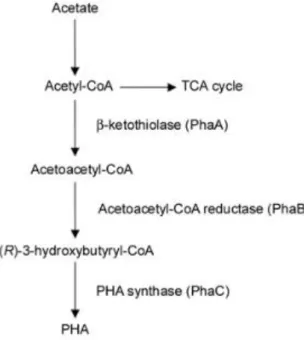
Mannoproteins are glycoproteins constituted by a central protein linked to two side chains of heteropolymer mannan, a long chain (usually up to 100 monomers) which is highly branched, and a smaller chain with usually no more than 5 monomers, which are linked to the central protein through asparagine and threonine or serine residues, respectively. Mannans and mannoproteins are produced in the endoplasmic reticulum from a nucleotide diphosphate mannose, GDP-Man. Mannosyl residues are obtained from GDP-Man by mannosyltransferases, which are then linked together in a linear form and bound to the proteins. The glycoproteins are then conducted to the cell wall by proper vesicles [3]. A scheme comprising the formation of yeast cell wall components is presented on figure 1.1.
Figure 1.1. Simplified scheme for the formation of yeast cell wall components.
Adapted from Freitas et al. [3].
Many different kinds of mannans are naturally produced, with different biological activity properties, including immunomodulator, antimicrobial, antiviral, antioxidant or antimutagenic activity. Remarkably so, phosphomannans (mannans with a high presence of diester phosphate linked side chains) have been proved to possess antitumoral effect, especially when chemically modified to include other substituents such as sulphates [14, 15].
4 Polyesters are polymers whose repetitive units are bound together by ester linkages [16, 17]. They are usually obtained by esterification of dicarboxylic acids and dihydroxyl alcohols, and esterification can occur on either end of the molecules, allowing for vast polymeric chains to be produced [18]. Trihydroxyl alcohols and tricarboxylic acids can also be used, with esterification occurring in multiple sites, in which case the resulting polymer has a chance to contain crosslinks in its structure [18]. Polyesters can be divided into aliphatic and aromatic. Aromatic polyesters are polyesters which contain aromatic residues in their structure, whether part of the original monomers or co-polymerized with it [19]. These aromatic residues are responsible for the polymers lack of biodegradability, as most enzymes’ active center cannot fit the cyclic carbon structure of the aromatic and therefore degrade the polymer [20]. The most notorious example of aromatic polyesters is polyethylene terephthalate (PET), commonly used in the food industry to produce plastic PET bottles [18].
Aliphatic polyesters are usually regarded as polymers with high biocompatibility and biodegradability/bioresorbability, and have therefore been proposed in numerous applications in the medical field, such as sutures, drug delivery vehicles, tissue engineering or implants [21, 22], as well as in food and agriculture fields as packaging materials. These polymers include polylactic acid (PLA) and poly(ε-caprolactone) (PCL) which are commercially available, but also other polymers with interesting properties such as polyhydroxyalkanoates (PHAs) [16].
Polyhydroxyalkanoates (PHAs) are naturally occurring aliphatic polyesters produced by some organisms, including bacteria, as a carbon and energy source. PHAs are synthetized intracellularly by a cascade of enzymatic reactions (figure 1.1). Upon production, PHAs are accumulated in the form of granules in the cytoplasm, surrounded by a monolayer of phospholipids and associated proteins [17, 23]. PHAs can be composed of numerous monomers, which lends co-polymers of these monomers a wide range of physical and thermal properties, including ones that resemble petrochemical-derived polymers currently used in the industry, such as polypropylene or polyethylene [23, 24]. Regarding monomer size, PHAs can be divided in three categories: short-chain length PHA (scl-PHA), with monomers composed of 5 or less carbon units, medium-chain length PHA (mcl-PHA), with monomers ranging from 6 to 14 carbon atoms, and long-chain length PHA, with monomers composed of over 14 carbon atoms. The most common PHAs currently studied are scl-PHA, in which 3-hydroxybutyrate (HB) and 3-hydroxyvalerate (HV) monomers are included [23]. Poly(3-hydroxybutyrate) (PHB) is a scl-PHA homopolymer composed only of HB monomers and is produced by, among others, Cupriavidus
necator (formerly Ralstonia eutropha H16) [24, 25]. This bacterium has also been described to
incorporate HV in this polymer, producing a co-polymer poly(3-hydroxybutyrate-co-3-hydroxyvalerate) (PHBHV). This co-polymer has increased mechanical properties when compared to PHB (such as improved ductility), similar to those of PLA, and is now commercially available [26].
5 Mixed cultures have also been used to produce PHAs. Advantages of this PHA production method include less control steps required for its production, such as sterilization or pH control, and an efficient valorization of wastes, which in turn may lower the overall production costs. Other steps may be necessary, however, such as transformation of the carbon source, as typical carbohydrate substrates may be converted in different products. In such cases, a simple fermentation of the carbon source could be used, yielding fatty acids which are preferential for PHA production [27]. Organisms that accumulate PHA as a carbon and energy stock are naturally selected from the mixed cultures due to their higher adaptability to nutrient variations, and the culture is constantly adapting to the changes in the medium. This is especially useful since mixed cultures PHA production was initially thought for waste water treatment systems, in which medium variability plays an important role in bacterial survival, and accumulated PHA is also a metabolic intermediate to the waste water treatment process by these bacteria [28]. PHAs produced by mixed cultures vary according to the feedstock supplied and may be homopolymers such as pure PHB, or co-polymers such as P(3-HB-co-3-HV) with reported HV contents as high as 82 % [28].
The incorporation of PHAs in the industry, however desirable their properties are, is still going at a slow pace due to their production costs, most notably the recovery of the polymers. In the laboratory, PHA can be obtained with high purity and yield, but most methods employed are undesirable at the industry level. PHA is usually extracted from the cells and purified through the use of organic solvents, such as chloroform, or using enzymatic or chemical cell wall disruption methods, which impair their large scale production due to economic and environmental reasons [23, 25].
Figure 1.2. Simplified PHA biosynthesis metabolic pathway.
6 Some hydrophilic polymers, such as polysaccharides, have the ability to form hydrogels under specific conditions. Hydrogels are polymeric structures composed of water-swollen polymers, with varying degrees of cross-links [29], whose network may contain up to thousands of times the weight of the dry polymer [30].
Hydrogels may be classified in multiple ways. As to their network structure, hydrogels may be classified as permanent/chemical gels, or reversible/physical gels. Permanent gels are those in which the network structure is made by covalent bonding of the cross-links and are usually not homogenous as different sites of the gel may contain more or less cross-links (and therefore less or more water particles, respectively). Reversible gels are held together by the action of secondary forces, such as ionic, hydrogen or hydrophobic forces, as well as molecular entanglements, the latter of which contributes to a non-homogenous gel [30]. When these gels are formed by the action of a multivalent ion of opposite charge of the polymer, an ionotropic hydrogel is formed. The stability of ionotropic hydrogels is very dependent on several factors, such as the valency of the ion used, which will determine the cross-links in the gel, as well as the concentrations of the polyelectrolyte and the multivalent ion. Furthermore, environmental conditions play an important role regarding the use of these gels, as the reversible bonds can be easily broken by changes in the physical conditions of the medium such as ionic strength, pH, temperature, pressure or solute competition [30].
Other methods of classification of gels exist, such as the number of different components in the gel matrix (simple, composed of a single component such as a polysaccharide; mixed, composed of two or more components such as two polysaccharides; or composites, in which the matrix is composed of two or more different components, such as a polysaccharide and a protein) or by the gelation procedure involved, including cold-set, heat-set, ionotropic, pH, or enzyme induced gelation methods [31].
Smaller sized particles (micro and nano) have been increasingly studied due to the different interesting properties shown when particles reach such scales, especially at nano-scale where size-dependent properties really come into play, as surface/volume ratio largely increases [32]. Biopolymers have been investigated to develop these novel sized structures (beads, spheres and capsules), in which different compounds could be encapsulated, including antioxidants, vitamins, pre- and probiotics for food applications [33], but also pharmaceuticals for the medical field [34].
The preparation of such particles should include the use of non-toxic solvents, be economically feasible and account for the properties desired for the matrix, including particle size and shape, surface charge and permeability to the encapsulated components. Various methods for the preparation of polymeric particles are currently used, including emulsification (blending two immiscible
7 liquids through agitation), extrusion (dropwise addition of a polymeric solution into an ionic solution), spray drying (hot air spraying of a solution for quick solvent evaporation), coacervation (electrostatic interaction of opposite charged biopolymers) and precipitation (based on a varying capacity of the solution to solubilize the biopolymer, using two solutions with different solubilization capacities for the polymer) [34].
Polymeric films are thin layers of a polymeric material or blend of materials [35]. Natural polymer films have been reported for their good mechanic or barrier properties and were proposed to be used in the food industry (in applications such as packaging) or the medical field (as topic drug delivery and wound healing systems). Such polymeric films are usually easy to manufacture, cost-effective and biocompatible, and sometimes display adhesive properties [36].
Regarding porous cell scaffolds for the medical field, several methodologies for film formation have been described, including solution casting and solvent evaporation, and particle leaching (consisting of a dissolved polymer with insoluble particles suspended on that specific solvent being cast on a surface for the evaporation of the solvent, and then washed with a different solvent to remove the particles) or emulsion freeze-drying (in which an emulsion of two immiscible liquids, one of which contains the dissolved polymer, is freeze-dried) [37]. Other techniques include gas foaming (in which a salt is used to release gas particles into the medium under certain conditions, such as temperature, followed by freeze drying) or 3D-printing of the scaffold (using CAD designs) [37, 38]. Among porous films for biological uses, PHA films (PHB and PHBHV) have been investigated and reported as suitable for constructing in vitro matrices [39]. Furthermore, PHB and copolymeric P(3-HB-co-4-HB) have been successfully blended with a polysaccharide (bacterial cellulose, BC) and the respective blends (PHB-BC and P(3-HB-co-4-HB)-(PHB-BC) have been tested in fibroblasts. The results show that the polyester-polysaccharide blends produce much better porous matrices for cell adhesion, increasing the number of adherent cells when compared to the polyester films alone [40, 41].
As mentioned above, various polymeric structures have been proposed for the medical and pharmaceutical fields and were proven quite adept to be used as drug delivery systems or tissue regenerating scaffolds. Among such structures, polysaccharide gel particles and polymeric films stand out [34, 36]. Mannans are hydrophilic and biocompatible components, with proven interesting properties such as immunomodulator, antimicrobial, antiviral, antioxidant, antimutagenic and antitumor activity [3, 14, 15]. PHAs are biocompatible polymers with diverse possible arrangements and interesting physical and thermal properties, comparable to those of commercially available polymers
8 Nevertheless, any polymer proposed for biological applications must be individually submitted to extensive and thorough tests, to evaluate its biocompatibility and cytotoxicity. In vitro tests are essential and determinative forefront tests than must be performed to assess a material’s biological compatibility. They represent the frontline safety barrier for compounds to reach human (and animal) applications, and though they are not representative of complete systems (such as organs or the full body), in vitro tests grant researchers relevant insights on how compounds will affect their targets in
vivo. As such, a proper choice of the cell models and the employed methodologies must be made in
order for in vitro tests to be representative.
As such, the objective of this thesis was to produce and extract these polymers from their producing organisms (mannans from the cell-wall of the yeast Komagataella pastoris and PHAs from the biomass of a mixed microbial culture) and use them to create different structures for biological applications. Mannans were tested for their gelling capacity and used to manufacture different gel particles using di- and trivalent cations following ionotropic gelation procedures. The produced gel particles were tested with different tumoral cell lines and a regular skin fibroblast cell line, in order to be potentially used as a drug delivery system for cancer. PHAs and mannans were used to produce polymeric films (both alone and blended) and their cytotoxicity and capacity to support cell adherence was determined using normal fibroblasts and a tumoral cell line, envisaging their use as drug delivery systems or wound dressings.
9
Komagataella pastoris (DSM 70877) was used to produce the polysaccharide in use, mannans. The
yeast was cultivated in medium K, with the following composition: KH2PO4, 28 g L-1; CaSO4.2H2O, 0.125 g L-1; MgSO4.7H2O, 2 g L-1; (NH4)2SO4, 13.5 g L-1. Medium K was supplemented with 2 mL L-1 of a trace mineral solution, with the following composition: CuSO4.5H2O, 2 g L-1; MuSO4.H2O, 3 g L-1; ZuCl2, 7 g L-1; FeSO4.7H2O, 22 g L-1; Biotin, 0.2 g L-1; H2SO4, 5 mL L-1. The pH was adjusted to 5.0 with NaOH 2 M.
The inoculum was prepared in four 500 mL shake flasks, adding 1 mL of cryopreserved yeast to each of them holding 200 mL of medium K supplemented with 45 g L-1 glycerol (Scharlau, 86-88 % w/w). The inoculum was incubated for 40 h at 30 ⁰C and 200 rpm in an orbital shaker (IKA KS 260 basic). Medium K and glycerol were sterilized separately by autoclaving (Uniclave 77, Portugal) at 121 ⁰C for 20 min, and the trace mineral solution was sterilized by filtration (Whatman, 0.2 µm).
The production assay was performed in a 10 L bioreactor (BioStat B-plus, Sartorius), with a starting volume of 8 L, using medium K prepared as described above (2.1.1.1.1.). The inoculum was added in a 10 % (v/v) ratio to the bioreactor.
The culture was grown in batch mode until the depletion of the carbon source (at around 24 h), at which point a fed-batch phase was initiated and kept for the remainder of the assay (26 h). For the fed-batch phase, 3430 g of glycerol were used, supplemented with 67 mL of trace mineral solution, at a constant flow of 144.64 g h-1. The assay was conducted with controlled temperature (30 ± 0.1 ⁰C), as well as controlled pH (5.0 ± 0.02), through the addition of either HCl 2 M or NH4OH 25 % (v/v). The dissolved oxygen concentration (pO2, %) was controlled by the automatic adjustment of the stirring (between 300 and 1500 rpm) by two six-blade impellers. The setpoint was 15 % of the air saturation. Periodic samples (10 mL) were retrieved from the bioreactor for biomass quantification.
Cell growth was monitored during the experiment by measuring the optical density at 600 nm (OD600nm) in a UV-Vis spectrometer (VWR V-1200). The absorbance of the samples was kept under
10 0.3 by dilution of the broth with deionized water.
Biomass quantification was performed gravimetrically. The cell dry weight (CDW) was determined by treatment of the acquired samples, namely by centrifugation (Sigma, 4-16KS) at8875 g for 15 min at 4 ⁰C to remove the supernatant, and subsequent washing of the pellet with deionized water. The washed biomass was afterwards deep frozen (80 ⁰C) and freeze dried (Telstar, Cryodos) for 48 h at -90 ⁰C. The CDW was defined by the resulting weight of the lyophilized pellet.
NaOH 2 M was added in a 1:1 (v/v) ratio to the culture broth for an alkali-heat treatment, as previously described by Freitas and co-workers [42]. The resulting mixture was heated up to 65 ⁰C, for 2 h under constant stirring (400 rpm). After cooling, the mixture was subjected to centrifugation (13131 g, 15 min, 4 ⁰C) for separation of cell-wall polymers.
Mannans were recovered from the supernatant (alkali soluble fraction) and dialyzed with a 12000 MWCO membrane against deionized water at room temperature (20 ⁰C) for 48 h. The dialysis was performed under regular water exchanges accompanied by pH and conductivity measurements, being stopped at neutral pH (between 6.0 and 7.0) and low conductivity (< 50 µS cm-1). The resulting dialyzed polymer was then freeze dried (Telstar, Cryodos) for 48 h at -90 ⁰C. The polymer was kept in closed sample containers (30 mL) with screw caps, at room temperature and sheltered from light and moisture until use.
Mannans were analyzed regarding their monomeric composition. Briefly, 5 mg of polymer were digested with 2 % trifluoroacetic acid (Sigma-Aldrich, 99 %) in deionized water at 120 ⁰C for 2 h. The resulting hydrolysate was diluted (1:5 v/v in deionized water) and filtered (0.2 µm nylon membrane, VWR) for high pressure liquid chromatography (HPLC).
HPLC analysis was conducted using a CarboPac PA10 4x250 mm + Aminotrap column (Dionex) equipped with an amperometric detector, using NaOH 18 mM at 1 mL min-1 as eluent, at 25 ⁰C. Glucose (Fisher Scientific, 99.5 %), mannose Aldrich, 99.0 %) and glucosamine (Sigma-Aldrich, 99.9 %) were used as standards in ultrapure water ranging from 1 to 100 µg mL-1, covering the concentrations of monomer contents of the mannans samples.
11 Protein content of the samples was determined by a protein precipitation method, using trichloroacetic acid (TCA) (Scharlau, 99.5 %). A mixture of 1 g of polysaccharide with 2 g of TCA was made in ultrapure water, and the solution was left at 4 ⁰C for 15 min. After the settling period, it was centrifuged (7155 g, 15 min, 4 ⁰C) and the protein pellet was separated from the supernatant. The supernatant was submitted to the same process until there was no visible pellet formation, after which it was dialyzed by the process described above (2.1.1.2.). The various pellets were put together as one, repeatedly washed with deionized water, and both pellet and supernatant were freeze dried as described in section 2.1.1.2. and sent for elemental analysis. Precipitated protein was quantified gravimetrically from the precipitation pellet, and total protein was estimated based on a 6.25 factor of total nitrogen found in the sample, a non-specific factor developed by Jones [43].
The moisture content of the polysaccharide was measured gravimetrically, by leaving a predetermined amount of mannans to dry in an oven (100 ⁰C) overnight. The polymer weight was then measured and left in open air until constant weight (48 h).
Mannans inorganic content was determined gravimetrically, by placing a predetermined amount of polymer (50 mg), previously dried at 100 ºC for water removal, in an oven (Nabertherm, LE060K1BN Compact Muffle Furnace) at 500 ⁰C overnight. The resulting powder was then weighted. The assay was done in triplicate.
For elemental analysis, a predetermined amount of polysaccharide was delivered as obtained from the purification step. Elemental analysis was performed regarding nitrogen, carbon, hydrogen and sulfur using an Elemental Analyzer (Thermo Finnigan-CE Instruments, Flash EA 1112 CHNS Series), using a Multiseparation Column (SS; 2 m; 6x5 mm). Combustion reactor temperature was of 950 ⁰C and sulfanilamide was used as standard, with a composition of 16.26 % N, 41.85 % C, 4.68 % H e 18.62 % S, using factor K as calibration.
12 Samples were prepared by diluting the polysaccharide (10 mg ml-1) and were then sent for analysis. Analysis was performed using an ICP (Ultima model, Horiba Jobin-Yvon, France) equipped with a 40.68 MHz RF generator and a Czemy-Turner sequential monochromator with 1.00 m and a nebulizer (MiraMist). Phosphorus Standard for ICP 1000 mg L-1 (Sigma-Aldrich, Trace-CERT) was used as standard, diluted in ultrapure water ranging from 1 to 100 mg L-1.
Deproteinized mannans were obtained using the protein quantification method described above (2.1.1.3.2.), by recovery of the protein-free supernatant followed by dialysis using a 12000 MWCO membrane (20 ⁰C, 48 h) and freeze drying (-90 ⁰C, 48 h). The polymer was kept in closed sample containers (30 mL) with screw caps, at room temperature and sheltered from light and moisture until use.
A phosphorylation procedure, adapted from DiLuzio [44], was applied to mannans, in an attempt to increase mannans content in phosphorous. Briefly, 1 g of mannans was dissolved in 50 mL DMSO and heated up to 100 ⁰C, at which point 10 mL of orthophosphoric acid (H3PO4, Sigma-Aldrich, 85 %) were added dropwise, under constant magnetic stirring (400 rpm). The temperature of the reaction was maintained for 6 h, after which the solution was allowed to cool at room temperature. After cooling, it was dialyzed using a 12000 MWCO membrane (20 ⁰C, 48 h) and freeze dried (-90 ⁰C, 48 h) to obtain the phosphorylated polymer. The polymer was kept in closed sample containers (30 mL) with screw caps, at room temperature and sheltered from light and moisture until use.
Polyesters, namely polyhydroxyalkanoates (PHAs), were extracted from previously produced mixed cultures dry biomass. Biomass with accumulated PHA was provided by Mariana Matos and Liane Meneses from the BioEng group. PHA production occurred in a three-step process: a first step consisting in the acidogenic fermentation of a fruit waste pulp (supplied by SumolCompal SA, Portugal) into organic acids and ethanol, used as carbon source for the production of PHAs; a second step consisting of a Feast and Famine feeding mode to select PHA accumulating bacteria; and a third step, where the selected microorganisms were fed the resulting carbon source from the acidogenic fermentation process in a pulse feeding mode (controlled by DO), under limiting nitrogen conditions to impair cell growth [28]. The third step was conducted in a 10 L bioreactor for the production of PHA
13 with 17 % HV content, and in 50 L bioreactors for the production of PHAs with 18, 27 and 38 % HV contents.
The extractions occurred using a Soxhlet extractor (250 mL extractor, Lenz-Laborglasinstrumente), in which a biomass filled cartridge (working as a thimble) was placed inside the main chamber of the apparatus (holding 5 g of dry biomass), and chloroform (Honeywell, 99.4 %) was used as solvent (250 mL). The extraction period was of 24 h at 90 ⁰C. After the extraction, the solution was recovered, containing both the polyester and membrane lipids dissolved in chloroform. The purification process consisted of pouring the solution dropwise in glacial ethanol (Carlo Erba, 99.9 %) in a 10 % (v/v) ratio to remove the lipidic fraction of the sample. Lipids dissolved in ethanol, while the polyester turned insoluble and precipitated. The extraction solvent/ethanol mixture was submitted to repeating freeze-thaw cycles (-20 ⁰C for 4 h) to ensure maximum precipitation of polyester. The polymer was then removed from the mixture and left to dry inside the fume hood at room temperature. PHAs with different compositions were obtained, namely co-polymers of hydroxybutyrate (HB) and 3-hydrovalerate (HV) with varying HV contents (ranging from 17 % HV to 56 % HV).
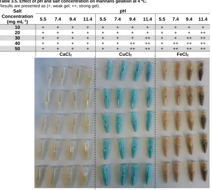
Gelation procedures were tested using mannans. In a preliminary phase, it was tested the effect of pH, temperature and salt addition, in various crossed experiments. Individual mannans solutions (50 mg ml-1) were submitted to different pH values (1.9, 5.6 and 9.6) at 4 ⁰C for 96 h, as well as pH 5.6 at autoclave temperature (121 ⁰C) for 20 min, followed by 96 h at 4 ⁰C. In a secondary experiment, different salts were added to fresh mannans solutions (50 mg mL-1) at the three tested pH values, namely FeCl2.4H2O (Sigma-Aldrich, 99.0 %), CuCl2.2H2O (Sigma-Aldrich, 99.0 %) and CaCl2 (PanReac AppliChem, 95 %) at 20 mg mL-1, and the solutions were kept at 4 ⁰C for 72 h.
In a second phase, fresh mannans solutions (10 % (w/w)) were set at pH 5.5, 7.4, 9.4 and 11.4, and for each pH different concentrations (from 10 to 50 mg mL-1) of the previously used salts were tested. Samples were kept at 4 ⁰C and evaluated periodically over 144 h.
In both phases, mannans solutions were prepared using deionized water and the pH was set using either HCl 2 M (Fischer Scientific, 37 %) or NaOH 2 M (EKA, 97 %).
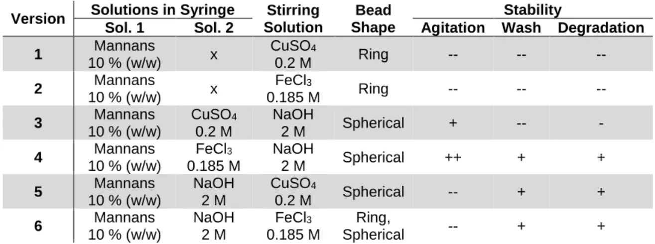
The third phase of the gelation procedures was the obtainment of gel structures. Six different versions of this experiment were performed, using either CuSO4.5H2O (Riedel-de Haën, 99.0 %) or FeCl3.6H2O (Sigma-Aldrich, 98.0 %), as follows:
1) Dropwise addition of mannans solution (10 % (w/w) in H20) to a CuSO4 50 mg mL-1 solution; 2) Dropwise addition of mannans solution (10 % (w/w) in H20) to a FeCl3 50 mg mL-1 solution;
14 3) Dropwise addition of mannans (10 % (w/w) in H20) and CuSO4 50 mg mL-1 solution to a NaOH
2 M solution;
4) Dropwise addition of mannans (10 % (w/w) in H20) and FeCl3 50 mg mL-1 solution to a NaOH 2 M solution;
5) Dropwise addition of mannans solution (10 % (w/w) in NaOH 2 M) to a CuSO4 50 mg mL-1 solution;
6) Dropwise addition of mannans solution (10 % (w/w) in NaOH 2 M) to a FeCl3 50 mg mL-1 solution.
The dropwise addition of the mannans solution (1 mL) was made using sterile needles (HenkeSassWolf, Fine-Ject, 23 G – 0.6 x 25 mm) into 30 ml of the cation solution. The structures obtained were left under stirring for 30 min and were then washed using deionized water (30 mL) for removal of excess salt and lowering of the pH value, controlled by measurement of pH (until neutral pH) and conductivity (until below 50 µS cm-1). The structures were then freeze dried (-90⁰C, 48 h) and weighted. Version 4 was also repeated twice, in which the washing process was changed to either centrifugation (7155 g, 10 min, 4 ⁰C) or dialysis against deionized water (20 ⁰C, 24 h). In either case, the structures were then freeze dried (-90 ⁰C, 48 h) and weighted.
For preparation of mannans films, 500 mg of the polymer were dissolved in 5 mL of deionized water, at room temperature, under magnetic stirring (300 rpm) until complete dissolution. Two other variants of the above were performed, one to which 50 mg of glycerol (Scharlau, 86-88% w/w) were added, and another to which 50 mg of polyethylene glycol 4000 (PEG) (Fluka, Sigma-Aldrich) were added. These two components were used as plasticizers, to account for the natural brittleness of the polymeric films. The solutions were cast on Teflon plates (50 mm), and the solvent was left to evaporate in an oven (Raypa) at 30 ⁰C for 120 h (figure 2.1 A).
In a second stage, 1 g of mannans was dissolved in 5 mL of deionized water, at room temperature, under magnetic stirring (300 rpm). This procedure was repeated, with the addition of 50 mg PEG, and both films were left to dry in an oven (Raypa) at 30 ⁰C for 120 h. A third variant was performed as was the first (1 g mannans in 5 mL deionized water) but was instead freeze dried (-90 ⁰C, 48 h) (figure 2.1 B). The first and the third variant of this assay were reproduced in plastic petri dish scale (Labbox, 90 mm diameter) as well as cell culture plates (VWR, 35 mm diameter) at 10 % (w/w). The first variant (petri dish) was then used to mimic some procedures developed for gelation purposes. One piece of film was placed in a FeCl3 50 mg mL-1 solution for 2 h, and then relocated to a NaOH 2 M solution for 2 h. Another piece was placed in a FeCl3 50 mg mL-1 solution for 2 h, and the final piece was placed inside a NaOH 2 M solution for 2 h. All three pieces of film were later washed with deionized water to
15 either lower pH (up until neutral pH) and/or conductivity (below 30 µS cm-1) (figure 2.1 C).
Also, films performed using iron gelling structures (2.2.1, version 4) dried in cell culture plates (both in an oven at 30 ⁰C and by freeze drying at -90 ⁰C for 48 h) were obtained (figure 2.1 D).
16
Figure 2.1. Film formation procedures.
(A) and (B) represent early stages of film formation, in which plasticizers (glycerol and PEG) were tested, and films were cast on Teflon plates. (C) represents version 1 and 3 of figure B (left and right arrows, respectively) being cast on both petri dish and cell culture plates, as well as the transformations employed on the petri dish film (30 ⁰C, 120 h). (D) represents films performed using iron gelling structures. Red “X” marks non-used films, whereas “Biological Assays” marks films used for posterior cell adherence assays.
In a later stage, films of both polyester alone and in conjunction with mannans were prepared, by dissolving both polymers and allowing the solvent to evaporate inside a desiccator (Schott, Vakuumfest) in the fume hood at room temperature. Firstly, two methods were tested, in which PHB was dissolved in chloroform and mannans were either dissolved in deionized water or suspended in the PHB solution in chloroform. The second method was used in later assays. Secondly, an assay for optimal PHB/mannans ratio was conducted, testing the two polymers at different ratios, namely 25/75 % (w/w), 50/50 % (w/w) and 75/25 % (w/w), the latter of which was chosen for the final tests.
The various PHA co-polymers obtained (150 mg), with HV contents comprehending 0 % (HB homopolymer), 17 %, 18 %, 27 %, 38 % and 56 % HV (w/w), were then dissolved in 10 mL of chloroform (alone – 100 % (w/w) PHA; or alongside mannans – 75/25 % (w/w) PHA/mannans ratio) under magnetic stirring (500 rpm) and allowed to dry inside a desiccator (Schott, Vakuumfest) in the fume hood, at room temperature.
17 Three adherent cell lines (colorectal carcinoma cell line – HCT116; ovarian carcinoma cell line – A2780; normal human skin fibroblasts) were purchased from American Type Culture Collection (ATCC®), and one adherent cell line (modified breast adenocarcinoma cell line – MCF7-GFP) was purchased from Cell Biolabs Inc (San Diego, CA, USA). HCT116 and normal fibroblasts were grown in Dulbecco’s Modified Eagle Medium (DMEM) (Gibco, Life Technologies, USA), supplemented with 10 % (v/v) FBS (Gibco, Life Technologies, USA) and 1 % (v/v) antibiotic/antimycotic solution (Invitrogen). MCF7-GFP was grown in DMEM (as above) supplemented with an additional 1 % (v/v) MEM non-essential amino acids (Gibco, Life Technologies, USA). A2780 was grown in Roswell Park Memorial Institute (RPMI) medium (Gibco, Life Technologies, USA) supplemented with 10 % (v/v) FBS and 2 mM glutamine. All cultures were maintained in an incubator (SANYO, CO2 Incubator, Electrical Biomedical CO., Osaka, Japan) at 37 ⁰C, with an atmosphere of 5 % (v/v) CO2 and 99 % (v/v) relative humidity (RH).
Cytotoxicity assays were performed for both mannans and its deproteinized version, in HCT116, A2780 and normal fibroblasts. The three cell lines were independently seeded in 96-well plates (VWR, Radnor, Pennsylvania, USA) at a density of 0.75 x 10⁵ cells per well and left to adhere for 24 h in an incubator at the conditions described above. After 24 h, seeding medium was replaced with fresh medium containing either regular or deproteinized mannans (0 – 2000 µg mL-1) or control components (0.4 µM of doxorubicin (DOX), 0.4 µM of dimethyl sulfoxide (DMSO), TCA 0.5 % (w/v)) and left for 48 h in the same conditions. After 48 h, both polysaccharide and control loaded media were replaced with fresh medium containing 20 % (v/v) Cell Titer 96 ® AQueous Non-Radioactive Cell Proliferation Assay Kit (Promega, Madison, USA) (composed by 3-(4,5-Dimethylthiazol-2-yl)-5-(3-carboxymethoxyphenyl)-2-(4-sulfophenyl)-2H-tetrazolium (MTS inner salt) and phenazinemethosulfate (PMS, coupling reagent)). After a new incubation for 45 min (under the conditions described above), absorbance measurement at 490 nm was performed for each well using Tecan Infinite F200 Microplate Reader (Tecan, Männedorf, Switzerland). Cell viability (%) is given by (Eq. 1):
Eq.1
A cell density of 0.75 x 10⁵ cells per well was achieved by Trypan Blue Exclusion method. Cells were centrifuged at 500 g and resuspended in 1 mL of appropriate medium, followed by dilution to 10 % (v/v) alongside Trypan Blue reagent (Sigma, St. Louis, USA) 20 % (v/v) in the same growth medium. This solution was inserted in a hemocytometer (Hirschmann, Eberstadt, Germany), and cell count was performed by microscopic observation of viable cells using an Olympus CXX41 inverted microscope (Tokyo, Japan). Cell density (cells mL-1) is given by (Eq. 2):
18 Eq. 2
Cell adherence assays were performed for previously described films using either normal skin fibroblasts or MCF7-GFP. Cell line MCF7-GFP was used due to microscopic observation constrains when using normal skin fibroblasts, as the films opacity cloaked the visualization of these cells. MCF7-GFP produces Green Fluorescent Protein (MCF7-GFP), whose fluorescence was used to overcome the visualization constraints. Mannans films (regular mannans films (dried at 30 ⁰C or freeze dried), iron gelling structure films (dried at 30 ⁰C or freeze dried), and films obtained from manipulation of mannans film to mimic gelation procedure methods (first and second pieces, both dried at 30 ⁰C)) were tested using regular fibroblasts. These films were prepared inside the cell culture plates (VWR, 35 mm), and sterilized using UV light for 30 min. A cell suspension of 0.75 x 10⁵ cells mL-1 was prepared and added to the films (1 mL per plate) and incubated for 24 h (at 37 ⁰C, 5 % (v/v) CO2, 99 % (v/v) RH) for cell adhesion. After 24 h, cell adhesion was verified using a fluorescence inverted microscope (Nikon Eclipse Ti), (Nikon, Intensilight C-HGFI).
PHA/mannans films were tested using both skin fibroblasts and MCF7-GFP. These films were prepared in separate glass vessels, sterilized in autoclave (121 ⁰C, 30 min) and inserted in cell culture plates. The following procedure was identical to that used for mannans films.
Cell viability assays were also performed with pure PHA films on adherent cells. MCF7-GFP were seeded on top of pure PHA films in cell culture plates (VWR, 35 mm) at a density of 0.75 x 10⁵ cells per plate, and incubated for 48 h at 37 ⁰C, with an atmosphere of 5 % (v/v) CO2 and 99 % (v/v) RH. After the 48 h incubation period, seeding medium was replaced with fresh medium containing 15 % (v/v) Cell Titer 96 ® AQueous Non-Radioactive Cell Proliferation Assay Kit (Promega, Madison, USA), and subsequently incubated for 1 h at the same conditions, after which absorbance measurement at 490 nm was performed for each plate using Tecan Infinite F200 Microplate Reader (Tecan, Männedorf, Switzerland).
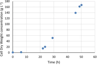
19 The bioreactor was operated in batch mode for the first 24 h, after which it was switched to a fed-batch mode for the remainder of the assay. Carbon source for the fed-batch mode was supplied at a constant rate of 116.84 mL h-1 (144.64 g h-1). During the exponential phase, yeast biomass grew at a specific cell growth rate of 0.17 h-1 similar to that found in literature for Komagataella pastoris grown in glycerol by Roca and co-workers [11], reaching a maximum CDW value of 166.88 g L-1 at 50 h of cultivation (Figure 3.1).
Mannans content was determined to be 37.75 % (w/w) of CDW and the volumetric productivity was calculated to be 1.25 g L-1 h-1. The growth yield was 0.42 gbiomass gglycerol-1, lower than that described for pure glycerol, 0.55 gbiomass gglycerol-1 as described by Roca and co-workers [11]. This discrepancy could be due to glycerol that was administered to the bioreactor but which ended up not being consumed by the yeast. However, since glycerol was not quantified at the end of the assay, this hypothesis cannot be confirmed. The product yield was of 0.13 gmannans gglycerol-1.
Figure 3.1. Cell Dry Weight over the course of the bioreactor assay.
Monomeric analysis of the produced mannans revealed a mannose content of 24.13 ± 2.04 % w/w. Traces of glucose (2.27 ± 0.02 % w/w) and glucosamine (0.78 ± 0.01 % w/w) were also detected. Glucosamine is a characteristic monomer of CGC and the fact that it was present in the sample could be proof that the extraction process was not 100 % effective, as purified mannans should not have any trace of it. This fact misleads the calculation of true monomeric content of the sample, both by underestimating the mannose content (which should be higher) and overestimating the glucose
0 20 40 60 80 100 120 140 160 180 0 10 20 30 40 50 60 Ce ll Dry W eight co n ce n tra tio n (g L -1) Time (h)
20 content (which may as well be part of the CGC fraction present in the sample).
Protein quantification was achieved by a protein precipitation method using TCA as a precipitation agent. The total protein precipitated was as high as 19.45 ± 0.53 % (w/w). This amount of precipitated protein, however, does not encompass all the initial protein in the sample, as discussed below (3.2.5). The precipitation process also depleted the sample of about half of its remaining polymeric content, as only 41.19 ± 1.73 % (w/w) of the initial sample was able to be retrieved, which had a mannose content of 24.13 ± 2.04 % w/w. This is most likely due to hydrolysis effect caused by the TCA, as the carbohydrate sidechains of the mannoprotein fraction of the sample were too small and therefore able to cross the pore of the dialysis membrane. It is also possible that non-mannoprotein mannans were hydrolyzed into smaller polysaccharides.
Moisture content of the mannans sample was determined to be 5.08 % (w/w).
Inorganic content of the mannans sample was determined to be 1.46 ± 0.09 % (w/w).
Elemental analysis was performed for mannans as obtained, deproteinized mannans as well as the deproteinization pellet. Results are displayed in table 3.1.
Table 3.1. Elemental analysis of mannans.
Element % (w/w)
Nitrogen Carbon Hydrogen Sulphur Mannans 6.90 ± 0.09 43.76 ± 0.01 6.86 ± 0.28 0
Deproteinized Mannans 2.09 ± 0.02 41.42 ± 0.06 6.63 ± 0.03 0
Protein Pellet 10.98 ± 0.06 57.45 ± 0.08 8.23 ± 0.26 0.30 ± 0.03
Results are presented as Mean ± SD (%).
The results show a total of 6.90 ± 0.09 % (w/w) nitrogen content, corresponding to a protein content of 42.71 ± 0.19 % (w/w). Protein quantification was performed based on a 6.25 factor as previously described (2.1.1.3.2).
21 Phosphate content was determined for samples of original, deproteinized and phosphorylated mannans as well as for the phosphorylation pellet. Results are shown in table 3.2.
Table 3.2. Phosphate and ammonia content of mannans. Phosphate % (w/w) Mannans 1.97 ± 0.17
Deproteinized Mannans 1.31
Phosphorylated Mannans 1.04 ± 0.04
Phosphorylation Pellet 0.59
Results are presented as Mean ± SD (%).
Regarding phosphate content, analysis was performed by ICP-AES). Phosphate content was determined to be 1.97 ± 0.17 % (w/w) in the original polymer. Phosphate content was determined due to its relevant role in numerous biological processes, as phosphorylated polysaccharides (both naturally occurring and transformed) have been described to have enhanced immunocompetence, anticoagulation and antineoplastic activity [45], as well as antitumor activity [3]. The content in phosphate as also been related to its activity, both immunochemical and serological [46, 47]. The phosphate content of the mannans in this study is within range from mannans obtained from different yeasts, namely C. albicans, which have reported values typically ranging from 0 to 2.4 %, although higher phosphate content values have been reported, almost up to 5 %, depending on cultivation conditions [48]. Other studies on P. holstii showed that certain fractions of polysaccharides may be even richer in phosphate contents (from 3 to 7.4 %) [49], although the complete mannans profile may not be present. Although many reported phosphomannans contain lower amounts of phosphate than the one in this study, phosphate content is a relevant factor for the desired properties, and thus an attempt to increase it was made (3.3.2).
As described above (3.2.2), deproteinized mannans were obtained from the supernatant of the protein precipitation method, with a yield of 41.19 ± 1.73 % (w/w) for recovered polymer and a yield of 69.49 ± 0.44 % for removed protein. From the results of elemental analysis (table 3.1), it is possible to see that the TCA precipitation method did not remove all the protein from the initial sample, but total protein precipitated was higher than the previously calculated (3.2.2), totalling 29.68 ± 0.32 % (w/w). The protein precipitation method employed, using TCA, has been described as a high yielding precipitation method, with precipitation yields above 90 % for removed protein [50], well above those described in this study. Moreover, polymer recovered in that study was higher than the one reported here, with a total loss of polymer of 19 %. This shows that the TCA method employed, while effective in removing