P
OLYMER-C
ERAMICN
ANOCOMPOSITESF
ORB
ONER
EGENERATIONDissertação apresentada à Universidade Católica Portuguesa para obtenção do grau de Mestre em Medicina Dentária
Por
Sónia de Lacerda Schickert
P
OLYMER-C
ERAMICN
ANOCOMPOSITESF
ORB
ONER
EGENERATIONDissertação apresentada à Universidade Católica Portuguesa para obtenção do grau de Mestre em Medicina Dentária
Por
Sónia de Lacerda Schickert
Sob orientação de Professora Doutora Ana Leite Oliveira
Acknowledgments
I owe a sincere and earnest thankfulness, in first place, to my Mentor, Dra. Ana Leite Oliveira, for believing in me when I needed the most, helping me achieve my professional goals and providing me a prospective future. Her example as a professional is one I hope to match someday.
I would like to thank deeply the department of Biomaterials of the Dentistry Faculty at the UMC St. Radboud University, for being so welcoming, so supportive and showing an endless availability for helping me whenever I needed. A special word of appreciation to Professor Sander Leeuwenburgh, Professor Fang Yang, Stefan Remmers and Pedro Babo.
To all the professors and professionals in the dentistry area that invested time in endless explanations, my sincere thank you.
My word of gratitude to the Portuguese Catholic University, all the employees and stuff for the help, support and patience.
I offer my sincere gratitude to my father, Vítor. The constant support, the motivational speeches and endless patience were essential for the conclusion of this thesis. His love, affection and understanding way of being makes him my number one pillar in life and knowing I can count on him for everything gives me courage to face the future.
A special “thank you” to the Schickerts, especially to Gerhard, who I believe helps me from above in all situations (and this dissertation was no exception) and tante Christa, for remembering me the value of family and always having a special comfort word to offer in the current difficult moments.
I thank deeply my friends, colleagues and supporters, Maria Inês Barbas, Mafalda Barros and Johnny Leite. Without them this study, the 5 years of the dentistry degree and life in general would be way more complicated.
Special thanks to my second family, Rita Mineiro Rodrigues and Filipe Rodrigues. In them I found a refugee and an endless support for accomplishing this dissertation and my personal and professional goals in life.
Finally, I would like to thank my mother, for creating the conditions that prove that I am capable of succeeding against all the odds.
Abstract
Bone is a very important structure in the human body. In the craniomaxillofacial complex, trauma and pathological situations such as maxillofacial tumors produce major bone defects that cannot be healed by physiological processes. As a response to this problem, many strategies have been studied throughout the years, with the purpose to find a material that reunites ideal properties for regenerating or substituting the needed bone and therefore guarantee its physiological and structural functions. Among this strategies, transplantation of bone within the same individual (autografts), from another human being (allograft) or from an individual of another species (xenograft) have been studied and are clinically applied nowadays. However, these techniques revealed to have drawbacks such as donor site morbidity, risk of disease infection and immunological reactions. In this context, new solutions are needed.
In the field of biomaterials, nanocomposites are considered a promising material for many outcomes, mainly because their nanometer-scale allows a superior structural performance, when compared to microcomposite materials. In the bone regeneration area, ceramic nanocomposites dispersed in a polymeric matrix are a promising solution, because they reassemble the physiological bone structure, adding unique biocompatible and stimulating properties to the already outstanding mechanical behavior.
The investigation of the different types of ceramic-polymeric nanocomposites, as well as the co-factors that can be added to enhance the biological response and their processing ability for the commercial level, is extremely relevant. This study intends to present a review of the published information about the in vivo and in vitro studies that have been performed in the last 5 years and their contribution for the development of an ideal nanocomposite material to be used in bone regeneration, particularly in the craniomaxillofacial context.
Resumo
O osso é uma estrutura muito importante no corpo humano. No complexo crâniomaxillofacial, situações como trauma e tumores maxillofaciais têm como resultado defeitos ósseos de grandes proporções que não são passíveis de ser curados por processos fisiológicos. Com o intuito de dar resposta a este problema, várias estratégias têm vindo a ser estudadas ao longo do tempo, para encontrar um material que reúna as propriedades ideias para a regeneração ou substituição do osso em défice e assim garantir o restabelecimento as suas funções fisiológicas e estruturais. De entre estas estratégias, a transplantação de osso no mesmo indivíduo (autotransplante), transplantação de osso entre seres humanos (alotransplante) e de osso de indivíduos de outra espécie (xenotransplante) têm sido estudadas e são clinicamente utilizadas hoje em dia. Contudo, estas técnicas têm mostrado possuir alguns inconvenientes como são a morbilidade da zona óssea dadora, risco de doença infeciosa e reações imunológicas. Neste contexto, novas soluções são necessárias.
Na área de biomateriais, os nanocompósitos são considerados um material promissor para vários fins, principalmente porque a sua escala nanométrica permite uma performance estrutural superior, quando comparada com materiais microscópicos. Em regeneração óssea, nanocompósitos cerâmicos dispersos numa matriz polimérica, apresentam-se como uma solução promissora, uma vez que simulam a estrutura fisiológica do osso, adicionando propriedades de biocompatibilidade e estimulação únicas ao já muito bom desempenho mecânico.
A investigação de diferentes tipos de nanocompósitos cerâmico-poliméricos e cofatores que possam ser adicionados para melhorar a resposta biológica, bem como técnicas de produção para uma possível escala comercial, é extremamente relevante. Este estudo tem como objetivo apresentar uma revisão da informação publicada sobre estudos in vivo e in vitro que têm sido efetuados nos últimos 5 anos e o seu contributo para o desenvolvimento de um material ideal.
Palavras-chave: Osso; regeneração; nanocompósitos; cerâmica; polímeros
Table of Contents
Acknowledgments ... VII Abstract ... IX Resumo ... XI Table of Contents ... XIII List of Abbreviations ... XV List of Tables ... XIX List of Figures ... XXI
I. Introduction ... 3
1. The Bone Biology ... 4
2. Bone Grafts ... 13
3. Matching bone through nanocomposites ... 21
II. Materials and Methods ... 31
Search Strategies ... 31
Study Selection ... 32
Inclusion and Exclusion Criteria ... 32
Data extraction and meta-analysis ... 33
III. Results ... 37 1. Year of publication ... 37 2. Study design ... 38 3. Country of Origin ... 39 4. Publishing Journal ... 40 5. Materials Selection ... 42 6. In vivo Studies ... 45 7. In vitro Studies ... 56 IV. Discussion ... 71 Materials Selection ... 71 In vivo Assays ... 77 In vitro Assays ... 81 Grow-factors Incorporation ... 87
Antibacterial Agents Incorporation ... 87
VI. Bibliography ... 95 VI. Annexes ... 109 Annex 1 – Research Diagram ... 111
List of Abbreviations
B
BG – Bioactive Glass
BMP – Bone morphogenetic protein BMP-2 – Bone morphogenetic protein two BMP-7 – Bone morphogenetic protein seven BMSc – Bone Mesenchymal stem cells BSE – Bovine spongiform encephalitis
C
CPC – Calcium phosphate cement
D
DBM – Demineralized bone matrix DNA – Deoxyribonucleic acid
E
ECM – Extracellular matrix e-PTFE – polytetrafluoroethylene
F
FDA – Food and Drug Administration
G
Gel – Gelatin
H
M
MSCs – Mesenchymal stem cells MWNT – Multi-walled carbon nanotubes
N
NCES – N- carboxyethyl chitosan nHA – Nano-hydroxyapatite
P
P66 – Aliphatic form of poyamide PA – Polyamide
PAA – Poly(aspartic acid) PAs – Peptide amphiphiles PCG – Polycaprolactone PCL – Polycaprolactone PEG - Polyethylene glycol PGA – Poly (glycolic acid) PHA – Polyhydroxyalkanoate PHB – Poly(3-hyroxybutyrate) PLA – Poly (lactic acid)
PLEOF – Poly (lactide – co – ethylene oxide fumarate) PLGA – Poly(lactide – co – glycolic acid)
PLLA – Poly(L – lactide acid) PP – Polypropilene
PU – Polyurethane PVA – Poly (vinyl alcool)
S
SBF – Stimulated body fluid SF – Silk fibroin
T
TCP – Tricalcium Phosphate TGF – Transforming growth factor THF – Tetrahydrofuran
TiO2 – Titanium dioxide
U
US – United States (of America) α-TCP – Alpha tricalcium phosphate β-TCP – Beta tricalcium phosphate
List of Tables
Table 1 - World Health Organization classification of benign and malignant bone tumors of the maxillofacial region ... 8 Table 2 - Components of bone ... 9 Table 3 - Influence of growth factors on graft incorporation and bone healing. 16 Table 4 - In vivo studies: osseous implantation ... 46 Table 5 - In vivo studies: non-osseous implantation ... 51 Table 6 - Results obained regarding the in vitro studies of nanocomposites to be used in bone applications in the maxillofacial acontext ... 58 Table 7- Summary of the four attributes in terms of similarity between animal and human bone ... 78
List of Figures
Figure 1 – Maxillofacial Trauma ... 7 Figure 2 – Bone hierarchical organization ... 10 Figure 3 – The nanoscopic detail of fibril ... 12 Figure 4 – Various applications of composite - polymers throughout the body 22 Figure 5 – The geometry of nanoscaled fillers ... 25 Figure 6 – Relevant papers published per year between January of 2009 and 2014 ... 37 Figure 7 – Amount of in vitro and in vivo studies ... 38 Figure 8 – Different countries of origin of the authors of the included studies . 39 Figure 9 – Journals with representation in the present thesis ... 40 Figure 10 – Types of journals publishing more articles about polymer-ceramic nanocomposites in the maxillofacial area ... 41 Figure 11 – Origin of polymers used in the considered articles ... 42 Figure 12 – Nanofillers that constitute the nanocomposites studied in the
considered articles ... 43 Figure 13 – Laboratory animals used in the in vivo studies ... 53 Figure 14 – Polymers chosen to integrate the nanocomposite is osseous and non-osseous studies ... 54 Figure 15 – Nanofillers that integrate the nanocomposite studied in both
osseous and non-osseous studies ... 55 Figure 16 – Studies using stem cells, osteogenic cells and Stimulated Body Fluid ... 56 Figure 17 – Polymers used as matrixes in the nanocomposites subjected to in vivo studies... 67 Figure 18 – Nanofillers used in the nanocomposite subjected to in vitro studies ... 68
I.
Introduction
Natural bone has a great healing capacity. Through times, this capacity has been studied in order to understand what the limitations are and how they could overcome. A well-known limitation of the natural healing process is related to the extension of the bone defect. When the lesion is larger than 2 mm, the bone structure is not capable of regenerating by itself.1 Situations such as diseases,
abnormalities or trauma which have as a consequence major bone defects have been extensively studied in order to find the best therapeutic solutions.2
In the craniomaxillofacial skeleton, bone defects can vary from small (few millimeters) periodontal defects, to large segmental defects that are originated mainly in major trauma, followed by surgical excision or cranioplasty.3 Such
structures, have a complex tree-dimensional structural need, which is difficult to fulfill. In cranial vault defects, the underlying brain needs permanent protection. Segmental jaw defects require restoration of mechanical integrity, temporomandibular joint function and inter-maxillary dental occlusion. Maintaining acceptable facial esthetics is another unique consideration in the treatment of facial defects that cannot be underestimated.3
Due to the constant need to heal, techniques to replace, restore, or regenerate bone were noticed as a major clinical quest in the fields of orthopedic, spinal, dental, cranial and maxillofacial surgery. The earliest report of a bone grafting procedure appeared in 1682, in a book by Job Janszoo van Meekeren, a surgeon in Amsterdam. In this document, the author reported a case in Russia, where the surgeon restored a cranial defect using a cranial bone graft from a dead dog.4 In 1881, Sir William MacEwen of Rothesay, from Scotland, published
the first case report of successful inter-human transfer of bone grafts.5 Since then,
many changes occurred due to centuries of subsequent research both in academia and industry.
Now-a-days new materials are being investigated, with the purpose of changing the goal of bone grafts from just a replacing role, to the complete bone restauration, allowing for the own bone to grow and regenerate itself. In this context, polymer-ceramic nanocomposites arise as important candidates for this
application, since bone itself is composed by polymer-ceramic nanocomposite structure.
1.
T
he Bone Biology
Bone is a calcified, living, connective tissue that forms the majority of the skeleton. Their basic constitution is an intercellular calcified matrix, containing collagen fibers and several types of cells within the matrix.6 Actually, bone is the
only mineralized (collagen mineralized with calcium phosphate) structure that contains living cell bodies on its structure. Dentin, for example, is composed of collagen mineralized with calcium phosphate but it does not contain cell bodies, only tubular extensions of cells.7 Other significant tissue mineralized with calcium
phosphate is the enamel.7 However, in this case, there are virtually no cells or
cell processes or, indeed, much too organic matrix.
There are two major types of bone: Cancellous (also called spongy bone) and Cortical.8 Cancellous bone forms the internal pours framework of bones. It
contains stem-cells-rich bone marrow which is essential for the growth of new connective tissue (e.g. muscle, cartilage, bone and tendons) and the production of blood cells. Cortical bone surrounds the cancellous bone and forms an outer shell, thus giving the bone shape and form. In load bearing bones, the cortical component is markedly thickened to form a strong shaft.
Bone is responsible for 5 basic functions:6 (a) to support all the body
structures, (b) to protect vital organs, (c) to be a reservoir of calcium and phosphorus, (d) to function as a lever on which muscles act to produce movement and (d) to host blood-producing cells.
In terms of development, all bones are originated in the mesenchyme either by intramembranous ossification, in which mesenchymal models of bone undergo ossification or endochondral ossification, in which cartilaginous models of bones form and undergo ossification.6 There are five distinct types of cells associated
with bone tissue formation and regulation: osteoprogenitor cells, osteoblasts, osteocytes, osteoclasts, and bone-lining cells.9 Osteoprogenitor cells (or bone
Osteoblasts are the cells that are responsible for the formation of new bone tissue. They start with secreting collagen followed by coating non-collagenous proteins, which are similar to a glue that has the ability to bind the minerals, mostly calcium and phosphate, from the bloodstream. Osteocytes are matured cells derived from the osteoblasts that are responsible for the maintenance of the bone tissue. They function as transporting agents of minerals between bone and blood. Osteoclasts are the largest cells found at the surface of bone mineral. They are responsible for the resorption of the bone tissue. They secrete acids or enzymes to dissolve the minerals as well as collagen from the matured bone. The dissolved minerals then re-enter the bloodstream and are carried to different parts of the body. Bone-lining cells are found along the surface of the matured bone and are responsible for regulating the transportation of minerals in and out of the bone tissue. They also respond to hormones by producing proteins that activate the osteoclasts. Together, these five types of cells are responsible for building the bone matrix with hierarchical structure, self-assembly ability and remodeling capability. All these processes must be in equilibrium to ensure a healthy bone tissue.6-9
When bone suffers an injury, it possesses natural healing capacity which allows for a successful regeneration. After damage, the bone adjacent structures cause a hematoma around the broken bone ends.10 Fibroblasts enter the site and
form granulation tissue, composed predominantly of type-III collagen. Then, two different processes occur in the proximal and the distal site. In the proximal, chondroblasts arise from the periosteum and produce hyaline cartilage. In the distal site, osteoblasts arise from the periosteum and produce woven bone (majorly constituted by type-I collagen).10 Both sites combine and form the
fracture callus. The woven bone then undergoes substitution and the hyaline cartilage undergoes endochondral ossification. These processes both produce lamellar bone. Osteoblasts and vascular channels penetrate the mineralized matrix and stronger trabecular bone is laid down. From here, final remodeling occurs by the deposition of compact bone by osteoblasts in resorption pits prepared by osteoclasts.10 This process, known as primary or direct osteonal
healing, is just possible if the gap between bone ends is less than 2 mm and absolute stability exists.
1.1. Craniomaxillofacial anatomy and physiology
The skull is divided in two major parts: The neurocranium and the viscerocranium.11 The neurocranium, constituted by the cranial bones form the
cranial cavity which encloses and protects the brain. In addition, the cranial bones stabilize the positions of the brain, blood vessels, lymphatic vessels and nerves through the attachment of their inner surfaces to meninges (membranes). The outer surface of cranial bones provide large areas of attachment for muscles that move various parts of the head. Underneath it, the skull base forms the floor of the cranial cavity and separates the brain from other facial structures.11,12
Anteriorly attached to the skull is the viscerocranium.
The viscerocranium forms the anterior part of the cranium and consists of facial bones surrounding the mouth, nose, and most of the orbits. They consist on the framework for the face and provide support for the entrances to the digestive and respiratory systems. Therefore, vital functions such as breathing, eating, talking, smelling, seeing and having equilibrium are ensured by this bones. Moreover, it is also an esthetically important region. Therefore, any change to its normal constitution and/or function seriously damages the individual’s life quality.12 In such situations, maxillofacial surgery applies different
technics for healing and restoring the normal function.
1.2. The need for maxillofacial surgery
Surgeries concerning the use of bone replacements or bone grafts in the craniomaxillofacial area are requested in two major situations: in case of trauma or when there is a pathological abnormality in the anatomy and physiology of this area (e.g. a congenital and developmental deformity or an acquired condition).
Causes of craniomaxilofacial trauma injuries vary from interpersonal violence to rodoviary accidents.13 Fractures of the maxilla are the result of
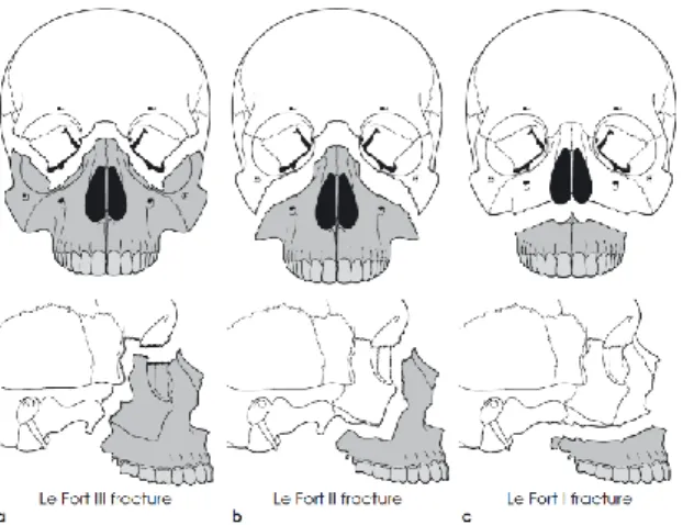
considerable force and are often associated with craniocerebral injury. The maxilla offers a high resistance against forces directed upwards but relatively little resistance if the impact is directed horizontally. Fractures may occur at three
levels, Le Fort I, II and III, and may be unilateral or bilateral (Figure 1-A). The palate may also be fractured in the midline. Mandibular fractures, which are the ones that happen more often,11,13-15 may happen in different sites of the mandible (Figure 1-B). The symptoms and signs of a fractured mandible include pain and swelling over the fracture site as well as trismus and malocclusion.
Congenital and developmental deformities of the cranial skeleton occur due to errors in the growth process while the fetus is in utero and tend to become more evident in the pubertal growth spurt.15 They have the particularity of being
really easy to diagnose, since the facial deformity is obvious, but extremely difficult to categorize. Since the skin drapes over the bony skeleton, most deformities are caused by bone abnormalities (soft tissue abnormalities are rare and when they occur, they are commonly secondary to skeletal abnormalities). According to Booth et al.,15 these abnormalities can be broadly subdivided into
craniofacial anomalies, clefting anomalies and orthognathic deformities. This last one involves the dentofacial anomalies that can be corrected by orthognathic surgery, repositioning parts of the facial skeleton to correct the deformity. Craniofacial and clefting anomalies, may require bone grafts to add bone tissue or realign it through complex maxillofacial surgeries.
Figure 1 – Maxillofacial Trauma
(A) Mandibular Fractures: Le Fort fracture I, II and III. (B) Incidence of fractures occurring at various sites of the mandible.15
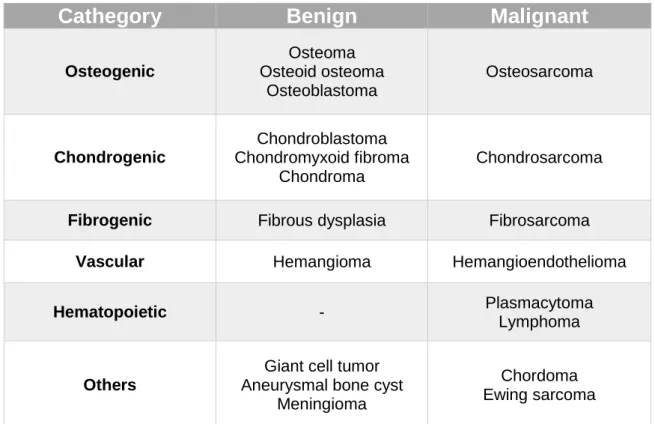
In the craniomaxillofacial complex, a high percentage of diseases affecting the bones are tumoral conditions.16 Bone tumors of the maxillofacial region may
arise from osteogenic, chondrogenic, fibrogenic, vascular, hematopoietic and other elements of the bone.17 Table 1 shows the World Health Organization
classification of benign and malignant bone tumors of the maxillofacial region.18
As a consequence to tumors excisions, a critical-sized bone defect remains, leading to noticeable deformity and dysfunction. These situations generally require reconstructing strategies such as the replacement with autogeneous or allogenic bone grafts, xenografts or alloplastic materials. The clinical results of these procedures are limited, since the craniomaxillofacial bones have a complex three dimensional structure and the replacement materials do not possess the necessary characteristics to fulfill their goal.19 Other request for bone
augmentation has to do with the loss of alveolar bone after teeth loss. The main purpose of creating new alveolar bone is generate a bone structure to anchor dental implants and dentures and allow the requalification of the orthognathic system.19
Table 1 - World Health Organization classification of benign and malignant bone tumors of the maxillofacial region 18
Cathegory
Benign
Malignant
Osteogenic Osteoma Osteoid osteoma Osteoblastoma Osteosarcoma Chondrogenic Chondroblastoma Chondromyxoid fibroma Chondroma Chondrosarcoma
Fibrogenic Fibrous dysplasia Fibrosarcoma
Vascular Hemangioma Hemangioendothelioma
Hematopoietic - Plasmacytoma
Lymphoma
Others
Giant cell tumor Aneurysmal bone cyst
Meningioma
Chordoma Ewing sarcoma
1.3. Bone as a Nanocomposite
Nanocomposites are defined as a heterogeneous combination of two or more materials in which at least one is at the nanometer-scale.20 A good example
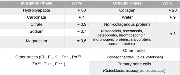
of a nanocomposite is bone tissue: it is composed of two major phases at the nanoscale level, namely the organic (protein) and the inorganic (mineral). These phases have multiple components which consist of, in decreasing proportions: minerals, collagen, water, non-collagenous proteins, lipids, vascular elements, and cells (Table 2).21 The mineral of bone is mainly composed of HA and the
organic part of bone is mainly composed of collagen. Here, collagen acts as a structural framework in which plate-like tiny crystals of HA are embedded to strengthen the bone. Bone collagen has a typical fibrous structure, whose diameter varies from 100 to 2000 nm. Similarly, HA in the bone mineral is in the form of nanocrystals, with dimensions of about 4nm by 50nm by 50nm.22,23The
primary role of minerals is to provide toughness and rigidity to the bone, whereas collagen provides tensile strength and flexibility needed.21 In other words, the
function of collagen fibers is to provide strength in tension and resistance in bending whereas the apatite crystals embedded between the nanofibers will resist to compression.24
Table 2 - Components of bone 21,23
(This composition can vary slightly from species to species and from bone to bone)
Inorganic Phase Wt % Organic Phase Wt %
Hydroxyapatite ≈ 60 Collagen ≈ 20
Carbonate ≈ 4 Water ≈ 9
Citrate ≈ 0,9 Non-collagenous proteins
(osteocalcin, osteonectin, oeteopontin, thrombospondin, morphogenic proteins, sialoprotein,
serum proteins) ≈ 3 Sodium ≈ 0,7 Magnesium ≈ 0,5 Other traces (Cl- , F- , K+ , Sr 2+, Pb 2+, Zn 2+, Cu 2+, Fe 2+) Other traces
(Polysaccharides, lipids, cytokines) Primary bone cells
1.4. Hierarchical structure
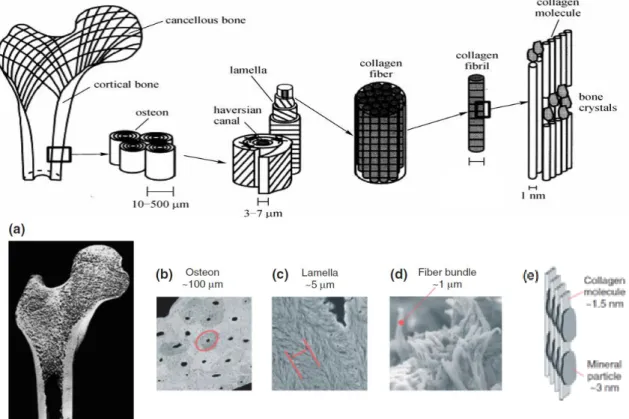
Because of its structural function, bone needs to be tough and resistant. The key to this strength is the complex structural hierarchy into which it is organized in a self-assembling mode. Thus, bone can be considered as an assemblage of various levels of hierarchical structural units elegantly designed at different scales, from nano to macro (Figure 2), to meet multiple functions.
At the macrostructure level, matured bone can be divided in two types: spongy bone and compact bone. Structurally they are different because they play different roles in bone. Spongy bone represents approximately 20% of the total amount of bone, whereas compact bone represents the other 80%. Spongy bone is very porous and light and has a high concentration of blood vessels (which are
Figure 2 - Bone hierarchical organization
(a) the macrosctructured level: bone is divided in compact and spongy bone; (b) the microstructured
level: osteons; (c) and (d) the mesostructured level: lamellae and its fibrillar composition (e) the nanoscopic level: the 2 major components, collagen and HA. 2,8
responsible for the transportation of nutrients, oxygen and body fluids). Compact bone, on the other hand, has a very dense structure and is less porous, not being able to host a large amount of blood vessels. The compact bone functions include supporting bone mechanically in tension, compression and torsion. Spongy bone functions mainly in compression.8
At the microstructural level, compact and spongy bone have different structural units. In compact bone, osteons or harvesian systems repeat themselves working as weight-bearing pillars, while spongy bone contains an interconnecting framework or trabeculae.8 Each osteons consists of concentric
layers, or lamellae, that surround a central canal, the haversian canal, that contains nerve and blood supplies. As the body ages, the number of osteons increase, resulting in a tissue completely filled with osteons, which are in direct contact with each other. When bone is compressed, osteons present a unique crack pattern, consisting of stacks of ark-shaped circumferential microcracks that propagate radially from the center of each osteon.8 As the compression level
rises, the radial cracks that exist in the neighboring osteons connect, forming a network of cracks throughout the tissue that redistributes the stress and preventing the catastrophic failure, despite the large amount of energy applied. Furthermore, this unique type of microcracking allows the material to maintain its high strength and resilience even in the inelastic regime of deformation. This example illustrates how the hierarchical organization of bone (in this case in the microstructural level) determines its unique mechanical resilience.8
The lamellae are made of one or more parallel fibrillar arrays varying in thickness from a few hundred nanometer to a few microns. The thicknesses and the main fibril direction in neighboring lamellae can vary leading to the formation of unique patterns that are often species specific and can even vary among different bone types in the same species. The spaces between the fibrils contain extrafibrillar mineral crystallites and noncollagenous molecules, such as glycoproteins and glycosaminoglycans that provide a viscoelastic medium isolating the collagen fibrils. In addition, this non-collagenous proteins have other important function: they form a supramolecular network based on the ionic cross-links between their acidic side chains and the calcium ions in the solution as well as the interactions between these acidic moieties and calcium ions on the surface
of mineral crystal. This ions based, reversible and sacrificial networks dissipate large amounts of energy under mechanical stress and reforms quickly when the load is off.8,23,25 In the hierarchical structure, the mesostructural level is of
particular importance because although its basic components are in the nanoscopic level, the chains that they form together have specific mechanical functions when joined. This can be considered as another good example of the benefits of bone’s hierarchical organization.
Finally, at the nanostructured level, the two major components (collagen fibers and nanocrystals of HA) are found (Figure 3) in higher proportions among other minor components such as non-collagenous proteins, molecules of water and lipids. The presence of water and lipids is essential for cellular functions: water plays and important role in fluid movement within bone, transports nutrients and waste products and also calcium and cytokine factors, and is an important determinant of the biomechanical behavior of bone while lipids play an important role in regulating the biomineralization process.2
Figure 3
Structurally, a collagen framework embeds plate-like tiny crystals of HA. This conformation is essential because the mechanical properties that are characteristic of the bone tissue exist do to the isolated and combined properties of its nano components. This means that, in the end, all boils down to high level of integration of the mineral and organic phases at atomic and molecular levels.7
A badly formed collagen has compromising effects on the structural and mechanical characteristic of bone and an example of this is Osteogenis
Imperfecta. In this disease, a mutation in the genes that are responsible for the
production of collagen type I can lead to an abnormal triple helix structure, compromising the whole bone structure.27 Moreover, Currey et al., 28 preconized
that one of the reasons why ionized radiation decreases the bending strength and the work to fracture of bone structures, is due to the radiation specific damaging of the collagen matrix in an irreversible way.
Nonstoichiometric carbonated hydroxyapatite is the specific type of HA found in bone tissues. The fact that it presents itself in plate-like crystallites with very small average dimensions is the reason for the unique mechanical behavior of its structure. In fact, Gao et al.29 demonstrated that crystals this small are
extremely tolerant to flaws and that this hydroxyapatite specific crystals have a strength of a perfect crystal. Other favorable characteristic is the high surface and bulk ratio of these crystallites that increase their chemical reactivity and consequently the interactions with the organic molecular components.30
2.
B
one Grafts
Bone grafting is a surgical procedure which entails replacement or reconstruction of missing or damaged bone with material from either patient's own body, an artificial or natural substitute.31 In order to accomplish its goal, bone
grafts should be osteoconductive, osteoinductive and osteogenetic:32 (A)
Osteoconduction is a term that means that bone grows on a surface. Therefore, an osteoconductive surface is the one that has ideal characteristics to permit bone growth, by stimulating the attachment, survival, migration and distribution of cells that are involved in bone formation such as mesenchymal cells, osteoblasts, osteoclasts, and vasculature; (B) Osteoinduction refers to the
process in which primitive, undifferentiated and pluripotent cells are somehow stimulated to develop into a bone forming cell lineage. It can be considered that osteoinduction is the process by which osteogenesis is induced; (C) Osteogenesis is the formation of bone in general. It can be produced by cells of the host or by living cells included in the graft.33,34
2.1. Classification of Bone Grafts
There are several categories of bone grafts which encompass a variety of materials, material sources, and origins.33 In order to present a more complete
classification, one will be used, adapted from Laurencin et al..35
a) Harvested bone grafts
Autografts
Allografts constitute 58% of the bone substitutes and are typically tissues harvested from the patient’s own bone tissue such as the iliac crest.2 They are
considered the gold standard for bone repair, because they offer minimum immunological rejection, complete histocompatibility and provide the best osteoconductive, osteogenic and osteoinductive properties. Autografts usually contain viable osteogenic cells, bone matrix proteins and support bone growth which are obtained from vascularized and non-vascularized cortical and autologous bone marrow grafts. They offer structural support to implanted devices and ultimately become mechanically efficient structures as they are incorporated into surrounding bone through creeping substitution.33 However,
they are limited in availability and often associated with donor-site morbidity and increased operative blood loss particularly when a large graft is required.36
Allografts
Allografts are tissues obtained from banked freeze-dried bones of human cadavers and represent about ≈34% of the bone substitutes. They are
osteoconductive and have fewer limitations on supply.35 However, allografts are
usually not osteoinductive or osteogenic and are associated with risks of immunological reaction or disease transmission. Furthermore they possess insufficient mechanical properties for load-bearing bone applications.2
Xenografts
Xenografts are harvested from one individual and transplanted into another individual of a different species. The common available xenografts are derived from coral, porcine, and bovine sources.37 Xenogenous bone grafts are a
theoretically unlimited supply of available material if they could be processed to be safe for transplantation in humans. A major concern with bovine-derived products is the potential transmission of zoonotic diseases and prion infections such as bovine spongiform encephalitis (BSE). Xenografts, similar to allograft, lose their osteogenic and partly osteoinductive properties during the processing to counteract their antigenic properties and prevent transmission of infection.38
Because xenografts produce poor clinical outcome, new insights in this same field have been investigated. For example, Keskin et al.,39 evaluated the
effectiveness of autologous bone marrow on the healing of bone defects filled with bovine-derived xenografts in rabbits. They concluded that when xenografts were combined with autogenous red bone marrow, the drawbacks of xenografts are slightly compensated, but even with this enhancement of biocompatibility, their origin and properties continue to arise unsolvable problems.
b) Bone Graft Substitutes
While harvested bone grafts use pre-existing bone, bone graft substitutes are biomaterials made of a variety of sources and using different strategies to promote bone growth in the host.35
Growth factor-based bone graft substitutes
Growth factors are secreted by a wide range of cell types to transmit signals that activate specific developmental programs, controlling cell migration, differentiation and proliferation.40 Therefore, integrating them in a biomaterial with
regeneration purposes is a great strategy for enhancing its biological properties, simulating host cells to adhere to the material and proliferate. Polymeric systems can be successfully used to administer small doses of factors at defined dose rates directly to target cells. In fact, biodegradable polymers are able to provide a controlled release of growth factor delivery through its degradation rate. In the bone regeneration field, osteoinductive growth factors have been reported.41,42
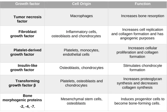
Table 3 specifies these factors, as well as its origin and specific function.
Table 3 - Influence of growth factors on graft incorporation and bone healing.43
Growth factor Cell Origin Function
Tumor necrosis factor
Macrophages Increases bone resorption
Fibroblast growth factor
Inflammatory cells, osteoblasts and chondrocytes
Increases cell replication and collagen formation and has
angiogenic purposes Platelet-derived growth factor Platelets, monocytes, endothelial cells Increases cellular proliferation and collagen
formation
Insulin-like
growth factor Osteoblasts, chondrocytes
Stimulates chondrocyte formation
Transforming growth factor β
Platelets, osteoblasts and chondrocytes
Increases proteoglycan synthesis and decreases
collagen synthesis
Bone morphogenic proteins
-2, -4, -7.
Mesenchymal stem cells, osteoblasts
Induces progenitor cells to become bone-forming cells.
Among this group of growth factors, BMPs are a group of high interest. They are members of the β-transforming growth factors (β-TGF) superfamily and have been identified as having different degrees of cellular activity, including cartilage or bone inducing properties. Lee et al., 41 demonstrated that the use of BMP-2
enhanced significantly bone regeneration in a critical size femoral defect rat model in amounts that are one order of magnitude lower than that required for healing in this animal. The presence of more mature bone in the new ossified tissue was noted when a low dose of BMP-2 was delivered using a biomimetic supramolecular system. In human patients, the failure to naturally regenerate bone seems to be related to the genetic changes in the BMP signaling system.44
The US Food and Drug Administration (FDA) has approved two recombinant proteins that are presently commercially available: recombinant human bone morphogenic proteins rhBMP-2 and rhBMP-7.31 Clinical orthopedic studies have
shown the benefits of these recombinant human BMPs but side effects such as swelling, seroma, and increased cancer risk, have been reported, probably due to high BMP dosage.42
Cell-based bone graft substitutes
These bone grafts combine living cells with biomaterial scaffolds ex vivo to allow the development of a three-dimensional structure. Autologous cells may be used in this approach via the isolation of a small number of differentiated adult cells or stem cells, followed by in vitro expansion to produce the material that will be reimplanted in the host posteriorly.40 Mesenchymal stem cells (MSC) are
capable of continuously replicate themselves, while a portion becomes committed to mesenchymal cell lineages such as bone, cartilage, tendon, ligament, and muscle, depending on the outer stimuli that they receive. They are considered to be a great tool for regenerative cell therapy and have been used since the 1980s, when Friedenstein et al.45 first reported their effectiveness on
developing engineered tissues.
Wong et al.,46 in a clinical trial performed with injectable cultured bone
marrow-derived mesenchymal stem cells was able to regenerate cartilaginous and osseous tissues in patients that suffered a high tibial osteotomy procedure.
Though this study, the authors proved the effectiveness of the mesenchymal cell based treatments.
Osteocel ® Plus, a commercially available example of cell-based bone graft is produced by NuVasive ® since 2005. It consists in an allograft cellular bone matrix containing mesenchymal stem cells (MSCs) and osteoprogenitor cells extracted from adult donor tissues, combined with Demineralized bone matrix (DBM) and cancellous bone.
Ceramic-based bone graft substitutes
These bone grafts have in its constitution ceramic based materials, such as calcium phosphate, calcium sulfate, and bioglass used alone or in combination. Bioceramics can be divided in 4 categories: they can be silicate-based, as are wollastonite (CaSiO3), bioglasses and diopside (Ca2MgSi2O6), phosphate-based,
as are hydroxyapatite and β-TCP, carbonate-based, as are the coral-ceramic (CaCO3) and finally sulfate-based, as is calcium-sulfate (CaSO4). The major
component of these ceramics determines its biological behavior towards natural bone.
OsteoGraf ® (DENTSPLY ®) and Osteoset ® (Wright Medical Technology ®) are two commercial examples of these materials. The first, Osteograft ®, uses hydroxyapatite as bone graft material in either a block or a particulate form, whereas Osteoset ® is a calcium sulfate tablet used for bone defect sites.
Polymer-based bone graft substitutes
These are bone grafts that use natural or synthetic, degradable or non-degradable polymers alone or in combination with other materials. Some commercially available examples include CORTOSS ® (Orthovita ®), which is a self-setting glass ceramic polymeric composite engineered specifically to mimic the characteristics of human bone and to provide fixation for vertebral compression fractures, among others.
2.2. Essential properties of bone grafts
The biological process leading to graft incorporation is very similar to that of fracture repair.3 Bauer et al. 47 summarized graft incorporation into five major
steps:
I. Hematoma formation, release of bone inducing factors and cellular recruitment;
II. Inflammation and development of fibrovascular tissue, connecting the graft to the adjacent bone
III. Vascular invasion of the graft;
IV. Focal resorption of the graft by recruited osteoclasts;
V. New bone formation, union between the graft and the surrounding bone, and graft remodeling.
In order to accomplish the incorporation stages properly, the bone graft has to have some basic characteristics. The primary requirements of any implant material or device are related to their biocompatibility aspects and in vivo function3,31,32,35,47. First of all, bone grafts should be biocompatible so that they
can integrate with the host tissue without any harmful immune response.48 In
order to enhance this property, modified implant surfaces may regulate the host response, attachment of cells and their functions.49 Furthermore, bone
regeneration strategies should be biodegradable with non-toxic degradation products that can be metabolized and excreted by the body, and with controllable degradation kinetics to match the rate of bone healing process so that the newly formed tissue compensates the mechanical and mass loss of the degraded matrices. Products that may result from corrosion, resorption, hydrolysis and enzymatic reactions, may direct local and systemic immune responses, affecting significantly the implant-host integration.50
A complete regeneration and functional restoration may be achieved when the bone graft is well integrated with the host, remodeled and replaced with native bone tissue. To ensure this process, the material should have a porous structure to enable the transport of oxygen and nutrients.51 Porosity is the percentage of
void space within a solid object.33,51 Although it is known that the ideal pore size
a consensus among researchers about its ideal size. It has been demonstrated that microporosity (pore size <10 μm) allows body fluid circulation whereas macroporosity (pore size >50 μm) provides scaffold (pore size-100-200 μm and porosity-60-65%) for bone-cell colonization.52 For instances, Gauthier et al.53
reported that the ideal macropore size for bone ingrowth is a pore size diameter of ~600 μm, because it provides better biological responses when compared to a smaller size (~300 μm) whereas Kuhne et al.54
demonstrated in an in vivo study performed in rabbits that the optimal size of the pores in a coralline hydroxyapatite bone substitute is ~500 μm. Besides porosity, surface roughness is also very important, since it promotes cellular adhesion, proliferation, and differentiation of anchorage-dependent cells.51
A bone graft substitute should have adequate mechanical properties to support the native forces usually experienced under loading.19 This is most critical
to protect the tissues and transmit the compressive and tensile force and mechanical cues across the defect to the regenerative cells. The degradation profile of the implanted graft should allow the mechanical load to be supported and gradually transferred to the newly forming tissue within the implant.55
To enhance the cell infiltration and consequently the regeneration of new bone tissue, osteoconductive materials such as hydroxyapatite, collagen, autogenic and allogenic bone can be used.1 Other effective solution can be the
use of osteoinductive materials, such as demineralized bone matrix (DBM). Kinard et al. 56 stands that DBM promotes formation of new bone when applied
to defect sites. That has to do with the fact that DBM possesses in its constitution BMPs, which, as already referred, are proteins with a huge osteoinductive capability. By using DBM in a biodegradable hydrogel, the authors were able to promote bone augmentation in rats, correlating directly the DBM dose with the amount of new-formed bone. Other osteoinductive responses may be produced by several growth factors, as referred before, always concerning the best host-response to the graft.43
3.
M
atching bone through nanocomposites
In order to replace the classical solutions to bone replacement, several approaches have been studied throughout the years. Therefore, and regarding the nanostructure of bone with its organic (collagen) and inorganic (HA) phase, research has been conducted to develop therapeutic materials with the same hybrid structure that combine the strength, stiffness and osteoconductivity of an inorganic component with the flexibility, toughness and resorbability of an organic phase. In this context, researchers have developed polymer-ceramic nanocomposites which have the advantages of both polymers (structural stability, strength, biocompatibility and desired shape) and ceramics (bioactivity and osteoconductivity), and are more close to natural bone.1,57
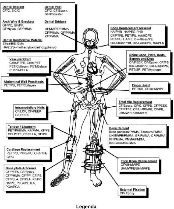
Polymers have been used (combined or individually) in different applications (Figure 4).58 They can be divided into two groups regarding their source: natural and synthetic. Cellulose, collagen, agarose, chitin or hyaluronan form the members of natural polymeric materials or so-called biological polymers.31
Natural polymers such as collagen have been used for bone tissue engineering purposes. In contrast to natural polymers, synthetic polymers are also used in the bone engineering field. Some examples of these polymers are poly-lactic acid (PLA), poly-glycolic acid (PGA), polyurethane (PU) and polycaprolactone (PCL), from which PLA and PLGA have receives the highest interest because of their biological properties and easy processability.58
Regarding their response when applied to living tissues, polymers can be biodegradable or non-biodegradable. Biodegradable synthetic polymers, like natural polymers, are resorbed by the body. To have the implant being resorbed by the body while the replacing tissue is regenerating is highly advantageous.
Figure 4 - Various applications of composite - polymers throughout the body 58
Legenda
CF: Carbon Fibers; C: Carbon; GF: Glass Fibers; KF: Kevelar Fibers; PMMA: Polymethylmethacrylate; PS: Polysuifone; PP: Polypropylene; UHMWPE: Ultra-high-molecular weight polyethylene; PLDLA: poly (L-DL- Lactide); PGA:
Poliglycolic acid; PC: Polycarbonate; PEEK: Polyethereutherketone; HA: Hydroxyapatite; PMA: polymethacrylate;
BIS-GMA: Bis-phenol A glycidyl methacrylate; PU: polyurethane; PTFE: Polytetrafluorethylene; PET: Polyetylenetephthale; PEA: Politethylacrylate; SR: Silicone rubber; PHB: Polyhydroxybutyrate; PEG: Polyethyleneglycol; PHEMA: Poly
These polymers do not require a second surgical event for its removal once it is no longer needed. This last characteristic can be a huge advantage in the bone field, because the bone can develop a phenomenon called “stress shielding”: when a bone fractures and it is fixed with a rigid, non-biodegradable stainless steel implant, it may not heal properly and can eventually re-fracture upon removal of the implant.59 Bone does not carry sufficient load during the healing
process, since this is carried mostly by the rigid stainless steel.59 However, an
implant prepared from a biodegradable polymer can be engineered to degrade at a rate that will allow for the slow transfer of the load to the healing bone.60
Poly(glycolic acid), poly(lactic acid) (PLA) and their copolymer poly(lactide-co-glycolide) (PLGA), polydioxanone, poly (ethylene oxide) and poly(trimethylene carbonate) are examples of polymers that have been used for this purpose. In addition, poly(ε-caprolactone) (PCL), polyanhydrides, poly(vinyl alcohol) (PVA), polyurethanes and recently polyhydroxykanoates (PHA) which are linear polyesters of microbiological origin, have also been investigated for bone regeneration.31,57
At the same time, non-biodegradable polymers have been used in bone tissue engineering due to their better mechanical properties and chemical stability than biodegradable polymers.57 In the bone field, these materials are mainly used
when the tissue cannot be regenerated due to large losses or for elderly patients with a less effective self-healing ability of the tissue. Currently, synthetic nonbiodegradable polymers used are for instances: polyethylene, polypropylene, polytetrafluoroethylene, poly (vinyl chloride), polyamide (PA), poly (methyl methacrylate), polycarbonate, poly (ethylene terephtalate), poly (ether ketone), acrylics and silicones.57
Ceramics such as calcium phosphates and calcium sulfates are clinically used as implant coatings or bone-void fillers because of their attractive biodegradable, bioactive and osteoconductive properties, and also because they have a notable ability to bond directly to bone.57 Calcium phosphates appear in
several different forms, being hydroxyapatite (Ca10(PO4)6OH2) and tricalcium
phosphate (Ca3(PO4)2) the most currently used.1 Hydroxyapatite (HA) is a
naturally occurring calcium phosphate that comprises up to 70% of the dry weight of bone. The synthetic form of HA is osteoconductive and can have a crystalline
structure similar to the HA in bone. HA synthesized at high temperatures is highly stable and slower to resorb than its endogenous form and may stay at the site of implantation for many years. Tricalcium phosphate (TCP) is a bioceramic that exists in alpha (α-TCP) and beta cristal (β-TCP) forms.61 α-TCP is the
high-temperature and β-TCP is the low-high-temperature polymorph of TCP.62
β-Tricalcium phosphate (β-TCP) has been successfully used in posterolateral spinal fusions, dental procedures and as a component of bio-resorbable screws.63 In fact, Ogose et al. 63 demonstrated that β-TCP appears to
be advantageous in comparison to HA for surgery involving bone tumors and exibits superior osteoconductivity. Calcium sulphate (CaSO4) is resorbed within
6 weeks – which is faster than both HA and TCP. It was already successfully used in tuberculosis. Calcium sulphate pellets have been used as autograft extender for instrumented short segment posterolateral spinal fusions for degenerative disease.64 Although the biological characteristics are favorable for
bone engineering purposes, the main drawbacks of these materials are their brittleness, low fracture strength, low mechanical reliability, lack of resilience and high density which makes them susceptible to catastrophic failure.
3.1. The structural organization of polymer-ceramic nanocomposites
Typically, the polymer-ceramic composites used for bone replacement and regeneration are systems in which a ceramic filler is dispersed within a polymer matrix. In nanocomposites, the dispersed filler material is at the nano dimension. One of the main reasons for using nanomaterials is the large surface to volume ratio, which increases the number of particle-matrix interactions, increasing the effects on the overall material properties.65,66 The interphase formed around
nanoparticles exhibits distinct properties from those of the bulk matrix.67 It can be
a region of altered chemistry, polymer chain mobility, degree of cure or crystallinity Therefore, by controlling the degree of interaction between the polymer and the nanofiller, the properties of the entire matrix can be controlled.68
Another important consideration is the dispersion of the filler,65 since a well
particle agglomerates decrease the material performance by the inclusion of voids that act as preferential sites for crack initiation and failure.65 Particles,
especially in the nano range (less than 100 nm) tend to agglomerate, or cluster, due to the dominant intermolecular van der Waals interactions between them.65
Therefore, when fabricating a polymer-nanocomposite scaffold, the technique to disperse nanoparticles into the polymer matrix is important, because it provides a good mechanical behaviour in vivo.
In order to simplify the discussion about nanofillers, Ajayan et al.68 grouped
nanofillers into three categories, according to their geometrical shape: fiber-like, plate-like and three-dimensional (Figure 5). This is a convenient way to discuss polymer-based nanocomposites, because the processing methods used and the properties achieved depend strongly on the geometry of the fillers.
a) Nanosphered and nanopatirculated composites
Nanoshperes or nanoparticles can be dispersed throughout a continuous matrix to induce porosity,69 to improve mechanical properties of a bulk scaffold
as a reinforcement phase70 or a crosslinking agent71 and as a drug delivery
vehicle.72 They can also be used as building blocks to establish scaffolds by a
bottom-up approach without a surrounding matrix material. Particulate building blocks can also be used to prepare 3D stand-alone scaffolds without surrounding material through rapid prototyping,73 random packing74 or directed assembly.75
Figure 5
Rapid prototyping uses a computer-aided design to produce 3D constructs with customizable architecture using nanospheres as building blocks.73
Nanopaticulated composites can be produced in a random packing technic or in a direct assembly. In a random packing technic, composite gels are putted up together in a structurally random way, which leads to poor integrity of these gel-based scaffolds resulting from weak interparticle interactions and consequently poor mechanical stability.74 To preserve the agglomeration of
nanosphere formulation after implementing these scaffolds, maintaining the particles together, crosslinkers and compatibilization agents have been investigated.75 On the other hand, directing the assembly by introducing
interparticle forces (such as electrostatic forces or hydrophobic interactions), can produce formulations with enhanced structural integrity and mechanical stability without the use of compatibilization agents or crosslinkers.75 Therefore, this
technique allows for a level of structural organization that ensures good mechanical properties. Clinically, these techniques have been used to produce injectable gels which act as scaffolds for bone regeneration in minimally invasive surgery.76
When dispersing nanoparticles in a polymeric matrix, two main alternatives can be considered: they can be dispersed in the polymeric matrix during the polymer synthesis or after the polymer synthesis being complete.65 By blending
the nanoparticles in pre-synthesized polymer matrixes, it is possible to have a full control over both the nanoparticles and the matrix. The preparation of most of the nanoparticles and nanomaterials can be divided into four main methods.65
I. The wet method, in which nanoparticles are prepared in a medium using a sol-gel technique, emulsion approaches or the intercalation polymerization;
II. The dry method, that includes preparing of nanoparticles using methods such as the abrasive, burning, gas impinging and collision approaches, high-energy ball milling processes;
III. The evaporating method, where nanoparticles are prepared using chemical vapor deposition or the gas deposition approach, like laser gas vapor deposition.
IV. The sedimentation method, for special nanocomposites that possess heavy weight selection particles.
In ceramic-polymer nanocomposite processing, wet and dry methods are used very often, being the sol-gel technique one of the most used. It consists in a series of hydrolysis and condensation reactions that transform a solution, which has colloidal particles dispersed in it, into a gel, where both liquid and solid are dispersed in each other, through low temperature methods.
b) Nanofiber composites
An important class of nanostructured biomaterials on which intensive research has been carried out is nano-fibrous materials, especially biodegradable polymer nanofibers. In this specific material, nanofibers are dispersed in a biodegradable polymer matrix. The large surface-area-to-volume ratio of nanofibers combined with their porous structures favours cell adhesion, proliferation, migration, and differentiation, as it was already discussed. There are three main techniques to produce nanofibers: phase separation, self-assembly and electrospinning.77 Nanofiber mats can be morphologically similar to
extracellular matrix (ECM) of the natural tissue, which is characterized by a wide range of pore diameter distribution, high porosity, effective mechanical properties, and specific biochemical properties.78
Phase separation, for instance, is a technique often used to produce nanofibrous scaffolds.77 This process is based on thermodynamic demixing of a
homogenous polymer solvent solution into a rich phase and a polymer-poor phase. Cooling the solution to a specific point causes this separation. However, usually there is lacking of interconnected macropores, so this technique is often combined with other scaffold fabrication techniques (e.g. particle leaching) to allow broader control over the scaffold architecture.79,80 With
this technique, nanofibers with a range of diameters of 50 to 500 nm are produced. Ma et al.81 have used this technique to produce a PLLA fibrous
structure from a PLLA/THF solution to be used in regeneration scaffolding materials that provide a better environment for cell attachment and function.
Self-assembly is the most complex technique and it allows the creation of nanofibers with very small diameters (a few to 100 nm). It basically consists on an autonomous organization of components that are able to assemble at the
molecular level.77,82 Being a natural process for several essential biological
components including nucleic acid or protein synthesis, self-assembly technology usually incorporates some specific biological components of the extracellular matrix (ECM), closely mimicking the ECM assembly process.80 In this process,
molecules require some specific configurations to be assembled into nanofibers. Molecules that meet this requirements are peptide-amphiphiles (PAs),80
oligopeptides, synthetic diblock/triblock copolymers83 and dendrimers.84 Both
self-assembly and phase separation have the disadvantage to create only short strands of nanofiber.80
Another example is electrospinning, which in contrast, represents the most reliable method to simply fabricate long continuous strands of nanofibers with a diameter ranging from nanometers to microns (50–1,000 nm). Besides being simple, it is relatively quick and cost-effective.85 This process involves subjecting
a polymeric solution to a high electric field, which overcomes the surface tension of the solution and drives the ejection of a polymer jet. The charged polymer solution or melt is ejected, dried and solidified onto a grounded substrate. The ejected polymer solutions repel each other during the travel to the grounded collector, which forms thin fibers after solvent evaporation. By controlling the spinning conditions, the resulting fibers can range from about 0.02 μm to about 20 μm.86 Many electrospun nanofibrous biomaterials have been investigated as
tissue regeneration scaffolds.78,86 Materials used in electrospinning can be
natural macromolecules such as collagen, chitosan, silk fibroin; synthetic biodegradable polymers such as PGA, PLGA, PLLA, PCL; and combinations of these natural and synthetic polymers. In addition, various substances (proteins, growth factors, and hydroxyapatite) can be incorporated into nanofibrous materials during electrospinning. Therefore, electrospun nanofibrous biomaterials have been explored to engineer various tissues. However, significant challenges still exist in using this technique to fabricate complex 3D scaffold shapes or to generate designed internal pore structures, limiting its potential for many tissue engineering applications.80
II. Materials and Methods
S
earch Strategies
In order to perform a bibliographic review as complete as possible, data was extracted and selected from scientifically relevant databases, in this case ScienceDirect and PubMed.
In a first stage, the following combination of words was used in both online data-bases: “Bone AND Regeneration AND Nanocomposite AND Polymer AND maxillofacial AND NOT review”. In the PubMed database, 5 results were found and in ScienceDirect, 33. It was then realized that this combination of words restricted too much the online search too much, so other MeSH terms were selected and integrated in the combination. The word “cranial” was selected (replacing the word “maxillofacial”), to provide articles about bone regenerative measures using nanocomposites among the whole cranial complex and not just the maxillofacial area. The MeSH term “augmentation” was added as well, in order to include articles about the use of regenerative techniques in alveolar bone augmentation. The different MeSH words and combinations, as well as the obtained results are included in results diagram (Annex 1) and can be summarized as follows:
“Bone AND Maxillofacial AND Regeneration AND nano composite AND polymer NOT review”, with the particularity of having the nanocomposite written separately (nano composite), obtained 5 results in PubMed and 73 in ScienceDirect;
“Bone AND Cranial AND Regeneration AND nanocomposite AND Polymer NOT review”, in which the word “Maxillofacial” was replaced by the word “cranial”, obtained 9 results in PubMed and 27 in Science Direct;
“Bone AND cranial AND regeneration AND nano composite AND Polymer NOT review”, the same search performed before, with nanocomposite written separately (nano composite), obtained 13 results in PubMed and 60 in ScienceDirect;