UNIVERSIDADE DE LISBOA Faculdade de Ciências Departamento de Biologia Vegetal
The role of SETD2 in transcription
and in DNA damage response
Sílvia Filipa Camoeira Gonçalves de Carvalho
Dissertação
Mestrado em Biologia Molecular e Genética 2012
UNIVERSIDADE DE LISBOA
Faculdade de Ciências Departamento de Biologia Vegetal
The role of SETD2 in transcription
and in DNA damage response
Sílvia Filipa Camoeira Gonçalves de Carvalho
Dissertação orientada pelo Doutor Sérgio Fernandes de Almeida
e pelo Prof. Doutor Rui Gomes
Mestrado em Biologia Molecular e Genética
ii
Content
Content ... ii
List of Figures ...iv
List of Abbreviations ... v
Original Articles ... vii
Sumário ... viii Resumo ...ix Abstract ... xiv Introduction ... 1 1. Chromatin structure ... 1 2. Chromatin remodelling ... 1 3. Transcription ... 2
3.1. The FACT complex is a major regulator of nucleosome dynamics ... 3
4. DNA damage response ... 4
4.1. DNA damage signalling ... 4
4.2. p53 activation during the DNA damage repair ... 5
5. Chromatin and cancer ... 6
5.1. Clear cell renal cell carcinoma ... 6
6. Aims of the study... 6
Results and Discussion ... 8
1. SETD2 role in transcription ... 8
1.1. Downregulation of SETD2 affects RNAPII turnover at the transcription site ... 8
1.2. SETD2 prevents widespread intragenic transcription initiation ... 10
1.3. SETD2 promotes FACT recruitment ... 12
2. SETD2 role in DNA damage response ... 13
2.1. ccRCC cell lines as an experimental model system to investigate the tumour suppressor role of SETD2 ... 13
2.2. SETD2 is required for DNA damage repair ... 14
2.3. SETD2 is required for ATM activation following DNA damage ... 16
2.4. SETD2 is needed for proper DNA damage signalling ... 17
2.5. SETD2 regulates p53 activity during DNA damage repair ... 18
2.6. The role of SETD2 in DDR is independent from H3K36me3 ... 19
iii
Material and Methods... 23
1. Cell culture ... 23
2. RNA interference ... 23
3. Western blot ... 23
4. Chromatin Immunoprecipitation ... 24
5. Fluorescence recovery after photobleaching (FRAP) ... 24
6. RNA-seq data analysis ... 25
7. Comet assay ... 25 8. Immunofluorescence ... 25 Acknowledgments ... 27 Bibliography... 29 Annexes... I Annex I ... I Annex II ... II
iv
List of Figures
Figure 1 SETD2 is a determinant of RNPII levels along the coding region of transcribed genes ... 9
Figure 2 Genome-wide analysis of intragenic transcription initiation induced by SETD2 depletion.... 11
Figure 3 SETD2 is required for FACT recruitment ... 12
Figure 4 SETD2 activity is reduced in mutant ccRCC ... 13
Figure 5 SETD2 activity is required for DSBs repair ... 15
Figure 6 SETD2 activity is required for ATM activation ... 16
Figure 7 SETD2 activity is needed for DSBs signalling ... 17
Figure 8 SETD2 is required for p53 activation ... 18
Figure 9 H3K36me3 levels do not change during the DDR ... 20
v
List of Abbreviations
ATM Ataxia telangiectasia mutated
ATMp ATM phosphorylated at Ser1981
ccRCC Clear cell renal cell carcinoma
ChIP Chromatin immunoprecipitation
CK2 Casein kinase 2
Ct Cycle threshold
DDR DNA damage response
DNA Deoxyribonucleic acid
DNA-PKcs DNA-dependent protein kinase, catalytic subunit
DSBs Double strand breaks
FACT Facilitates chromatin transcription
FBS Fetal bovine serum
FRAP Fluorescence recovery after photobleaching
h Hours
H2Bub Histone H2B ubiquitinated
H3K36me3 Histone H3 lysine 36 trimethylation
MDM2 Mouse Double Minute 2
ITI Intragenic transcription initiation
min Minutes
MRN Mre11, Rad50 and Nbs1
MUT Mutant
p21 Protein 21
p53 Protein 53
p53p Protein 53 phosphorylated at Ser15
RCC Renal cell carcinoma
RNAi RNA interference
RNAPII RNA polymerase II
RNAPII-GFP RNA polymerase II fused to green fluorescent protein RT-qPCR Real time quantitative polymerase chain reaction
vi
s Seconds
SDS-PAGE Sodium dodecyl sulphate polyacrylamide gel electrophoresis
SETD2 SET domain containing 2
SPT6 Suppressor of Ty 6 homolog
SPT16 Suppressor of Ty 16 homolog
siRNA Small interfering RNA
SSRP1 Structure specific recognition protein 1
Ser Serine
U2OS Human osteosarcoma-derived cells
VHL Von Hippel-Lindau
WT Wild-type
x Times
vii
Original Articles
Part of the results presented in this thesis were published in:
Histone methyltransferase SETD2 coordinates FACT recruitment with nucleosome dynamics during transcription. Carvalho S, Raposo AC, Grosso AR, Rino J, Martins FB, Carmo-Fonseca M, de Almeida SF. 2012, Submitted in revised form to Nucleic Acids Research.
Splicing enhances recruitment of methyltransferase HYPB/Setd2 and methylation of histone H3 Lys36. de Almeida SF, Grosso AR, Koch F, Fenouil R, Carvalho S, Andrade J, Levezinho H, Gut M, Eick D, Gut I, Andrau JC, Ferrier P, Carmo-Fonseca. 2011, Nature Structural & Molecular Biology, Vol. 18, pp. 977–983.
viii
Sumário
As moléculas de DNA estão hierarquicamente compactadas em cromatina, de forma a poderem ser contidas dentro do núcleo da célula. Os vários processos que requerem o acesso ao DNA superam esta barreira através da modificação pós-traducional de histonas e da actividade de complexos remodeladores de cromatina. Estes têm vindo a ser reconhecidos como intervenientes no processo de tumorigénese. Recentemente, identificou-se a metiltransferase de histonas SETD2 como um novo supressor tumoral, desempenhando um papel importante no carcinoma de células renais de células claras (ccRCC). A principal função da SETD2, a trimetilação da lisina 36 da histona H3 (H3K36me3), é uma característica da transcrição, contudo o seu papel ainda não foi completamente elucidado. Os resultados obtidos demonstram que a H3K36me3 recruta o facilitates chromatin transcription (FACT) para a cromatina, este chaperone de histonas permite a modificação da estrutura da cromatina durante a transcrição, impedindo a RNA Polimerase II de aceder e iniciar a transcrição em regiões intragénicas. Nesta tese, propomos que a função de SETD2 não está confinada à transcrição, ao demonstrarmos que esta é necessária para a activação de um dos principais reguladores da resposta a lesões no DNA (DDR), a ATM (ataxia telangiectasia mutated). Esta permite a sinalização e reparação de quebra de cadeias duplas do DNA através da fosforilação de vários substratos tais como a histona H2AX. De facto, na ausência de SETD2 verificou-se uma deficiente fosforilação de histona H2AX e também uma redução na actividade da proteina p53. Em conjunto, os dados apresentados sugerem que o papel supressor de tumores da SETD2 poderá estar relacionado com a regulação do padrão da iniciação de transcrição e da DDR. A ausência de actividade de SETD2 poderá levar consequentemente a alterações da expressão genética e instabilidade genómica, podendo, desta forma, contribuir para a tumorigénese, em particular ao desenvolvimento de ccRCC.
Palavras-chave: SETD2; Iniciação de transcrição intragénica; Resposta a lesões no DNA; FACT; ATM; ccRCC.
ix
Resumo
Introdução
A célula eucariota guarda a sua informação genética em moléculas de DNA, cujo tamanho é muito maior que o compartimento celular que as contém, o núcleo. De forma a acomodar-se dentro deste, as moléculas de DNA estão hierarquicamente compactadas em conjunto com proteínas, conjunto o qual se designa por cromatina. Contudo, existem vários processos, como a transcrição e a resposta a lesões no DNA (DDR), que requerem acesso ao DNA, e, para tal, é necessário uma remodelação da estrutura da cromatina através de modificações pós-traducionais de histonas e da actividade de complexos remodeladores de cromatina. O chaperone de histonas facilitates chromatin transcription (FACT) tem um papel fundamental na remodelação dos nucleossomas. Durante a fase de elongamento transcricional da RNA Polimerase II (RNAPII) o FACT remove dímeros de histonas H2A-H2B dos nucleossomas e reposiciona-os após a passagem da RNAPII. Para além do papel na transcrição, o FACT também actua na DDR, ao participar na troca da histona H2A pela sua variante H2AX e ao interagir com factores de reparação de DNA.
Recentemente, vários remodeladores da cromatina e alterações epigenéticas têm vindo a ser reconhecidos como intervenientes no processo de tumorigénese, como é o caso particular do carcinoma de células renais de células claras (ccRCC) em que a metiltransferase de histonas SETD2 foi implicada como um novo supressor de tumor. No entanto, o mecanismo de supressão tumoral em que a SETD2 participa ainda não foi desvendado. Sendo que, o seu papel conhecido é a trimetilação da lisina 36 da histona H3 durante a transcrição. Em leveduras, esta modificação da histona H3 recruta um complexo de desacetilação de histonas, prevenindo, desta forma, a iniciação de transcrição intragénica (ITI). Contudo, este mecanismo não é conservado em mamíferos. Este trabalho tem como objectivo investigar qual o mecanismo em que intervém a SETD2 que possa justificar o seu papel como supressor de tumores. Em concreto, pretende-se investigar o mecanismo pelo qual a SETD2 regula a a transcrição em células humanas e o seu papel na DDR.
x Resultados
O papel da SETD2 na transcrição
Para estudar o impacto da SETD2 na transcrição, o nível de SETD2 foi reduzido por interferência de RNA (RNAi) e analisou-se a distribuição da RNAPII por imunoprecipitação de cromatina (ChIP). Os resultados revelam que a depleção de SETD2 leva a um aumento de RNAPII nos exões internos dos genes estudados. A análise da dinâmica da RNAPII através da técnica de microscopia recuperação de fluorescência após fotodegradação (FRAP) demonstrou que quando os níveis de SETD2 são depletados por RNAi a recuperação de fluorescência da RNAPII é mais rápida, indicando uma maior mobilidade da RNAPII no local de transcrição. Em conjunto, estes dados sugerem que a SETD2 tem um papel essencial na prevenção do acesso de RNAPII a regiões intragénicas das unidades transcricionais. Como tal, a SETD2 poderá ter um papel importante na prevenção de ITI em células humanas, em semelhança com o que acontece em leveduras. Para estimar a incidência de ITI após a depleção de SETD2, estimámos o número de transcritos iniciados em regiões intragénicas através de uma análise de dados de sequenciação do transcriptoma total de células com e sem depleção de SETD2 por RNAi. Esta análise mostrou que a diminuição dos níveis de SETD2 leva a um aumento da quantidade de transcritos cujo início de transcrição se encontra a jusante do primeiro exão anotado, indicando uma maior incidência de ITI.
De seguida, tentámos perceber como é que a SETD2 previne a ITI. Apesar de em leveduras a H3K36me3 regular a desacetilação de histonas como um meio de prevenir iniciação de transcrição em regiões intragénicas, em mamíferos, a inactivação da H3K36me3 não afecta os níveis de acetilação das histonas. De modo a investigar qual o mecanismo dependente de SETD2 que impede a ITI em células humanas analisou-se o recrutamento de diferentes chaperones de histonas em genes activamente transcritos. Esta análise revelou níveis reduzidos do chaperone de histonas FACT após depleção de SETD2. Níveis insuficientes de recrutamento de FACT para a cromatina provocam alterações na densidade de nucleosomas e, consequentemente, leva a ITI.
Deste modo, os dados apresentados nesta tese permitem sugerir que a SETD2 impede a ITI por estimular o recrutamento de FACT para os genes transcritos.
xi O papel da SETD2 na resposta a lesões no DNA
Tendo em conta que o FACT tem um papel activo na resposta a lesões no DNA, a descoberta da modulação do recrutamento do complexo FACT pela SETD2 sugere que a DDR possa ser outro processo no qual a SETD2 actue. De forma a explorar o papel da SETD2 na DDR, estabeleceu-se um modelo experimental com linhas celulares de ccRCC em que a SETD2 apresenta actividade de metiltransferase (SETD2 wild-type) e células de ccRCC contendo mutações com perda de função (SETD2 mutante).
Primeiro, verificou-se a dinâmica da reparação de DDR em função da actividade de SETD2. As lesões no DNA foram induzidas através do tratamento das células com etoposido e as quebras de cadeia dupla do DNA quantificadas recorrendo à técnica de comet assay em condições de pH neutro. Este ensaio permitiu verificar que as lesões são induzidas de forma semelhante em células com SETD2 wild-type e células com SETD2 mutante. No entanto, a reparação das lesões revelou-se mais eficiente nas células SETD2 wild-type, que reparam 42% da lesão em 6h enquanto que as células com SETD2 mutante reparam somente 24% no mesmo período de tempo. Estes resultados sugerem que a SETD2 é requerida para uma reparação adequada das quebras de cadeia de DNA.
Sendo a ATM (ataxia telangiectasia mutated) um dos principais reguladores da resposta a lesões no DNA (DDR), prosseguiu-se à análise da sua actividade através da medição dos níveis da forma fosforilada de ATM (activa) e dos seus níveis proteicos totais após indução de lesões no DNA com etiposido. Os resultados demonstraram que nas células com SETD2 wild-type a ATM é eficazmente activada após lesão do DNA, enquanto que em células mutantes não se verificou activação de ATM. Entre os substratos que a ATM activa através da fosforilação encontra-se a histona H2AX. A fosforilação da histona H2AX é um dos primeiros eventos da DDR e tem um papel fundamental na sinalização de DSBs. De forma a verificar-se se a fosforilação da histona H2AX está comprometida em células com SETD2 mutante, os seus níveis foram avaliados por western blot e imunofluorescência após a indução de DSBs. Estas experiências revelaram que, de acordo com a actividade reduzida de ATM, os níveis de histona H2AX fosforilada estavam comprometidos em células com SETD2 mutante.
Outro evento regulado pela ATM com relevância na DDR é a activação da proteina 53 (p53), reconhecida como um supressor de tumor que se encontra frequentemente mutado em diversos tipos de cancro. Curiosamente, apesar da sua via se encontrar afectada, as mutações de p53 são infrequentes em ccRCC. Tendo isto em conta, averiguámos os níveis proteicos de p53 total e p53 activo (através da medição dos níveis de p53 fosforilado) por western blot
xii após a indução de lesões de DNA com etoposido. Em células SETD2 wild-type a quantidade de p53 total aumenta progressivamente após a lesão do DNA, voltando a diminuir quando a lesão é reparada. Em contraste, em células com SETD2 mutante os níveis de p53 total não aumentam ao longo do intervalo de tempo analisado após adição de etoposido. A actividade biológica do p53 é dependente dos seus níveis de fosforilação e da sua habilidade de se ligar a elementos de regulação transcricional no DNA de genes alvo, como é o caso do p21 (proteína 21). De forma a investigar-se se a actividade de p53 durante a DDR requer a actividade de SETD2, mediram-se os níveis de p53 fosforilado e os níveis proteicos de p21 total, cuja transcrição é activada pelo p53. Os resultados destas experiências revelaram que células com SETD2 wild-type possuem actividade normal de p53, revelada pelo aumento da seus níveis de fosforilação e pelo aumento dos níveis de p21 após a indução das lesões no DNA. Em contraste, a actividade de p53 está comprometida em células com SETD2 mutante pois os níveis de ambas as proteínas analisadas, p53 fosforilado e p21, não aumentam após a lesão do DNA.
Dado o papel proeminente de SETD2 como uma metiltransferase de histonas, procedemos à análise dos níveis de H3K36me3 durante a DDR em células SETD2 wild-type e SETD2 mutante e verificámos que em ambas as situações os níveis de H3K36me3 não alteram ao longo da DDR, o que sugere que a SETD2 actua na DDR por um mecanismo independente da sua função de metilação de histonas.
Conclusão
Este estudo demonstrou que durante a transcrição a actividade da SETD2 promove o recrutamento de FACT para a cromatina, permitindo a reconstituição dos nucleossomas após a passagem da RNAPII. Desta forma, a SETD2 permite a manutenção da integridade da cromatina e impede a iniciação da transcrição intragénica.
Os resultados apresentados nesta tese sugerem ainda que a função de SETD2 não está confinada à transcrição. De facto, os dados aqui apresentados revelam que a SETD2 é requerida por diferentes mecanismos envolvidos na DDR, tais como a activação da ATM (ataxia telangiectasia mutated) e a sinalização de lesões no DNA, através da fosforilação da histona H2AX. Em células contendo mutações com perda de função da SETD2, a reparação de lesões do DNA mostrou-se severamente comprometida. A observação de que também a activação da proteina p53 na DDR depende da actividade da SETD2 revela um possível mecanismo de supressão de tumores em que esta metiltransferase participa.
xiii Em conjunto, o papel da SETD2 em regular a iniciação da transcrição e a DDR poderá estar na base do seu papel supressor tumoral recentemente descrito no contexto de ccRCC. Estudos adicionais irão certamente clarificar a extensão exacta do impacto de mutações do SETD2 em mecanismos com potencial tumorigénico.
xiv
Abstract
DNA molecules are hierarchically packaged into chromatin, in order to fit inside the eukaryotic nucleus. However, several physiological processes require access to the DNA template buried within the complex chromatin structure. To overcome this barrier, histone post-translational modifications and chromatin remodelling complexes, grant the different transcription or DNA repair factors access to the hidden DNA. Several chromatin remodelers are being recognised as important players in tumorigenesis. For instance, the histone methyltransferase SETD2 was recently reported as a tumour suppressor in clear cell renal cell carcinoma (ccRCC). SETD2 is responsible for the trimethylation of lysine 36 of histone H3 (H3K36me3), which is a hallmark of transcription elongation. However, its precise role is not yet clear. We aim at clarifying the role of SETD2 in transcription. Our results show that SETD2 activity recruits the facilitates chromatin transcription (FACT) complex onto chromatin, which in turn will restore chromatin compaction after transcription and prevent the accessibility of RNA Polymerase II to the gene body, preventing intragenic transcription initiation. Furthermore, we propose that SETD2 activity is not confined to transcription but is also important during the DNA damage response (DDR). We demonstrate that SETD2 is required for the activation of the major regulator of DDR ATM (ataxia telangiectasia mutated). ATM activity allows a proper double strand break signalling, through histone H2AX phosphorylation, and DNA damage repair, processes that are impaired in SETD2 mutant cells. Moreover, the lack of functional SETD2 leads to a reduced activity of the p53 protein. Altogether, the data presented in this thesis suggest that SETD2 regulates the pattern of transcription initiation and the accuracy of the DNA damage signalling and repair. Loss of function mutations in SETD2 would most likely change the gene expression profile and introduce instability into the genome. Ultimately, it could contribute to tumorigenesis, in particular, to ccRCC.
Keywords: SETD2; Intragenic transcription initiation; DNA damage response; FACT; ATM; ccRCC.
1
Introduction
1. Chromatin structure
The eukaryotic cell stores its genetic information in DNA molecules, whose length is far greater than the size of the compartment in which it is contained, the nucleus. In order to fit inside the nucleus, DNA molecules are hierarchically packaged into chromatin. Such compaction is achieved through the action of DNA interacting proteins designated histones. The fundamental repeating unit of chromatin is the nucleosome, which is composed of two copies of histones H2A, H2B, H3 and H4 assembled in an octameric core with 146 base pairs of DNA wrapped around it. Nucleosomes are separated by linker DNA and its associated with histone H1 forming a “beads-on-a-string” fibre that represents the first level of chromatin organisation, which subsequently forms structures of higher compaction order (1-3). This repeating unit shows variations in its composition due to the exchange of canonical histones by histone variants (4) and post-translational modifications of histones (5).
Chromatin compaction is therefore a convenient way of packaging the DNA inside the relatively small nuclear compartment. However, several physiological processes - such as transcription, DNA damage response and replication (6-8) - require access to the DNA template buried within the complex chromatin structure. To overcome this physical barrier, fine-tuned mechanisms of chromatin remodelling, grant the different transcription, replication or DNA repair factors access to the hidden DNA.
2. Chromatin remodelling
Chromatin plasticity and dynamics are modulated by two major mechanisms: histone post-translational modifications and chromatin remodelling complexes (6-9).
The four core histones that compose the nucleosome are predominantly globular, except for their unstructured N-terminal tails, which prone to a wide array of post-translational modifications (5, 6, 10). These modifications are dynamic and can influence chromatin structure directly, by affecting the histone charge and weakening the interaction with DNA, or by signalling the recruitment of other non-histone proteins that can modify chromatin (5).
2 These histone-modification readers are extrinsic modulators of the chromatin structure and belong to two main groups: histone chaperones and ATP-remodelling complexes (6).
Histone chaperones prevent nonspecific interactions between the negatively charged DNA and positively charged histones. They promote the ordered assembly of the nucleosomes without ATP consumption. The function of histone chaperones is intimately coupled with the activity of ATP-remodelling complexes. These complexes use the energy of ATP to break histone-DNA contacts during chromatin disassembly and to reposition histones during chromatin assembly. Chromatin remodelling complexes can lead to transient histone eviction or nucleosome sliding to different regions (6).
The activity of the two different classes of chromatin modifiers is not mutually exclusive. In fact, there is a crosstalk between histone modifications and chromatin remodelers. Histone modifications can signal the recruitment of histone chaperones, as well as chromatin remodelers are able to promote the spreading of specific histone modifications (9).
3. Transcription
Transcription is a fundamental and tightly regulated step in gene expression. This process begins with the formation of the pre-initiation complex followed by RNA Polymerase II (RNAPII) elongation and release of the newly synthesized RNA transcript during termination (11). Several transcription factors require access to the DNA template, and so, chromatin has to be made accessible by the activity of the different chromatin modifiers (6). Nucleosomes, which could hamper transcription initiation by occupying key regulatory DNA sequences in the promoter and transcription start sites of genes, are typically excluded from these regions. Conversely, the coding regions of almost all genes are occupied by nucleosomes that need to be evicted and reassembly back to chromatin during RNAPII elongation (12-14).
Modification of histones plays a key role in the process of nucleosome eviction (10). Notably, there are dozens of different histone modifications, which disclose the existence of several distinct mechanisms of chromatin tailoring. Many of these histone marks are typically found on actively transcribed genes. However, only recently, the outcome in chromatin-templated processes provided by the combination of distinct histone modifications (histone code) started to be deciphered (15). For instance histone H2B ubiquitylation (H2Bub) is found on active genes and signals the recruitment of histone chaperones that facilitate RNAPII
3 elongation (16). Our lab has been studying another histone modification - histone H3 lysine 36 trimethylation (H3K36me3) - which is a hallmark of transcription elongation that is present in the body of active genes (9) and plays a key role in coupling RNAPII elongation to splicing (17-18). We have recently found that splicing is a determinant of H3K36me3 by promoting the recruitment of the histone methyltransferase SET domain containing 2 (SETD2) (18). An important role of H3K36me3 is the recruitment of a histone deacetylase complex, which hypoacetylates histone H3 and H4 after RNAPII transcription elongation (19-21). This mechanism, found only in yeast, prevents transcription initiation from within the body of the gene, downstream from the canonical transcription start site. In mouse, Setd2 depletion also promotes intragenic transcription initiation, however, the mechanism is not known but does not involve histone hypoacetylation (22). Intragenic transcription initiation (ITI) implies that portions of DNA are accessible to transcription factors and permit the assembly of the pre-initiation complex (23-24), which equals to say that they are nucleosome depleted or, at least, nucleosomes are in an alternative and more permissive conformation (14). It is, therefore, plausible to hypothesize that SETD2 depletion could somehow alter the activity of the histone chaperones or chromatin remodelers that operate to maintain the appropriate nucleosome occupancy and conformation in the wake of RNAPII elongation.
3.1. The FACT complex is a major regulator of nucleosome dynamics
Histone removal and their subsequent reassembly onto nucleosomes during transcription involve several histone chaperones, such as the facilitates chromatin transcription (FACT) and the suppressor of Ty 6 homolog (SPT6) (9). The human FACT complex is a heterodimer comprised of suppressor of Ty 16 homolog (SPT16) and structure specific recognition protein 1 (SSRP1) that facilitates the removal of histone H2A-H2B dimers from nucleosomes ahead of RNAPII and their reassembly in the wake of elongation (25-29). However, the mechanism by which FACT is targeted to transcribed chromatin templates remains elusive. FACT has been shown to interact with both H2A-H2B dimers and H3-H4 tetramers (28) and its binding to nucleosomes is impaired upon proteolytic removal of the histone H3 tail (30-31). Moreover, there is indication that FACT activity is influenced by several post-translational modifications of histone tails, including methylation (32). This raises the hypothesis that SETD2 and FACT could cooperate to regulate nucleosome dynamics and integrity during RNAPII elongation. In fact, previous studies showed that
4 depletion of the FACT complex results in ITI (33-35), the same phenotype observed upon depletion of SETD2 yeast homologue. Interestingly, beyond its pivotal role in transcription, FACT was also implicated in another DNA-related process, the DNA damage response, by exchanging histone H2AX (36) and recruiting DNA repair factors (37). Therefore, if SETD2 activity influences the recruitment and/or activity of FACT, it is plausible to assume that this methyltransferase would also impinge in the DNA damage response pathway.
4. DNA damage response
DNA is continually damaged by environmental agents and metabolic products such as ultraviolet light, free radicals and mutagenic chemicals (38). The cellular response to these aggressions, termed DNA damage response (DDR), consists in a coordinated series of events that allow DNA damage recognition, signalling and repair. During the DDR, the chromatin structure is tailored to enable the access of repair factors to the damaged DNA (8, 39). In 1991, Smerdon described the “access, repair and restore” model, which reflects the role of chromatin structure during this process (40). According to this model, once the DNA damage is detected, the compacted chromatin will first become more accessible to enable the DNA recruitment of the DNA repair machinery. Chromatin alteration is followed by repair of the lesion and by restoration of the chromatin organization. More recent studies have provided further insight into this mechanism, by demonstrating that histone post-translational modifications and chromatin remodelling complexes are key regulators of DNA accessibility (41) and that chromatin components can promote DNA damage signalling and repair (39).
4.1. DNA damage signalling
The damaged DNA must be recognised and signalled rapidly and efficiently in order to prevent the propagation of genomic abnormalities. DNA damage signalling consists in a coordinated series of events that require the activity of proteins classified as sensors, transducers, mediators and effectors (8).
After DNA damage the lesion is recognised by sensor proteins, which promote the recruitment and activation of transducers kinases. The Mre11, Rad50 and Nbs1 (MRN) complex is a sensor that is activated by DNA double strand breaks (DSBs) (42) and
5 contributes to the recruitment of the ataxia telangiectasia mutated (ATM) transducer kinase (43). With the help of mediator proteins, other kinases, collectively termed effectors, are activated spreading the signal through the entire nucleus (8). For instance, ATM phosphorylates the histone at Ser139 (γH2AX), which spreads megabases away from the lesion site and potentiates the recruitment of more MRN-ATM to the DNA damage site (44).
Histone H2AX is a histone H2A variant (45) found in nucleosomes throughout the entire genome (46). It differs from the canonical H2A in its ability to be rapidly phosphorylated at serine 139 of its carboxyl-terminal extension in response to DNA DSBs (47). Moreover, FACT is the major histone chaperone involved in the exchange of canonical histone H2A by the H2AX variant and is also able to shuttle H2AX-H2B dimers to and from nucleosomes (36). Nevertheless, the importance of this H2AX-related FACT activity for the DDR signalling pathway remains to be unveiled. Notably, recent studies showed that, during the DDR, FACT relocates to sites of DNA damage where it interacts with the repair factors Ku complex (composed of Ku70 and Ku86) and DNA-dependent protein kinase catalytic subunit DNA-PKcs (37).
4.2. p53 activation during the DNA damage repair
After the recognition and signalling of the DNA damage a cascade of events takes place in order to repair the DNA lesion (38-39).
Apart from the damage signalling, ATM regulates several cellular responses to DNA damage and has a wide range of different substrates, such as p53 (protein 53) (48-49). ATM phosphorylation of p53 at Ser15 contributes to p53 stabilization by preventing its degradation by MDM2 (mouse double minute 2) and promotes other post-translational modifications required for its activation (50). Therefore, this phosphorylation is required to increase the cellular abundance of p53 and its function in the context of a DDR. During the DDR p53 functions as a transcription factor that enhances the transcription of several factors that aid in the DNA lesion repair and halt the cell cycle progression, preventing cells to replicate damaged DNA (49). The copying of still unrepaired DNA could have dramatic effects to the cell and lead to further mutations and genomic instability, which are hallmarks of cancer.
6
5. Chromatin and cancer
The description and interpretation of genomic abnormalities in cancer cells have been the focus of cancer research for decades (51-52). Recently it was shown that both genetic and epigenetic alterations contribute to the initiation and progression of cancer (53). For instance, misregulation and mutations of many histone modifying enzymes are being identified as key features of tumorigenesis (54). Still, how these different epigenetic modifications lead to tumour initiation and/or progression remains poorly understood.
5.1. Clear cell renal cell carcinoma
Clear cell renal cell carcinoma (ccRCC) is the most prevalent subtype of renal cell carcinoma (RCC) (55), and is characterized by the inactivation of the VHL (Von Hippel-Lindau) tumour suppressor gene (56-57). However, compelling evidence suggests that ccRCC might be more genetically complex than previously thought. Indeed, the intriguing observation that inactivation of VHL alone does not generate any RCC phenotype (58), uncovered the need for additional alterations to further propel ccRCC development. Recently, a genome-wide analysis of ccRCC disclosed alterations in additional genes, including somatic mutations in the SETD2 gene (57). This finding led to the classification of this histone modifier as a novel tumour suppressor (59-60), which acts through a mechanism that is yet to be disclosed.
6. Aims of the study
Chromatin remodelling is crucial to many cellular processes. The regulation of transcription and DNA damage response are two of such processes that require the fidelity of several chromatin remodelling events. However, we are still far from understanding the exact mechanisms by which these events are signalled and executed.
In attempt to shed some light on the tumour suppressor mechanism mediated by SETD2, we conceived this study to investigate the role of SETD2 in transcription and in the DNA damage response. How SETD2 affects intragenic transcription initiation in human cells and how it impacts in the DDR are two outstanding questions that will be tackled herein. I
7 hypothesize that the recruitment of the histone chaperone FACT by SETD2 may be an important mechanism to maintain the integrity of the transcribed chromatin. Furthermore, I hypothesize that regulation of transcription initiation and DDR by SETD2 are important tumour suppressor mechanisms that can be relevant to understand the reason why SETD2 mutations are frequently found in ccRCC.
8
Results and Discussion
1. SETD2 role in transcription
1.1. Downregulation of SETD2 affects RNAPII turnover at the transcription site
The classical interpretation of the histone code states that H3K36me3 is a marker of productive RNAPII elongation, owing its correlation to the transcription levels and its presence in the body of actively transcribed genes (6). Findings in yeast state that RNAPII levels are significantly increased in the middle to late coding region of the SCC2 gene in a SETD2 homologue deletion strain (61). Nevertheless, to date, no mechanistic basis for this link and for SETD2 impact on RNAPII transcription elongation in human was proposed.
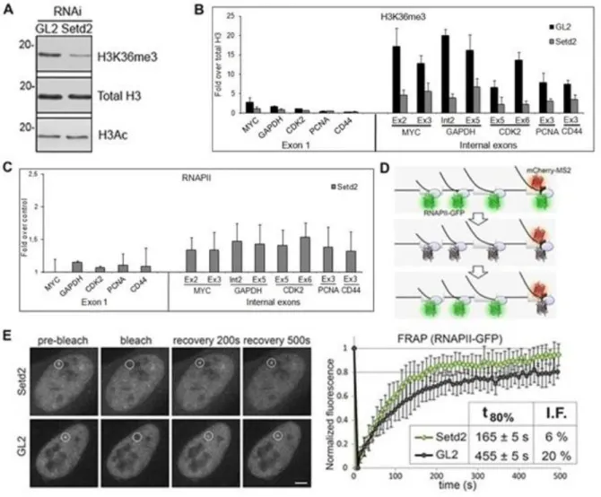
To study the impact of SETD2 in transcription of human genes, we reduced the levels of SETD2 in HeLa cells by RNA interference (RNAi; Figure 1A). Three different synthetic small interfering RNA duplexes targeting SETD2 were designed and tested and two of them were alternatively used throughout this study. As a negative control, we used the GL2 duplex, which targets firefly luciferase (62).
Chromatin immunoprecipitation (ChIP) was carried out using antibodies directed against total histone H3 or H3K36me3, followed by real-time quantitative PCR (RT-qPCR) with primers targeting the indicated exonic regions of five randomly selected genes (Figure
1B). Total histone H3 ChIP signal was used to normalize H3K36me3 levels, thus correcting
for differences in nucleosome density. We have previously reported that the level of H3K36me3 in the first exon is low and increases significantly in the second and subsequent internal exons (18). Accordingly, when compared to control GL2 treated cells, downregulation of SETD2 causes a marginal reduction in H3K36me3 signal associated with first exons (Figure 1B); in contrast, the level of H3K36me3 on internal exons is drastically reduced in SETD2 depleted cells (Figure 1B).
Next, we carried out ChIP analysis with an antibody that binds to RNAPII largest subunit independently of the phosphorylation status of its C-terminal domain (Figure 1C). The results, depicted as fold-change relative to control GL2 treated cells, reveal that SETD2 depletion does not alter RNAPII occupancy on the first exon, but leads to a higher density of
9 RNAPII on internal exons (Figure 1C), similarly with what happens in yeast (61). The high levels of RNAPII along the body of the gene could be due to increased accessibility of RNAPII to the DNA template downstream the promoter region, leading to intragenic transcription initiation.
Figure 1 SETD2 is a determinant of RNPII levels along the coding region of transcribed genes
(A) Total protein extracts of HeLa cells transfected with control and SETD2 siRNAs were analysed by western blot with the indicated. Molecular weight (kDa) markers are shown on the left. (B, C) ChIP analysis with antibodies that recognize H3K36me3 (B) and RNAPII (C). H3K36me3 ChIP data are normalized against total histone H3 ChIP, and RNAPII ChIP signals are shown as fold change over the RNAPII ChIP signal obtained on the control cells. Means and standard deviations from five independent experiments using two distinct siRNAs targeting SETD2 are shown. (D) Schematic representation of the MS2 system. Dimers of MS2 coat protein fused to mCherry bind to MS2 stem loops inserted in the third exon of the β-globin gene, while transcription is carried out by α-amanitin resistant RNAPII-GFP. (E) FRAP of U2OS cells expressing multiple MS2-tagged human β-globin genes and transfected with siRNAs targeting GL2 and SETD2. A circular region corresponding to the transcription site is bleached and fluorescence recovery is subsequently monitored. Scale bar: 5 μm. Normalized fluorescence recovery curves measured at the transcription site for Setd2 depleted (n = 19) and GL2 depleted (n = 18) cells from four individual experiments. t80% denotes the time required to recover 80% of the initial fluorescence. I.F. indicates the percentage of the immobile fraction of bleached RNAPII-GFP molecules at the site of transcription. Error bars represent standard deviations.
10 Next, we sought to explore the dynamic behavior of RNAPII in response to different levels of SETD2 in living cells using fluorescence recovery after photobleaching (FRAP). Human osteosarcoma–derived cells (U2OS) were engineered to stably express tetracycline-inducible β-globin transgenes integrated in tandem in the genome, as previously reported (63). Upon transcription activation, the site of transcription is detected by a red fluorescent protein (mCherry) fused to the MS2 protein that binds to an array of six MS2-binding sequences inserted in the third exon of β-globin (Figure 1D). Simultaneously, the cells express α-amanitin resistant RNAPII fused to green fluorescent protein (RNAPII-GFP). Incubation with α-amanitin causes degradation of the endogenous RNAPII, whereas the resistant RNAPII-GFP remains active. The FRAP analysis consists of bleaching the RNAPII-RNAPII-GFP fluorescence at the transcription site, and subsequently monitoring the fluorescence recovery by time-lapse microscopy (Figure 1E). While in control (GL2) cells the transcription site takes 455 ± 5s to recover 80% of the initial RNAPII-GFP fluorescence intensity, in SETD2 depleted cells RNAPII-GFP fluorescence recovers significantly faster (165 ± 5 s; Figure 1E). Since recovery of RNAPII-GFP fluorescence at the transcription site results from recruitment of unbleached RNAPII-GFP through transcription initiation, the faster recovery observed is consistent with the view that H3K36me3 might act as a deterrent of RNAPII accessibility to the gene. Moreover, the fraction of RNAPII molecules immobilized at the transcription site decreases from approximately 20% in control cells to 6% in SETD2 depleted cells (Figure
1E).
Altogether, the enrichment of RNAPII density and dynamics on internal exons suggest that H3K36me3 may play an essential role in transcription of human genes by preventing the access of RNAPII to regions within the gene body. Thus, SETD2 might have an important function in preventing transcription initiation from cryptic promoter-like sequences within gene body in human cells, similarly to what occurs in yeast (19-21).
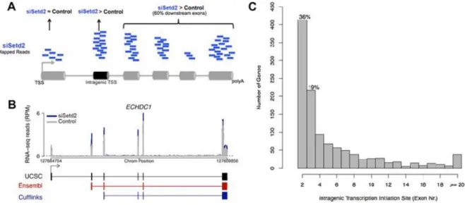
1.2. SETD2 prevents widespread intragenic transcription initiation
To estimate the genome-wide incidence of ITI upon SETD2 depletion, we analysed high-throughput transcriptome sequencing (RNA-seq) data from human mesenchymal stem cells transfected with control or SETD2 targeting siRNAs (64). To identify genes with ITI, we compared exonic read counts in depleted and control cells (Figure 2A). First, we selected all transcripts with higher read counts for at least one exon in SETD2 depleted cells; next, we
11 discarded transcripts with higher read counts for the first annotated exon (likely corresponding to up-regulated full-length mRNAs), and transcripts with higher read counts for isolated internal exons (possibly corresponding to alternative splicing events). Poly(A)+ spliced isoforms were reconstructed using algorithms (TopHat and Cufflinks), which do not depend on gene annotation (65-66). An example of a reconstructed isoforms is depicted in
Figure 2B. This analysis shows that downregulation of SETD2 led to increased levels of
transcripts that are normally spliced and polyadenylated but start downstream of the first annotated exon (Figure 2B). We found evidence for intragenic transcription initiation in 1139 genes among 10772 active protein-coding loci (11%). This analysis also showed that most (55%) intragenic transcription initiation in SETD2 depleted cells starts at the second or third expressed exons (Figure 2C) and at least 13% start at annotated alternative promoters. Taking into account that alternative transcription is emerging as a major contributor to transcriptome diversity (67), this observation raises the possibility that SETD2 may play a regulatory role in modulating alternative promoter usage.
Figure 2 Genome-wide analysis of intragenic transcription initiation induced by SETD2 depletion
(A) Schematic representation describing the constraints for identification of genes with intragenic transcription initiation in SETD2 depleted cells. Genes showing significantly higher read counts for exons downstream of the first 21annotated exon in SETD2 depleted cells were selected. The intragenic transcription initiation site is defined as the first exon with significantly higher read counts for which at least 60% of the downstream exons also show significantly higher read counts. (B) RNA-seq profiles for the gene ECHDC showing intragenic transcription initiation after SETD2 knockdown (blue) relative to control (gray). Dashed line represents the minimum threshold 5reads/100bp (normalized Reads Per Million). UCSC transcript annotation (black) compared with annotated alternative promoter from Ensembl (red) and Cufflinks transcript reconstruction (blue). (C) Histogram of intragenic transcription initiation site location (exon number).
12
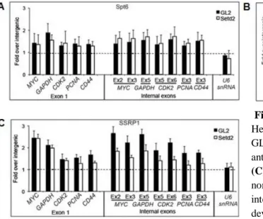
1.3. SETD2 promotes FACT recruitment
Next, we aimed to understand how SETD2 is able to prevent intragenic transcription initiation. Although in yeast, H3K36me3 directs histone deacetylation as a mean to prevent transcription initiation from within the gene body, the mammalian SETD2 inactivation doesn’t affect the acetylation levels on histones (22). Previous studies demonstrated the importance of other factors, such as the histone chaperones FACT and SPT6, in restoring the normal chromatin structure upon transcriptional elongation and preventing ITI (33-35). Considering this, we attempted to determine whether these different factors act through a common repression mechanism in human cells, and so, we reduced the levels of SETD2 in HeLa cells by RNAi, and analysed by ChIP the occupancy of SPT6 and FACT subunits (SPT16 and SSRP1) along five genes (Figure 3). Relative to control (GL2 treated) cells, the density of SPT6 across the analysed genes is not affected by SETD2 depletion (Figure 3A). In contrast, significantly lower levels of FACT subunits SPT16 and SSRP1 are detected over internal exons in SETD2 depleted cells (Figure 3B; 3C). There were no significant differences over first exons, consistent with our previous observation that H3K36me3 levels are low in this region (18). As a control, we analysed the human U6snRNA gene set, which is transcribed by RNA polymerase III (68) and lacks enrichment for H3K36me3 (69). As expected, the density of FACT subunits SPT16 and SSRP1 on the U6snRNA gene was not altered by SETD2 downregulation (Figure 3B; 3C).
Figure 3 SETD2 is required for FACT recruitment
HeLa cells were transfected with siRNAs targeting GL2 and SETD2. ChIP analysis was carried out using antibodies against SPT6 (A), SPT16 (B) and SSRP1
(C). For each antibody, the ChIP signals were
normalized against the ChIP signal of a nontranscribed intergenic region. Graphs depict mean and standard deviation from at least four independent experiments using two distinct siRNAs targeting SETD2.
13 Taken together, these results suggest that SETD2 activity enhances the recruitment of the FACT complex onto the active chromatin templates, permitting nucleosome reassembly after transcription elongation and preventing RNAPII transcription initiation from within the gene body. Additional data from our lab analysing the dynamics of nucleosomal histones in the presence or absence of SETD2 confirmed this view (data not shown).
2. SETD2 role in DNA damage response
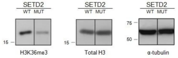
2.1. ccRCC cell lines as an experimental model system to investigate the tumour suppressor role of SETD2
SETD2 was recently reported as a tumour suppressor in ccRCC (57, 59, 60) and in human breast cancer (70-71). In order to explore the mechanism by which SETD2 thwarts tumorigenesis, we first sought to establish an adequate experimental model system. For that, we used three different ccRCC cell lines. Two of them – ccRCC FG2 and ccRCC MF – have a mutation in SETD2 that impairs the activity of the protein, whereas the other one – ccRCC JW – has a mutation that affects an amino acid which is not located in a known functional domain (59). To test the activity of SETD2 in these cell lines, western blot was performed using antibodies directed against histone H3, H3K36me3 and tubulin. Histone H3 and α-tubulin were used as loading controls. As previously reported (59), the levels of H3K36me3 were reduced in the mutant SETD2 cell lines FG2 and MF when compared with the JW cell line (Figure 4).
Figure 4 SETD2 activity is reduced in mutant ccRCC
Total protein extracts of SETD2 wild-type (WT) and mutant (MUT) ccRCC cells were analysed by western blot with antibodies directed against H3K36me3, histone H3 total and α-tubulin. Molecular weight (kDa) markers are shown on the left. The figure depicts the result of one experiment from three independent experiments performed with similar results.
Therefore, these cell lines constitute an appropriate experimental model to investigate the role of SETD2 in the DNA damage response. The following experiments were performed
14 in the three mentioned ccRCC cell lines. The non-functional SETD2 cell lines (FG2 and MF) showed similar results in all the experiments made and will be designated as mutant SETD2 cell lines. For the sake of simplicity, I will present only the results corresponding to one of these cell lines in addition to the JW cell line that exhibited wild-type behaviour and is therefore referred to as SETD2 wild-type.
2.2. SETD2 is required for DNA damage repair
Our finding that SETD2 activity modulates the recruitment of FACT to chromatin discloses a role of SETD2 on other processes that require chromatin remodelling and DNA accessibility such as: the DNA damage response.
Recent studies showed that FACT interacts with the DNA repair factors Ku complex and DNA-PKcs at the DNA damaged site (37). FACT is also involved in the phosphorylation of p53 via its association with the protein kinase CK2 (37) and is recognised as the major histone chaperone that assists the exchange of histones H2A and H2AX on nucleosomes. Furthermore, the histone variant H2AX plays a key role in DNA damage signalling and is also important in DNA damage repair by acting as a docking site for many DNA repair protein complexes (72, 73). Owing to its role in FACT recruitment (Figure 3) we hypothesized that SETD2 is required for a proper DNA damage repair.
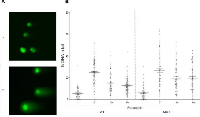
In order to test this hypothesis, we assessed the dynamics of DNA damage repair by performing neutral comet assays in SETD2 wild-type and mutant ccRCC cells. This technique allows a specific measurement of the amount of DSBs in a given cell. ccRCC cell lines were treated with the DNA damaging agent etoposide, which induces DSBs, and harvested immediately after inducing DNA damage (0 min time point) and at two time points following DNA damage (3h and 6h after treatment with etoposide). Then, neutral comet assays were performed: ccRCC cells embedded in a thin agarose gel on a microscope slide underwent electrophoresis to allow the smaller fragments of damaged DNA to migrate away from the nucleus. After staining with DNA-specific fluorescent dye, the gel was analysed under the microscope. The resulting image resembles a comet with a distinct head and tail, wherein the head is composed of intact DNA, while the tail consists of damaged DNA (Figure 5A). The amount of DNA damage is then estimated by the extent of DNA liberated from the head of the comet.
15 Regarding the effect of etoposide on the different ccRCC cells, no differences in the amount of DSBs after etoposide treatment were found between wild-type and mutant SETD2 cell lines (p=0,317; Figure 5B), indicating that SETD2 does not affect the efficacy of the drug nor the cell’s susceptibility to DNA damage. Notwithstanding, in wild-type cells, DNA damage was repaired with faster kinetics than in mutant cells (Figure 5B). While wild-type cells repaired 42% of the damaged DNA 6 hours after etoposide addition, the mutant cells repaired only 24% in the same period of time (Figure 5B). Statistically, the difference between the amount of DNA damage immediately after etoposide treatment and 3 hours after damage is considerably higher in SETD2 wild-type than in mutant cells (p=1-10 and p=0,0005). Furthermore, compared with the 3 hours time point, the amount of DNA repaired after 6 hours is statistically significant in SETD2 wild-type (p=0,04) but not in mutant ccRCC cells (p=0,971). Altogether, these results suggest that SETD2 is a determinant of DNA damage repair dynamics.
Figure 5 SETD2 activity is required for DSBs repair
(A) Neutral comet assay was performed with ccRCC cells with no treatment (-) and with etoposide treatment
(+). The resulting image resembles a comet with a distinct head (intact DNA) and a tail (damaged DNA). (B) Neutral comet assay was performed in SETD2 wild-type (WT) and mutant (MUT) ccRCC following different time points after inducing the DNA damage with etoposide Etoposide treatment is depicted as (-) for no treatment, (‘) for minutes and (h) for hours after DNA damage induction. The graph depicts means and standard error of means of % DNA in tail of all cells of at least three independent experiments performed with similar results.
16
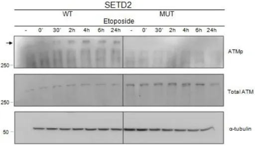
2.3. SETD2 is required for ATM activation following DNA damage
ATM is a protein kinase that is a central mediator of responses to DNA DSBs in cells. This kinase is activated in the vicinity of a DNA lesion and is recruited to the DNA lesion site by MRN complex, where it is fully activated (74-75). The activated ATM can then phosphorylate several substrates that participate in the DDR (76-77).
In order to investigate if the deficient kinetics of DDR in SETD2 mutant cells is accompanied by improper ATM activation, we analysed ATM protein and phosphorylation levels (ATMp) by western blot, after inducing DNA damage with etoposide (Figura 6). In SETD2 wild-type cells ATM became active (i.e. phosphorylated) shortly after DNA damage (Figure 6). In contrast, no phosphorylated ATM was observed in SETD2 mutant cells at any of the time points analysed (Figure 6), even though the levels of total ATM protein were similar to those of wild-type cells (Figure 6). Altogether, these results suggest that SETD2 is required for ATM activation during the DDR.
Figure 6 SETD2 activity is required for ATM activation
Total protein extracts of SETD2 wild-type (WT) and mutant (MUT) ccRCC cells were analysed by western blot, with antibodies directed against ATMp, total ATM and α-tubulin, following different time points after inducing the DNA damage with etoposide. Etoposide treatment is depicted as (-) for no treatment, (‘) for minutes and (h) for hours after DNA damage induction. Molecular weight (kDa) markers are shown on the left. The figure depicts the result of one experiment from three independent experiments performed with similar results.
17
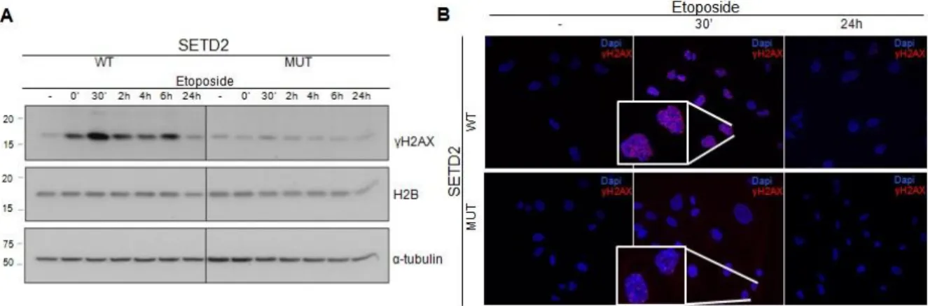
2.4. SETD2 is needed for proper DNA damage signalling
Once activated, ATM phosphorylates factors that are needed for an efficient DDR, including histone H2AX and p53. To investigate whether the lack of ATM activation translates into improper DDR signalling, we measured the levels of γH2AX in etoposide-challenged cells. ccRCC cell lines were incubated with etoposide, washed and harvested after different periods of time in fresh medium (0 min; 30 min; 2h; 4h; 6h; 24h). The levels of γH2AX in functional and non-functional SETD2 cell lines were then measured by immunofluorescence and western blot (Figure 7). Antibodies directed against γH2AX were used in both techniques and antibodies against histone H2B and α-tubulin were used as loading controls. In agreement with the lack of ATM activation, these experiments revealed that phosphorylation of histone H2AX is compromised in ccRCC cells with mutant SETD2 (Figure 7). The highest levels of γH2AX occurs 30 min after recovery of the DNA damage in both cell lines and decreases with time in both cell lines (47, 78). However, this dynamics is only evident when SETD2 is functional (Figure 7A). In mutant SETD2 cell lines, γH2AX levels did not rise properly after DNA damage (Figure 7). These data suggest that SETD2 is required for H2AX phosphorylation during the DDR.
Figure 7 SETD2 activity is needed for DSBs signalling
The DNA of SETD2 wild-type (WT) and mutant (MUT) ccRCC cells was damaged with etoposide and the
γH2AX levels were measured by western blot and immunofluorescence. (A) Total protein extracts of ccRCC
cells were analysed by western blot with antibodies against γH2AX, histone H2B and α-tubulin. Etoposide treatment is depicted as (-) for no treatment, (‘) for minutes and (h) for hours after DNA damage induction. Molecular weight (kDa) markers are shown on the left. The figure depicts the result of one experiment from three independent experiments performed with similar results. (B) ccRCC cells were immunostained with an antibody directed against γH2AX in SETD2 WT and MUT cells following etoposide treatment. Etoposide treatment is depicted as (-) for no treatment, (‘) for minutes and (h) for hours after DNA damage induction. The figure depicts the result of one experiment from three independent experiments performed with similar results.
18
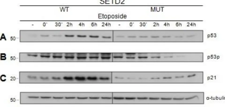
2.5. SETD2 regulates p53 activity during DNA damage repair
The findings reported herein suggest that impaired SETD2 impinges on different cellular mechanisms, such as ATM activation during the DDR. A prominent example of an ATM-dependent DDR-related mechanism is the p53 pathway (50). p53 is known as one of the major players in cancer due to its activity in cell cycle arrest, apoptosis, senescence, angiogenesis and DNA damage repair (79-80). Interestingly, the p53 pathway is repressed in RCC, although p53 mutations are infrequently detected in this cancer (81-83). Xie et. al demonstrated that SETD2 interacts with p53, regulating its transcriptional activity (84). We therefore aimed at investigating whether p53 activity is impaired in ccRCC cells with reduced SETD2 activity.
To this end, the p53 protein levels were analysed by western blot, after inducing DNA damage with etoposide (Figure 8A). Activation of the p53 function after DNA damage involves an increase in the overall levels of p53 protein (85-86), which we observed in the wild-type SETD2 cells (Figure 8A). The amount of p53 protein increased progressively until 6 hours after DNA damage and reduced 24 hours after DNA damage (Figure 8A). In contrast, in SETD2 mutant ccRCC cells, p53 levels did not increase as in wild-type cells (Figure 8A).
Figure 8 SETD2 is required for p53 activation
Total protein extracts of SETD2 wild-type (WT) and mutant (MUT) ccRCC cells were analysed by western blot, with antibodies directed against p53 (A), phospho-p53 (B), p21 (C) and α-tubulin, at different time points after inducing the DNA damage with etoposide. Etoposide treatment is depicted as (-) for no treatment, (‘) for minutes and (h) for hours after DNA damage induction. Molecular weight (kDa) markers are shown on the left. The figure depicts the result of one experiment from three independent experiments performed with similar results.
19 The biological activity of p53 is dependent on its phosphorylation and ability to bind to transcriptional regulatory elements in the DNA of target genes. p21 (protein 21) is one of such genes regulated by p53 (87-89). To investigate if the p53 activity during the DDR requires an active SETD2, we checked both the p53 phosphorylation and p21 protein levels by western blot after inducing DNA damage with etoposide (Figure 8B; 8C). The results revealed that p53 activity is impaired in SETD2 mutant cells when compared to wild-type cells, evidenced by the low levels of p53 phosphorylation observed throughout the time period following DNA damage (Figure 8B). A similar result was observed for the p21 protein, which was considerably higher in SETD2 wilt-type cells than in their mutant counterparts (Figure 8C). The α-tubulin protein was also immunobloted to control the loading of the different samples (Figure 8).
Overall, these results suggest that SETD2 activity is essential for p53 activation during the DDR.
2.6. The role of SETD2 in DDR is independent from H3K36me3
The post-translational modification of histones plays an important role in essentially all DNA-associated processes, including the DDR (7, 10). Moreover, there is evidence showing that relaxation of chromatin structure shepherds the DDR activation (90). Given the prominent role of SETD2 as a histone methyltransferase, we wondered whether the H3K36me3 histone mark mediates the SETD2 role in the DDR pathway.
Once again, western blot was performed following ccRCC treatment with etoposide. The cells were harvested at different time points after treatment (0 min; 30 min; 2h; 4h; 6h; 24h). As expected, H3K36me3 levels are lower in mutant than in wild-type SETD2 cells (Figure 9). Notably, however, in both cell lines, the amount of H3K36me3 observed in all time points after DNA damage did not change when compared to non-damaged cells (Figure
9). Total histone H3 and α-tubulin served as loading controls.
These results suggest that SETD2 operates in the DDR by a mechanism that is independent of its histone methyltransferase activity. Actually, Xie et. al have demonstrated that SETD2 has other activities beyond its histone modifier function (84). Further experiments are needed in order to identify the exact mechanism by which SETD2 impinges in the DDR and to clarify if H3K36me3 participates in any step of this process.
20
Figure 9 H3K36me3 levels do not change during the DDR
Total protein extracts of SETD2 wild-type (WT) and mutant (MUT) ccRCC cells were analysed by western blot, with antibodies directed against H3K36me3, total histone H3 and α-tubulin, at different time points after inducing the DNA damage with etoposide. Etoposide treatment is depicted as (-) for no treatment, (‘) for minutes and (h) for hours after DNA damage induction. Molecular weight (kDa) markers are shown on the left. The figure depicts the result of one experiment from three independent experiments performed with similar results.
21
Conclusion and Outlook
The overall goal of this study was to define the role played by SETD2 in transcription and in the DDR.
Previous studies had already implicated SETD2 in transcription elongation, however the precise role played by this methyltransferase remains elusive. This study demonstrated that SETD2 drives the recruitment of the FACT complex onto chromatin during transcription, which in turn will restore chromatin compaction and inhibit the accessibility of RNAPII within the body of the gene, preventing ITI. Importantly, if not suppressed, ITI can produce truncated proteins with anomalous functions (91-92). These could range from proteins with deletions of the N-terminal domain to completely new proteins encoded by alternative open reading frames or different splicing patterns (92). Notably, intragenic transcription initiation was reported in a number of tumours, including ccRCC, although the underlying mechanism was not fully understood. Moreover, several cancer-related genes show alternative transcription initiation (91, 93, 94, 95). For instance, p53 isoforms displaying alternative transcription initiation were previously found in ccRCC cells (96).
An important finding presented in this thesis is that SETD2 activity is not confined to the role played during transcription, but is also fundamental during the DDR. Here, it is shown that the absence of SETD2 impairs DNA damage signalling and repair. SETD2 activity is required for the post-translational activation of ATM, which in turn permits a proper DSBs recognition and DDR signalling through the phosphorylation of the histone H2AX. The comet assays further revealed that SETD2 is required for the DDR. Importantly, reduced ATM activity in SETD2 deficient cells is translated in reduced activity of p53.
Altogether, the data presented in this thesis suggest that SETD2 may promote tumorigenesis by altering the pattern of transcription initiation and the accuracy of the DNA damage signalling and repair. This would consequentially change the gene expression profile and introduce instability into the genome. Ultimately, it could contribute to tumorigenesis, in particular, to ccRCC (Figure 10).
Further studies aimed at disclosing the tumorigenic potential of the cellular processes on which SETD2 impinges will further clarify its tumour suppressor role and may pave the way for novel therapeutic interventions for ccRCC.
22
Figure 10 Proposed mechanism of SETD2 action as a regulator of transcription initiation and DDR
The inactivation of SETD2 impairs FACT recruitment to chromatin, which leads to alterations in the chromatin structure and to a higher accessibility of RNAPII within the gene body, resulting in ITI and deregulation of gene expression. Furthermore, the inactivation of SETD2 leads to a reduced ATM activity, which will compromise the DSBs signalling and repair, leading to genomic instability. Both of this pathways played by SETD2 can have catastrophic consequences in the cell and, ultimately, can lead to tumorigenesis.
23
Material and Methods
1.
Cell culture
HeLa cells were grown as monolayers in Dulbecco’s modified Eagle medium – DMEM (Invitrogen), supplemented with 10% (v/v) FBS and 100 U/ml penicillin-streptomycin. U2OS cells were grown in DMEM high glucose (Invitrogen), supplemented with 10% (v/v) FBS, 1% (v/v) L-glutamine, 200 µg/ml G418 and 1 µg/ml Puromycin. All ccRCC cell lines - RCC JW, RCC FG2 and RCC MF (Cell Lines Service) - were cultured in McCoy’s 5A (modified) GluteMAXTM medium (Invitrogen) supplemented with 10% (v/v) FBS and 100 U/ml penicillin-streptomycin.
To induce DNA damage, RCC cells were treated with etoposide 50 µM for 15 minutes and harvested immediately after the treatment and 30 min, 2h, 4h, 6h and 24h pos-treatment.
2.
RNA interference
To reduce the levels of SETD2 by RNAi, we used siRNA duplexes were used. GL2 siRNA, which induces effective silencing of firefly luciferase (62), was used as an unspecific siRNA control. HeLa cells were plated at a confluence of 50-60% and reverse transfected using OptiMEM (Invitrogen) and Lipofectamine RNAiMAX (Invitrogen) according to the manufacturer’s protocol. At 24 h after the first transfection, cells were re-transfected with siRNA duplexes and harvested on the following day.
The siRNA sequences are listed in Annex I.
3.
Western blot
Whole cell lysates of the different cell lines were prepared, resolved by SDS-PAGE and transferred onto a nitrocellulose membrane as described previously (97). Incubations with primary antibodies were followed by incubations with the appropriate secondary antibody
24 (Bio-Rad) and by detection using enhanced luminescence substrate (Amersham/GE Healthcare).
The following primary antibodies were used: rabbit polyclonal antibodies anti-histone H3 (ab1792, Abcam), anti-H3K36me3 (ab9050, Abcam), anti-ATM (PC116, Calbiochem), anti-ATMp (200-301-400s, Rockland), anti-α-tubulin (T5168, Sigma-aldrich), anti-γH2AX (05-636, Milipore), anti-H2B (ab1790, Abcam), anti-p53 total (sc-263, Santa Cruz), anti-p53p (ab38497, Abcam), anti-p21 (sc-397, Santa Cruz).
4.
Chromatin Immunoprecipitation
ChIP experiments were performed on HeLa cells and were followed by qRT-PCR, as described previously (18). Briefly, cells were treated with formaldehyde 1% to crosslink proteins to DNA and were harvested subsequently. The cellular extracts were sonicated 5x 20s at an amplitude of 10 microns, in Soniprep 150 (Sanyo), to share DNA into 200-400 base pair fragments. After incubation with specific antibodies and protein A sepharose beads (Sigma-Aldrich), protein-DNA crosslink was reverted and DNA from immunoprecipitated samples was extracted with Chelex 100 (Bio-Rad) as described previously (98). The following antibodies were used: anti-histone H3 (ab1791, Abcam), anti-H3K36me3 (ab9050, Abcam), anti-RNAPII (sc-899, Santa Cruz), anti-SPT6 (ab49066, Abcam), anti-SPT16 (sc-28734, Santa Cruz), anti-SSRP1 (609702, Biolegend).
RT-qPCR was carried out in the 7000 Real-Time PCR System (Applied Biosystems), using iTaqTM SYBR Green Supermix with Rox (Bio-Rad). The relative occupancy of the immunoprecipitated protein at each DNA site was estimated as follows: 2Ct (input)-Ct(IP), where Ct (input) and Ct (IP) are mean threshold cycles of qRT-PCR done in duplicate on DNA samples from input and specific immunoprecipitations. The primers sequences are presented in Annex II.
5. Fluorescence recovery after photobleaching (FRAP)
Human osteosarcoma–derived cells (U2OS) were engineered to stably express tetracycline inducible β-globin transgenes integrated in tandem in the genome, as previously reported (63).