DEPARTAMENTO DE QUÍMICA E BIOQUÍMICA
STUDY OF THE MOLECULAR MECHANISM
USED BY THE PEPTIDIC VECTOR PEP-1 TO
INTRODUCE PROTEINS INSIDE CELLS
Sónia Troeira Henriques
DOUTORAMENTO EM BIOQUÍMICA (Especialidade de Biofísica Molecular)
DEPARTAMENTO DE QUÍMICA E BIOQUÍMICA
STUDY OF THE MOLECULAR MECHANISM
USED BY THE PEPTIDIC VECTOR PEP-1 TO
INTRODUCE PROTEINS INSIDE CELLS
Sónia Troeira Henriques
Tese orientada pelo Prof. Doutor Miguel Augusto Rico Botas Castanho
DOUTORAMENTO EM BIOQUÍMICA (Especialidade de Biofísica Molecular)
Prefácio
O objecto de estudo desta tese foi o mecanismo de acção de um péptido vector, pep-1, capaz de introduzir proteínas dentro de células. Para a realização do trabalho usaram-se essencialmente técnicas espectroscópicas e de microscopia. Do trabalho realizado resultaram os seguintes artigos já publicados:
1) Henriques S.T., Castanho M.A.R.B. (2004) Consequences of nonlytic membrane perturbation to the translocation of the cell penetrating peptide pep-1 in lipidic vesicles, Biochemistry, 43, 9716-9724.
2) Henriques S.T., Castanho M.A.R.B. (2005) Environmental factors that enhance the action of the cell penetrating peptide pep-1. A spectroscopic study using lipidic vesicles, Biochim. Biophys. Acta, 1669, 75-86.
3) Henriques S.T., Costa J., Castanho M.A.R.B. (2005) Translocation of β-Galactosidase mediated by the cell-penetrating peptide pep-1 into lipid vesicles and human HeLa cells is driven by membrane electrostatic potential,
Biochemistry, 44, 10189-98.
4) Henriques S.T., Costa J., Castanho M.A.R.B. (2005) Re-evaluating the role of strongly charged sequences in amphipathic cell-penetrating peptides. A fluorescence study using Pep-1, FEBS Lett., 579, 4498-502.
5) Henriques S.T., Melo M.N, Castanho M.A.R.B. (2006) Cell-Penetrating Peptides and Antimicrobial Peptides: how different are they?, Biochem J, 399, 1-7.
6) Henriques S.T., Melo M.N, Castanho M.A.R.B. (2007) How to address CPP and AMP translocation? Methods to detect and quantify peptide internalization in
vitro and in vivo, Mol Memb Biol, 24, 173-184.
7) Henriques S.T., Quintas A., Bagatolli L.A., Homblé F., Castanho M.A.R.B. (2007) Energy-independent translocation of cell-penetrating peptides occurs without formation of pores. A biophysical study with pep-1, Mol Memb Biol, 24, 282-293.
8) Henriques S.T., Castanho M.A.R.B. (2007) Translocation or membrane disintegration? Implication of peptide-membrane interactions in pep-1 activity, J
Pep Science, no prelo.
Como a maior parte do trabalho experimental relativo ao estudo do pep-1 já foi publicado em revistas internacionais, optou-se pela sua apresentação com recurso aos artigos científicos e pela redacção da tese em língua inglesa.
O manuscrito está dividido em oito capítulos. O primeiro capítulo apresenta uma introdução geral ao tema da internalização de macromoléculas dentro das células, com particular destaque para os péptidos vectores, vulgarmente designados por cell
penetrating peptides (CPPs) e ao pep-1 como objecto de estudo. No segundo capítulo
estão descritos estudos de interacção péptido-membrana realizados com modelos de membrana, os quais foram publicados em dois artigos (artigos 1 e 2, ver lista acima). A totalidade destes estudos foi realizada no laboratório de biofísica da FCUL sob orientação do Prof. Dr. Miguel Castanho onde foram empregues metodologias biofísicas. No terceiro capítulo é apresentado um artigo (artigo 7) onde se explorou a possibilidade de formação de poro induzida pelo pep-1. A parte experimental foi realizada essencialmente em laboratórios estrangeiros sob a orientação do Dr. Fabrice Homblé (Université Libre de Bruxelles, Bruxelas, Bélgica) e do Dr. Luís Bagatolli (Southern Denmark University, Odense, Dinamarca) onde se aplicaram técnicas não disponíveis no laboratório de biofísica da FCUL tais como: espectroscopia de infravermelho; equipamento para a realização de medidas de electrofisiologia; equipamento para preparar vesículas unilamelares grandes por aplicação do método de electroformação e microscopia confocal. No capítulo quatro estão apresentados os estudos realizados com linhas celulares de Humano, os quais foram publicados em dois artigos (artigos 3 e 4). Os estudos in vitro foram realizados no laboratório de biofísica na FCUL e os estudos em linhas celulares foram realizadas em colaboração com a Dr. Júlia Costa (ITQB, Oeiras). No capítulo cinco são integrados os resultados e apresentadas as conclusões. Uma comparação do mecanismo de acção do pep-1 com o mecanismo de outros péptidos vectores é apresentada. No capítulo seis estão os anexos onde se encontram três artigos de revisão que foram publicados no decurso do trabalho doutoral e relacionados com o pep-1 e os CPPs em geral (artigos 5, 6 e 8).
deste péptido ocorre ao nível da membrana ou se dentro da célula. Como o PrP(106-126) possui uma estrutura anfipática semelhante à do pep-1, a possibilidade do PrP(106-126) atravessar a membrana por um mecanismo semelhante ao do pep-1 foi testada. Os resultados obtidos serão apresentados em dois artigos, ainda em preparação. No capítulo sete está um dos artigos ainda não publicado onde se incluem os resultados obtidos nos estudos com o péptido PrP(106-126). Por último, o capítulo oito compreende a bibliografia.
Gostaria de agradecer a todos aqueles que de alguma forma contribuíram para a realização dos trabalhos aqui apresentados.
Agradeço em primeiro lugar ao Professor Miguel Castanho pela oportunidade para a realização do doutoramento no seu laboratório, pela sua dedicação e empenho, pelos ensinamentos, pela sua formação pessoal e profissional, e sobretudo pela amizade.
Agradeço ao Departamento de Química e Bioquímica e ao Centro de Química e Bioquímica da Faculdade de Ciências da Universidade de Lisboa pelas facilidades concedidas para a realização do trabalho. Agradeço à Fundação para a Ciência e Tecnologia pelo apoio financeiro concedido (bolsa SFRH/BD14337/2003).
Agradeço à Salomé pelo exemplo de trabalho e dedicação, pelo companheirismo nos momentos bons e nos momentos mais difíceis, pelas discussões científicas e em especial pela amizade partilhada ao longo do percurso académico. Agradeço também à Virgínia pela boa disposição, pela amizade diária, pelos momentos de folia partilhados e pelo ombro amigo nos momentos menos alegres.
Agradeço a todos os colegas de grupo que passaram pelo laboratório de biofísica, incluindo os elementos estrangeiros, pela amizade, pelo apoio e pelos momentos de diversão. Quero agradecer em particular ao David Pina pela sua disponibilidade, pela simplicidade e por me ter alargado os horizontes. Agradeço ainda à Marta e ao Henri pela lufada de ar fresco que trouxeram ao laboratório.
Agradeço ao Professor Manuel Prieto e a todo os elementos do seu grupo no Instituto Superior Técnico pelas discussões científicas, pelas facilidades concedidas e pelos momentos de diversão fora das horas de trabalhos. Quero agradecer em particular ao Fábio pela simpatia e disponibilidade constantes que sempre demonstrou.
Agradeço à Doutora Júlia Costa por me ter recebido no seu laboratório. Agradeço os ensinamentos, a orientação, as discussões científicas e as facilidades
concedidas. Quero ainda agradecer a todos os elementos do grupo, em especial à Catarina Brito, à Vanessa Morais, ao Victor Sousa e ao Rui Almeida, por me terem recebido e pela ajuda no laboratório.
Ao Doutor Fabrice Homblé pela boa disposição, pela orientação no trabalho experimental realizado em Bruxelas na Université Libré de Bruxelles e ao Doutor Erik Goormaghtigh pelas dicas no infravermelho. Agradeço ainda ao meu tio Adérito Henriques pelo alojamento e pela companhia durante o estágio realizado em Bruxelas.
Agradeço ao Doutor Luís Bagatolli por me ter recebido no seu laboratório na Dinamarca, pelos seus ensinamentos e pela boa disposição. Aos restantes elementos do grupo pela simpatia com que me acolheram.
Agradeço à Doutora Marie-Isabel Aguilar por me ter recebido no seu laboratório na Monash University, Austrália. Agradeço ainda a todos os elementos do grupo que me integraram, em especial à Sharon Unabia pela simpatia e disponibilidade, ao Doutor Leonard Pattenden pela ajuda com o SPR e pelo entusiasmo, e ao Kristopher Hall pelas dicas no AFM.
Agradeço ao Doutor Cláudio Soares (ITQB-UNL) pela ajuda com o Pymol e ao Doutor Nuno Santos (IMM-FML) pela utilização do espectrofluorímetro. Agradeço ao Doutor Alexandre Quintas pela ajuda na obtenção e tratamento dos espectros de CD.
Agradeço ao Instituto de Odivelas pelo papel determinante na minha formação pessoal e académica e pelo alojamento durante o doutoramento. Agradeço ainda a todas as alunas universitárias que fizeram parte da residência do Instituto e que foram a minha família nos dias de semana.
Por fim quero agradecer à minha família, em especial mãe e irmãos, e ao Nuno, pela compreensão, pela dedicação e pelo amor que sempre me destinaram, o que me encorajou semana após semana. Depois dos momentos bons e momentos menos bons é a eles que dedico esta tese.
Contents
Prefácio ...iii Contents ...vii Resumo...xi Abstract ...xvii Keywords ...xixAbbreviations and Symbols ...xxi
Chapter 1. Introduction ... 1
1.1. Strategies to introduce macromolecules inside cells ...3
1.2. New strategy to introduce macromolecules inside the cells using peptides as vectors...7
1.3. How do CPPs translocate across cell membrane?...10
1.4. Pep-1 a new peptide carrier ...14
1.5. Main goals of the project ...16
Chapter 2. Pep-1 and model membranes ... 19
2.1. Introduction ...21
2.1.1. General membrane remarks ...21
2.1.2. Membrane phase behaviour...22
2.1.3. Membrane asymmetry ...24
2.1.4. Model membranes used in spectroscopy studies ...26 2.2. Interaction of Pep-1 with model membranes followed by
2.2.1. Environmental factors that enhance the action of the cell
penetrating peptide pep-1. A spectroscopic study using lipidic
vesicles ...29
2.2.1.1. Motivation and methodologies ...29
2.2.1.2. Declaration on authorship of published manuscript: Environmental factors that enhance the action of the cell penetrating peptide pep-1. A spectroscopic study using lipidic vesicles ...33
2.2.2. Consequences of nonlytic membrane perturbation to the translocation of the cell penetrating peptide pep-1 in lipidic vesicles...46
2.2.2.1. Motivation and methodologies used...46
2.2.2.2. Declaration on authorship of published manuscript: Consequences of nonlytic membrane perturbation to the translocation of the cell penetrating peptide pep-1 in lipidic vesicles ...51
Chapter 3. Does pep-1 form pores? ... 61
3.1. Introduction ...63
3.2. Energy-independent translocation of cell-penetrating peptides occurs without formation of pores. A biophysical study with pep-1 ...68
3.2.1. Motivation and methodologies ...68
3.2.2. Declaration on authorship of published manuscript: Energy independent translocation of cell-penetrating peptides occurs without formation of pores. A biophysical study with pep-1 ...79
Chapter 4. Pep-1 and mammalian cells ... 95
4.1. Introduction ...97
4.1.1. Endocytic routes for cell entry...97
4.2.1. Translocation of β-Galactosidase mediated by the
cell-penetrating peptide pep-1 into lipid vesicles and Human HeLa
cells is driven by membrane electrostatic potential. ...103
4.2.1.1. Motivation and methodologies ...103 4.2.1.2. Declaration on authorship of published manuscript:
Translocation of β-Galactosidase mediated by the cell-penetrating peptide pep-1 into lipid vesicles and Human HeLa
cells is driven by membrane electrostatic potential. ...108 4.2.2. Re-evaluating the role of strongly charged sequences in
amphipathic cell-penetrating peptides. A fluorescence study using
pep-1. ...121
4.2.2.1. Motivation and methodologies ...121 4.2.2.2. Declaration on authorship of published manuscript:
Re-evaluating the role of strongly charged sequences in amphipathic cell-penetrating peptides. A fluorescence study
using pep-1. ...123
Chapter 5. Conclusion... 129
5.1. The overall mechanism – the importance of the lipidic membrane
and electrostatic interaction on pep-1 uptake. ...131 5.2. Towards a new phase regarding CPP mechanisms: the coexistence
of translocation mechanisms ...136
Chapter 6. Annex I ... 141
6.1. Translocation or membrane disintegration? Implication of
peptide-membrane interactions in pep-1 activity. ...143
6.1.1. Declaration on authorship of published manuscript: Translocation or membrane disintegration? Implication of
peptide-membrane interactions in pep-1 activity. ...144 6.2. Cell-Penetrating Peptides and Antimicrobial Peptides: how
6.2.1. Declaration on authorship of published manuscript: Cell-Penetrating Peptides and Antimicrobial Peptides: how different are
they?...152 6.3. How to address CPP and AMP translocation? Methods to detect
and quantify peptide internalization in vitro and in vivo? ...160
6.3.1. Declaration on authorship of published manuscript: How to address CPP and AMP translocation? Methods to detect and
quantify peptide internalization in vitro and in vivo?...161
Chapter 7. Annex II... 175
7.1. Neurotoxicity of PrP(106-126) revisited. A biophysical study with
model membranes...177
7.1.1. Declaration on authorship of the manuscript: Neurotoxicity of PrP(106-126) revisited. A biophysical study with model
membranes...179
Resumo
A introdução de genes e de proteínas dentro de células é vista como uma possível ferramenta no estudo de processos celulares, bem como no tratamento de doenças genéticas. Esta possibilidade é no entanto limitada pela membrana celular, a qual constitui uma barreira para a entrada de moléculas hidrófilas. Para a possível entrada de moléculas com interesse farmacológico, ou com qualquer outra finalidade, é necessário utilizar um meio de transporte capaz de introduzir moléculas activas dentro de células. Nos últimos anos, têm sido desenvolvidas diferentes estratégias no sentido de contornar a barreira da célula. Como por exemplo: o uso de vectores virais; uso de lipossomas; electroporação ou microinjecção. Os primeiros, apesar de muito eficientes, devem ser evitados devido a reacções imunogénicas. Os restantes apresentam menos imunogenicidade mas são menos eficientes, apresentam uma baixa especificidade com dificuldades em atingir o alvo celular, elevados níveis de toxicidade e de consumo de tempo. Isto traduz-se numa incapacidade para atingir os efeitos bioquímicos desejados.
Algumas proteínas citoplasmáticas, quando adicionadas extracelularmente, são capazes de entrar na célula por um mecanismo não tóxico. A capacidade de internalização destas proteínas deve-se à existência de sequências peptídicas ricas em aminoácidos básicos na proteína. Estas sequências catiónicas, frequentemente designadas por cell-penetrating peptides (CPPs), quando acopladas a outras proteínas, funcionam como vectores para a introdução de macromoléculas dentro das células. O mecanismo usado por estes péptidos revelou-se não tóxico e não invasivo. A introdução da proteína ao invés do gene tem ainda a vantagem de conseguir modificar o fenótipo de uma forma rápida e eficiente e com um maior impacto para futuras aplicações. Na maioria dos CPPs o complexo CPP/macromolécula é obtido pela ligação covalente entre CPP e a macromolécula.
Os CPPs mais estudados são a penetratina (16 resíduos de aminoácidos) e o TAT (13 resíduos de aminoácidos), os quais são provenientes da proteína pAntp (proteína de transcrição da Drosophila) e da proteína Tat (proteína de transcrição do HIV-1), respectivamente. Estes péptidos foram os primeiros identificados com propriedades de vector. Depois da descoberta do possível potencial destes péptidos, um grande número de outros péptidos têm sido relatados com propriedades semelhantes. Actualmente esta família inclui péptidos provenientes de diferentes fontes: uns derivados de proteínas ou de toxinas, outros são sintéticos e há ainda péptidos que resultam da quimera de duas ou mais sequências provenientes de diferentes fontes. De um modo geral os CPPs podem ser descritos como: pequenos péptidos (não mais do que 35 aminoácidos), solúveis em água, não tóxicos, capazes de translocar através da membrana celular por um mecanismo independente de receptores e de transportar consigo moléculas hidrófilas.
Apesar das muitas possíveis aplicações destes péptidos o mecanismo usado pelos mesmos é tema de debate na literatura. A elucidação da estratégia usada por estes vectores é importante para uma futura distribuição de macromoléculas com interesse biológico em organismos vivos.
Inicialmente foi proposto um mecanismo independente de receptores (sequências L e D foram internalizadas com a mesma eficiência, o que exclui a intervenção de um receptor para mediar a entrada dentro da célula) e independente de endocitose. A possível contribuição da endocitose foi avaliada pela eficiência de internalização a 37ºC e a 4ºC por meio de observações microscópicas. Eficiências semelhantes para as duas temperaturas sugerem que a entrada dos CPPs é independente das vias endossomais (a baixas temperaturas a produção de ATP é inibida e os processos celulares dependentes de energia, tal como a endocitose, são inibidos). Estes resultados sugerem que a translocação dos CPPs envolve a interacção directa do péptido com a membrana, onde as interacções péptido-lípido apresentam um papel central no processo. A observação da translocação directa de CPPs em modelos de membrana, compostos apenas por lípidos, veio apoiar esta hipótese de translocação.
Contudo, observações recentes sugerem que a localização dos CPPs dentro das células a 4ºC é artificial e resulta de procedimentos de fixação das células para poderem ser visualizadas ao microscópio. A possível explicação para esta localização artificial prende-se com a grande afinidade dos CPPs para a membrana, devida à carga positiva
adsorvidos à superfície da membrana mesmo que não sejam internalizados. Após a fixação, um processo agressivo para as células, o péptido aparece no citoplasma ou no núcleo. Depois desta observação foi realizada uma reavaliação do processo de internalização e houve uma tendência geral para aceitar que o mecanismo fisiológico mais relevante para a internalização dos CPPs é a endocitose. Todavia, resultados contraditórios têm sido publicados e diferentes grupos apoiam uma ou a outra hipótese.
O péptido anfipático pep-1 (Ac-KETWWETWWTEWSQPKKKRKV-cisteamina) inclui-se na família dos CPPs. Este péptido apresenta uma grande eficiência para a introdução de proteínas, péptidos e anticorpos dentro células de uma forma não tóxica e não imunogénica. O pep-1 oferece vantagens relativamente a outros CPPs pois em vez de ligações covalentes entre o péptido e a macromolécula, a formação do complexo CPP/macromolécula é mediada por interacções electrostáticas e hidrófobas.
À semelhança do que acontece com os outros CPPs no início deste projecto havia incerteza no possível mecanismo de translocação usado por este péptido. Para uma aplicação mais generalizada deste transportador, e de outros pertencentes à mesma classe, a elucidação do mecanismo de acção é determinante. Os principais objectivos deste projecto são: a elucidação do mecanismo de translocação do pep-1; avaliar a sua qualidade como vector e comparar a seu modo de acção com os demais do grupo.
A estrutura primária do pep-1 pode ser dividida em três domínios: um domínio hidrófobo rico em resíduos de Trp (Ac-KETWWETWWTEW); um domínio hidrófilo rico em aminoácidos básicos (KKKRKV-cisteamina) e um espaçador entre os dois anteriores (SQP) aumentando a flexibilidade e a integridade dos outros dois domínios. Em condições oxidantes há uma ligação persulfureto entre duas moléculas de péptido devido ao grupo cisteamina na extremidade C. Tendo em conta a anfipaticidade do pep-1 é de esperar que apresente uma elevada afinidade para a membrana celular que poderá ser relevante para a sua actividade biológica. Este foi o ponto de partida para a realização dos nossos estudos.
O trabalho experimental foi desenhado no sentido de crescente complexidade. Numa fase inicial avaliou-se a interacção do pep-1 com modelos de membranas simples compostos apenas por fosfolípidos. Estes modelos lipídicos são simples mas permitem modular as propriedades da membrana, como a fluidez e a carga da membrana, a força iónica e o pH. Em seguida foram realizados estudos em cultura de células humanas
onde se testou a capacidade vector do pep-1 quando complexado com uma proteína. Pela utilização de diferentes metodologias aplicando essencialmente técnicas biofísicas (e.g. espectroscopia de fluorescência e espectroscopia de UV-vis, dicroísmo circular, espectroscopia de infravermelho e electrofisiologia) e técnicas microscópicas (epifluorescência e microscopia confocal) foi possível obter resultados que integrados indicam que o pep-1 é capaz de translocar através da membrana celular por um mecanismo não mediado por endocitose.
Resumidamente, foi possível verificar que a o mecanismo de translocação do péptido é iniciado pela partição do pep-1 na membrana celular. As interacções péptido-lípido são inicialmente governadas por interacções electrostáticas entre o domínio hidrófilo e os grupos polares dos fosfolípidos que compõem a membrana celular. Subsequentemente, dada a proximidade com a membrana, o domínio hidrófobo insere-se na bicamada, adquirindo uma conformação em hélice α. Aquando da ininsere-serção, o domínio hidrófobo induz destabilizações locais na membrana mas sem indícios para a formação de poro. A distribuição assimétrica das cargas na membrana celular (relativamente à composição lipídica e a gradiente electroquímico) é responsável pela existência de um potencial transmembranar (negativo dentro da célula) o qual promove a translocação do pep-1 da camada externa para a camada interna da membrana. Este processo é facilitado pelos distúrbios locais promovidos pela inserção do péptido na membrana e pelas interacções electrostáticas entre a molécula de péptido carregada positivamente e o interior da membrana carregada negativamente. Em contacto com o meio intracelular, ambiente redutor em contraste com as condições oxidantes fora da célula, o péptido perde afinidade para a membrana tornando-se mais estável dentro da célula. O processo torna-se praticamente irreversível e o equilíbrio é sempre deslocado no sentido extracelular → intracelular o que facilita a internalização do péptido bem como das macromoléculas associadas.
A translocação ocorre por um processo mediado pela afinidade do pep-1 para a membrana onde as interacções de natureza electrostáticas e hidrófoba são determinantes no processo. Pelos estudos in vivo foi possível confirmar que este é o único mecanismo com relevância fisiológica, não se tendo identificado indícios para a endocitose como possível via alternativa.
igual para todos os elementos desta família de péptidos e cada caso deverá ser analisado independentemente.
No decurso do nosso trabalho experimental usou-se o pep-1 não derivatizado, fazendo-se uso das suas propriedades fluorescentes intrínsecas. No entanto, um pep-1 derivatizado com uma sonda fluorescente foi também estudado e comparado com o péptido original. O péptido modificado apresentou uma menor afinidade para a membrana bem como uma menor eficiência de penetração nas células. Verificou-se ainda que, a pequena percentagem de péptido que entra na célula fá-lo por endocitose, ao contrário do pep-1 original. Estes resultados sugerem que o mecanismo de internalização utilizado depende especificamente do péptido, das condições experimentais e da sua afinidade para a membrana a qual modula a concentração de péptido particionado na membrana celular. Para baixas concentrações a endocitose é o mecanismo mais provável, ao passo que concentrações elevadas de péptido na membrana activam o processo físico, mais rápido que a endocitose.
Esta hipótese baseia-se na observação de que alguns péptidos, dependendo das condições e em especial da concentração em solução, apresentam uma internalização mediada maioritariamente ou por endocitose (para baixas concentrações) ou pela penetração directa através da membrana (para altas concentrações). A suportar esta hipótese está também a relação entre afinidade e partição observada em vários estudos.
É de referir que, quando os CPPs são internalizados por um mecanismo mediado por endocitose, a macromolécula acoplada ao vector só atingirá o seu alvo final se o CPP conseguir escapar dos endossomas. Para que isto ocorra o CPP terá de atravessar a membrana dos endosomas o que pressupõe um processo físico. O gradiente de pH, que existe entre os endosomas (pH =5) e o citoplasma (pH=7.4), é um possível potenciador da passagem de CPPs através da membrana, em semelhança ao que acontece quando a translocação é mediada pelo potencial transmembranar. CPPs cuja internalização ocorra maioritariamente por um mecanismo mediado por endocitose revelaram-se menos eficientes.
O trabalho aqui apresentado revela os princípios que modulam a translocação do pep-1. A internalização deste vector ocorre por um processo físico, rápido e com elevada eficiência. A aplicação do pep-1 como vector é mais vantajosa que o uso de vectores virais porque não induz imunogeneicidade e é mais eficiente que outros métodos não virais como a electroporação, a microinjecção ou mesmo outros CPPs.
Com este projecto é possível uma orientação mais focada para a utilização de um meio eficaz que consiga ultrapassar a barreira da membrana celular e introduzir fármacos dentro de organismos vivos.
Abstract
The introduction of genetic material or proteins to originate a defined biochemical effect inside the cell has a remarkable potential for the treatment of Human diseases, however this is hampered by the cell membrane barrier for the entry of hydrophilic macromolecules. A possible strategy to overcome the membrane barrier was proposed after the discovery of basic peptidic sequences with ability to pass trough the membrane in a non-toxic and non-immunogenic manner. These peptides are commonly designated as cell-penetrating peptides (CPPs). Pep-1 is a CPP and has been successfully used to introduce several macromolecules biologically active inside cultured cells. The main goal of this thesis is to clarify the mechanism used by this peptide to pass through the membrane and to confirm if its efficiency as a carrier to introduce proteins inside cells. The interaction with membranes was followed in vitro with model membranes: large unilamellar vesicles; planar lipid membranes, giant unilamellar vesicles and supported bilayers. HeLa cells were used to follow the translocation of pep-1 associated with a protein. Fluorescence and UV-Vis spectroscopy methodologies, CD and ATR-FTIR spectroscopy, electrophysiological measurements and fluorescence microscopy were used to carry on the experimental work. It was shown that pep-1 is able to interact with and to destabilize the lipidic bilayers without evidence for pore formation at variance with other CPPs that use an endosomal pathway. Although all the evidences show that pep-1 translocates by a physically-mediated mechanism promoted by transmembrane potential with no evidences for the use of endosomal routes as an alternative pathway. Differences between this particular pep-1 and other CPPs can be related with the affinity for membrane. Peptides with higher affinity for membrane have more propensities to be internalized by a non-endocytic mechanism. Lower affinity for membranes favours endocytic uptake.
Keywords
Drug delivery Cell-penetrating peptides Translocation Membrane Permeability Transmembrane potentialAbbreviations and Symbols
AMPs Antimicrobial peptides
ATR-FTIR Attenuated total reflection Fourier transform infrared BLMs Black lipid membranes
CD Circular dichroism CF Carboxyfluorescein Chol Cholesterol
CLIC Clathrin and dynamin-independent carriers CPPs Cell-Penetrating Peptides
CTxB Cholera toxin B
DPPC Dipalmitoyl-sn-glycero-3-phosphocholine DPPS 1,2-Dipalmitoyl-sn-glycero-3-phosphoserine EK Potassium Nernst potential
FITC Fluorescein isothiocyanate
FRET Fluorescence resonance energy transfer GEEC Early endosomal compartment
GUVs Giant unilamellar vesicle
IW Fluorescence intensities in the absence of lipid
IL Fluorescence intensities in presence of lipid
IR Infrared
IRE Internal reflection plate
K0 Extracellular potassium concentration
Ki Intracellular potassium concentrations KP Partition coefficient
Lβ Lamellar gel phase
Lo Liquid ordered phase
LDL Low density lipoprotein LUVs Large unilamellar vesicles MAPs Model amphipathic peptides MLVs Multilamellar vesicles
MUG 4-methylumbelliferone-β-D-galacto-pyranoside NBD 7-nitro-2-1,3-benzoxadiazol-4-il
NLS Nuclear localization signal NMR Nuclear magnetic resonance ONPG o-nitrophenyl-β-galactopyranoside
pAntp Drosophila antennapedia transcription protein PC Phosphatidylcholine
PE Phosphatildylethanolamine Penetratin CPP from pAntp
PLMs Planar lipid membranes
POPC 1-palmitoyl-2-oleoyl-sn-glycero-3-phosphocholine POPG 1-palmitoyl-2-oleoyl-sn-glycero-3-phosphoglycerol PS Phosphatidylserine
Rh Rhodamine
SM Sphingomyelin
SUVs Small unilamellar vesicles
Tat HIV-1 transcriptional activator protein TAT CPP from Tat
TB Trypan blue TRITC Tetramethylrhodamine 4-MU 4-Methylumbelliferone 5-NS 5-Doxyl-stearic acid 16-NS 16-Doxyl-stearic acid β-Gal β-Galactosidase
Chapter 1
Chapter 1.
Introduction
1.1. Strategies to introduce macromolecules inside cells
The introduction of genetic material or proteins to originate a defined biochemical effect inside cells has shown tremendous potential as a biological tool for studying cellular processes and is challenging for the treatment of human diseases (see reference [1] and references therein).
Hydrophilic macromolecules that lack specific membrane receptors are not able to cross plasma membranes due to hydrophobic nature of the core of lipid bilayers. An efficient cellular delivery system is of first importance for an insertion of macromolecules of pharmacological interest or other biological potential. Over the last years, several strategies have been developed to introduce material inside the cells for a variety of purposes such as: protein structure/function studies; DNA insertion to modulate expression of proteins; and drug delivery with therapeutic purposes. Some examples of approaches that have been used are: viral vectors; liposomes-based delivery; electroporation and microinjection [2].
Viruses are really efficient infecting a host cell and introducing their DNA or RNA genomes, this observation led to the use of viral particles as vectors to introduce exogenous genetic material to cells. The development of viral vectors for long-term gene delivery to mammalian cells has been the main goal of viral vectorology since early 1980s. Adenoviruses and retroviruses have been used in a broad host range and for multiple applications (see references [3-5] and references therein). These viral associated vectors are efficient, although the possible pathogenic effects against the host organism, the high costs of production, the limit size of the genetic material that can be packaged for viral gene therapy and a lack of desired tissue selectivity has hampered a more extensive and global therapeutic application. So non-viral alternatives have gathered much attention [2, 6].
Non-viral strategies which are potentially less immunogenic have been developed to overcome viral vectors limitations. Microinjection is an example; this approach involves the transfer of macromolecules into a living cell using micropipettes (Figure 1.1).
Figure 1.1. Microinjection approach to introduce
material inside cells. Macromolecules are injected inside cells by the use of micropipettes; this procedure has to be performed cell by cell. Source:
http://transgenese.crchul.ulaval.ca/transgeniques.htm
This technique has no cell-type restriction and there is no apparent limitation on the size or type of macromolecule that can be injected [7]. However, because this technique can only be performed one cell at a time with individual glass micropipettes, it is time consuming, requires specific technical skills and is practical only when treating a small number of cells [8]. There is also a limitation to the cell types that can be readily used for microinjection [8]. An efficient method for transforming a larger number of cells at once should be improved for a more generalized application.
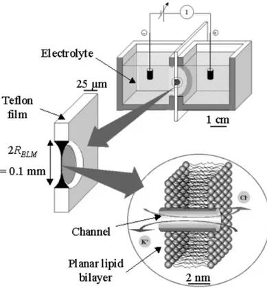
potential is above a threshold, transient pores are formed which allow cellular uptake of hydrophilic molecules (see Figure 1.2) (see reference [10] and references therein for possible applications). This technique can be applied to many cells at once, however it is a non-specific strategy and can become toxic, since molecules can enter or exit the cell without control during transient membrane destabilizations [2]. A large cell death percentage may occur [8].
Figure 1.2. Electroporation
approach to introduce material inside cells. An electric field induces transient pore formation which enables the entry of material.
A) Electron micrographs of cells
before and after brief electric pulses; opening pores and resealing of membrane is observed. B) Illustration of material entrance during electropermeabilization. Source:
http://www.inovio.com/technology/ electroporation.htm
In 1987 Felgner et al. reported the use of cationic liposomes as an efficient and effective system to deliver DNA [11]. Upon mixing DNA with cationic liposomes, DNA is condensed into small particles called lipoplexes, the process involving an initial rapid association of polycationic liposomes and polyanionic DNA through electrostatic interaction (see reference [2] and references therein). This gene therapy strategy is not immunogenic, does not have size restriction and different types of nucleic acids can be delivered ([6]). It is inexpensive and is relatively easy to use in large-scale [6]. Liposomal carriers have been successfully used to deliver genetic material into cells and can be internalized by endosomal vesicles through endocytosis or by direct fusion with the cell membrane. In endosomal route genetic material may stay enclosed within endosomes and fail to access the cytoplasm and nucleus, leading to inefficient low gene
A
Before electric pulse During electric pulse After electric pulse
of gene expression [6]. The uncertainty in the pathway for cell entry, the low efficiency of delivery [6], a possible toxicity related to gene transfer by lipoplexes [2] and potential interference with lipid metabolism [8] hamper a more general application of cationic liposomes as a vehicle to introduce material in vivo. Different strategies have been developed in order to improve transfection efficiency of liposomal carriers, some of them involving direct injection [6, 12].
Figure 1.3. Cationic liposomes can
be used to introduce genetic material inside the cell. Due to electrostatic interactions between negatively-charged DNA and cationic liposomes, a good delivery system can be obtained. Cationic liposomes are internalized by endocytosis. The escape from endosomes can hamper an efficient delivery.
A non-invasive administration with a better efficiency for a good transfection and application in vivo is of particular interest to the future development of gene therapy [6].
Nucleous Cell
Endocytosis
1.2. New strategy to introduce macromolecules inside the cells
using peptides as vectors
All the non-viral strategies above mentioned are more focussed and improved for genetic material delivery and all of them have serious drawbacks which result in a transient and varied gene expression with a consequent incapacity to reach the final desired effect [1].
The fact that some intracellular proteins when added to extracellular medium are able to pass through the membrane inspired a new approach. This phenomenon was first observed with Tat (HIV-1 transcriptional activator protein) [13] and pAntp (Drosophila antennapedia transcription protein) [14]. The ability of these proteins to cross the membranes is due to basic amino acid sequences and the minimal peptidic sequences necessary for the translocation to occur within Tat [15] and pAntp [16] were elucidated. Conjugated molecules such as peptides [17] or proteins [18] coupled to these basic peptides can be delivered into cells; this observation made these basic sequences very attractive and a new class of vectors, initially denoted as Protein Transduction Domains (PTDs) [19] and more recently re-baptized as Cell-Penetrating Peptides (CPPs) [20], emerged.
The CPP derived from pAntp has 16 amino acid residues [16] and is commonly known as penetratin. The Tat (48-60) fragment which include the whole basic regions of the protein and its Nuclear Localization Signal (NLS) is the most efficient in internalization [15].
This strategy prospects a substantial improvement in cellular delivery. The greater advantage of this strategy when compared with the above referred is the possibility to introduce proteins in a non-toxic and non-invasive way. Internalization of proteins instead of genes can modify the phenotype in less than 2 hours in an efficient manner envisaging a fast and effective strategy for drug delivery with a major impact on future treatments [21, 22]. This new approach opens new possibilities for the development of vaccines and protein therapies for cancer and infectious diseases [23].
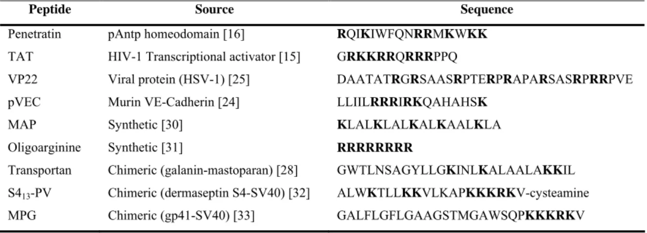
After the discovery of the potential of Tat peptide (TAT) and penetratin as vectors a larger number of peptides (Table 1.1), including peptides from protein sequences (pVEC [24] and VP22 [25]), synthetic peptides (such as model amphipathic
fusion of sequences from different sources, as transportan [28] and MPG [29]) have been shown to translocate across cellular membrane.
Table 1.1. Source and amino acid sequence of some CPPs. Positively-charged amino acids are
highlighted.
Peptide Source Sequence
Penetratin pAntp homeodomain [16] RQIKIWFQNRRMKWKK
TAT HIV-1 Transcriptional activator [15] GRKKRRQRRRPPQ
VP22 Viral protein (HSV-1) [25] DAATATRGRSAASRPTERPRAPARSASRPRRPVE
pVEC Murin VE-Cadherin [24] LLIILRRRIRKQAHAHSK
MAP Synthetic [30] KLALKLALKALKAALKLA
Oligoarginine Synthetic [31] RRRRRRRR
Transportan Chimeric (galanin-mastoparan) [28] GWTLNSAGYLLGKINLKALAALAKKIL S413-PV Chimeric (dermaseptin S4-SV40) [32] ALWKTLLKKVLKAPKKKRKV-cysteamine
MPG Chimeric (gp41-SV40) [33] GALFLGFLGAAGSTMGAWSQPKKKRKV
This ability is part of the biological function of many of these peptides, although this does not automatically mean that they can be used as a carrier. In order to distinguish between translocating peptides able to deliver cargoes and others unable to do it, a CPP can be defined as a short (no more that 35 residues) water soluble, non-toxic, peptide able to efficiently translocate through cellular membrane by a mechanism independent from a chiral receptor, able to deliver hydrophilic macromolecules into cells and eventually originating a defined biochemical effect inside the cell [34].
Besides proteins [18, 19, 35-37], several other hydrophilic macromolecules have been efficiently coupled to these CPPs and delivered inside the cells, such as: peptides [17, 38-40]; antisense oligonucleotides [29, 41-44]; SiRNA [45, 46] and plasmid DNA [47, 48]. Nanoparticles [49] and liposomes [50] have also been internalized by means of CPP. Introduction of cargo macromolecules into primary and transformed cultured cells with efficiency close to 100% can be obtained.
The application of CPPs to deliver macromolecules inducing a specific biochemical effect has been efficiently used in vivo in rats with a construct of penetratin associated to a peptide nucleic acid [44]. A caveolin-1 scaffolding domain peptide
biological effects were detected [51]. The capacity to pass blood brain barrier mediated by a CPP strategy was also shown by a CPP-dexorubicin construct [52] and also with a construct of TAT-β-Galactosidade [23]. This last CPP-cargo construct can also reach mice organs via systemic circulation and transduces in a variety of murine tissues, including liver, kidney, heart, muscle, lung, spleen and brain [23]. This unspecificity can limit the application of these CPP constructs when a single organ or tissue is the target.
1.3. How do CPPs translocate across cell membrane?
The exact mechanism underlying the translocation used by CPPs to pass through cellular membrane still awaits complete understanding; the information available in the literature is scarce and controversial. A mechanism independent on receptors was proposed based on studies with reverse and D-enantiomer sequences, which have similar translocation efficiency as the original peptide [27, 31, 53]. A mechanism independent of endosomal pathway was also purposed supported by the observation that internalization of such peptides is similar at 4ºC and 37ºC [15, 16, 27, 53, 54] (at low temperatures ATP production is inhibited and energy-dependent cellular processes, like endocytosis, are inhibited or greatly diminished).
Recent observations suggesting that the cell localization observed for CPPs is artifactual and occurs during cell fixation for immunochemistry and cell imaging [55], raised the questioned if the translocation of such peptides is really dependent on endocytosis. An artificial cell localization was first reported in 2001 with the structural protein VP22 [55], which also possesses vector properties and can be used to transport other proteins [25]. VP22 binds to surface which allow the protein to remain attached to cells during washing. In the cell fixation process the protein is released and co-localized with nucleus due to affinity of VP22 to DNA [55]. The artifactual cell localization by fixation was confirmed by comparison of the localization of the Histone H1, which does not have cell-penetrating properties, before and after cellular fixation [56]. Biased localization of soluble proteins, during preparation of cells for immunofluorescence, was formerly reported in 1992 [57]. The possibility of an artifactual cell localization was further confirmed in 2003 by Thorén et al [58] and a re-evaluation of the translocation mechanism of several CPPs has been done is the last 4 years.
By the re-evaluation of the translocation mechanism it is clear that penetratin [58, 59], TAT [60-64] and oligoarginine [60, 65-67] among other CPPs, can cross the plasmatic membrane by endocytosis. A tendency to accept this pathway as the main mechanism for internalization of all the CPPs has been generalized in the literature; however, there is no consensus in the specific endocytic pathway used for the uptake of
raft-dependent pathway involving macropinocytosis [68], clathrin-mediated endocytosis [69, 70] or caveolae-dependent endocytosis [62, 71]. Controversial results are published the dissimilarities being due to different experimental conditions in particular with different cellular lines, labelled-peptides and protein-conjugated peptides, which can inhibit some pathways while favouring other.
The involvement of the basic residues in peptide translocation was tested and it was found that the deletion or substitutions of a single basic amino acid residue dramatically reduced TAT uptake [72]. The importance of the overall charge of the peptide was further confirmed by Wender et al., and it was identified that nonaarginine (R9) is more efficiently internalized than Tat fragment [73].
These results suggest that the first interaction with membrane, prior to endocytosis event, appears to be governed by electrostatic interactions between the basic amino acids within CPPs sequence and biological membranes. The heparan sulfate (HS) proteoglycans at the cell membrane were proposed to act as receptors for penetratin [63, 74-76], Tat peptide [63, 70] and also for oligoarginine [66].
The biological activity exhibited by CPPs is consistent with the cargoes macromolecules reaching the cytosol. In a picture where the endosomal pathway emerges as the physiological uptake of CPPs, the escape from endosomes is mandatory for the potential use of CPPs as a strategy to deliver macromolecules with biological relevance inside the cell; this raises the question by which mechanism the internalized CPPs reach the final target?
An escape from endosomes due to acidification was proposed for penetratin, TAT and oligoarginine [61, 65]. This hypothesis is supported by Granslund and co-workers results, where penetratin was encapsulated in liposomes. In the absence of pH gradient no penetratin escape takes place, while when a pH gradient exist (5.5 inside, 7.4 outside), there is a fraction escaping from the liposomes and this occurs without membrane lysis neither pore formation [77]. In opposite to this it was verified that a higher membrane disturbance induced by poliarginines occurs with pH in the average 7.5-9.5 and an escape of endosomes prior to their acidification was suggested [66].
If a mechanism able to permeate membrane for endosomal escape exists, this can also suggest a similar mechanism to pass cell membranes. Direct observation of some CPPs translocation in model membranes systems [78, 79] support the existence of
a possible energy-independent mechanism, governed by peptide-membrane interactions, to cross the membrane. Furthermore, higher amounts of oligoarginine peptide were located in cytosol at 4ºC than at 37ºC. A possible explanation for this phenomenon is that when incubation is held at 37ºC, oligoarginine release in the cytoplasm can be difficult due to endosome entrapment [67], at 4ºC the existence of an alternative pathway which operates when endosomal pathways are inhibited can more easily locate oligoarginine in cytosol. A possibility of an alternative pathway to endocytosis was proposed [80] shortly after the discovery of Tat protein capacity to internalize inside cell.
A translocation dependence on a negative transmembrane potential was observed in vitro with liposomes [78] and in vivo into HeLa cells [81] for some CPPs. Terrone et al. suggested that a part of the peptide can transverse through the membrane by a mechanism dependent on transmembrane potential (negative inside) and other part is internalized by endosomal pathway. When in endosomes the membrane potential may facilitate translocation of CPPs from endosomal lumen to the cytoplasm, considering that the endocytic compartment exhibit a significant transmembrane potential (lumenal side positive) [78].
Even with direct observation of CPP internalization in membrane model systems, a broadly acceptance that the main cellular internalization mechanism of CPPs involves endocytosis, compromise the usage of these peptides for drug delivery. When entering in the cell via endocytosis the molecules can become entrapped in the endosome and ultimately end in the lysosome, where degradation processes take place. Thus even if efficient cellular uptake via endocytosis is observed, the delivery of intact peptide/protein or other macromolecule is compromised by insufficient endosomal escape and subsequent lysosomal degradation. An inefficient escape from endosomes was verified by the observation that after TAT-Cre peptide uptake the majority of the complex remained entrapped in endosomes even after 24h [68]. One possible solution is to complement CPP with a membrane destabilizing agent (e.g. viral fusogenic peptide or membrane-destabilizing peptide) to improve CPP-mediated protein transfection as has been proposed by Wadia et al. [68]. A markedly enhanced CPP-cargo construct escape from endosomes was verified when a fusogenic peptide was associated with the
in which six Arg residues where added to N-terminus make the CPP more efficient to internalize and to target a PNA with a high efficiency [82].
For a generalized application of these peptides as a vehicle with pharmaceutical relevance or other biological/scientific applications, the elucidation of the translocation mechanism is of first importance. The experiments to study CPP internalization are frequently carried on with TAT and Penetratin. In the last years, peptides from diverse sources have been identified with CPP properties, beside the ability to enter cells and the presence of basic amino acid residues, these peptides can vary in size, nature, biological function, secondary structure, hydrophobicity and amphipathicity (see reference [34] and references therein). The mechanism by which CPPs pass through the membrane is far from being completely understood. Taking into account all the information available in the literature regarding the translocation mechanism and the varied nature of the different peptides belonging to the CPP family, a careful and special attention to this topic should be taken and a simplistic comparison with other CPPs should be carefully done.
1.4. Pep-1 a new peptide carrier
In most CPPs so far referred, the CPP-macromolecule complex is obtained due to covalent link between the CPP and the cargo molecule. The strategy to couple cargoes to CPPs can be obtained by production of a CPP-cargo by chemical peptide synthesis, which is generally restricted to short cargoes (e.g. 10-20 amino acids); larger protein cargoes can be produced by recombinant DNA technologies with expression in bacteria and their purification prior to use [83]. Another approach is to synthesize the CPP and the cargo molecule independently and posterior linkage by a reactive residue included in either CPP and cargo molecules (e.g. activated cysteine at CPP N-Terminal with a free cysteine included in the cargo molecule). This approach enables coupling of large proteins (see reference [83] and references therein). After cellular uptake, the reducing environment in the cytosol cleaves the disulfide linkage and frees the cargo molecule from the CPP [26], avoiding interferences with cargo biological function or its cellular localization. All these strategies to couple a cargo have limitations, are time consuming and require specific technical skills; moreover a CPP-cargo may induce desnaturation of the protein due to covalent link to the CPP.
A new approach based on an amphipathic peptide named pep-1 (Chatiot®) has been proposed for protein delivery. Pep-1 delivers a broad range of protein directly into mammalian cells maintaining their biological activity in different cellular lines with lack of toxicity and lack of sensitivity to serum [84]. The advantage of this peptide in comparison with CPPs above referred is that this peptide forms a non-covalent complex with the macromolecule to be delivered, avoiding complicate procedures with covalent links above referred. This complex is easily obtained by mixing the peptide with the macromolecule during about 30 minutes and a complex, stable in physiological buffer, is obtained and ready to use onto cells. The size and the nature of the cargo protein to be delivered can influence the number of pep-1 molecules required to obtain an efficient complex. After cellular uptake the complex dissociates in the cytosol leaving the macromolecule biologically active and free to proceed to its target organelles, while pep-1 localizes in the nucleus and does not affect the final cargo location [84]. The efficiency of translocation can vary in the range 60-95%, depending on the cell type and
Pep-1 has been efficiently used to introduce several peptides [84-87], proteins [88-93] and biologically active antibodies [85, 94-99] inside cultured cells from different sources. It is noteworthy the capacity to deliver macromolecules in neuronal cells [88, 93, 95, 96], and also plant cells converted into protoplasts [92] producing the expected biochemical effect in the correct localization inside the cell. The biological activity is generally maintained after translocation of the delivered macromolecule, for instance: 1) enzymatic activity was detected after enzyme internalization [84, 90, 91]; 2) pep-1 was efficiently employed to deliver an antibody to clarify the intracellular trafficking of a membrane-anchored enzyme [98], which indicates that after internalization, the antibody diffuses throughout the cells and interacts with the target protein and thus, allows the identification of the subcellular compartment that harbours the target protein. The use of pep-1 in animals has also been reported [100, 101], where pep-1/cargo complexes were intratracheally instilled, for instance Aoshiba et al. have used pep-1 to deliver Caspase-3 to the lungs of mice; alveolar wall apoptosis and emphysematous changes were detected as a result of actively internalized Caspase-3 [100].
Altogether these published reports strengthen the use of pep-1 as a vehicle for intracellular deliver of macromolecules with biological interest such as pharmaceutical agents.
1.5. Main goals of the project
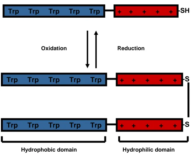
Pep-1 (acetyl-KETWWETWWTEWSQPKKKRKV-cysteamine) is an amphipathic peptide with three domains: (i) a Trp-rich “hydrophobic” domain (Ac-KETWWETWWTEW), responsible for “hydrophobic interaction” with proteins and cell membranes; (ii) a hydrophilic domain (KKKRKV-Cys) derived from a nuclear localization signal (NLS) of Simian Virus 40 (SV-40) large T antigen, required to improve solubility; and (iii) a spacer domain (SQP) which improves the flexibility and the integrity of the other two domains [84]. A cysteamine group is present in the C-terminal and an acetyl group caps the N-terminus. In oxidizing conditions a disulfide link is formed between two peptide molecules by the cysteamine group (Figure 1.4).
The term amphipathicity refers to molecules with both hydrophilic and hydrophobic parts [102] and is dependent on the relative abundance of hydrophobic and hydrophilic domains within a peptide. One possible quantitative measure of amphipathicity is the hydrophobic moment [103]. It is possible to distinguish between primary amphipathicity and secondary amphipathicity in peptides. Peptides with primary amphipathicity are made of a hydrophobic and a hydrophilic domain. A peptide with secondary amphipathicity has hydrophobic and only hydrophilic residues however a separation of hydrophilic and hydrophobic parts is only achieved with the peptide secondary structure [102]. The simplest and most common secondary structure in peptides is the amphipathic α-helix [102].
Cell membranes are composed of amphipathic bilayers, therefore an increasing hydrophobic moment of model peptide results in a significant increase in the permeability and haemolytic activities of these peptides in target membranes, which implies a better peptide-membrane affinity (see reference [103] and references therein). Antimicrobial peptides (AMPs) activity is known to be dependent on peptide-membrane interaction and on the capacity to induce membrane leakage. For AMPs the capacity to interact with membranes and the degree of amphipathicity are correlated. For these peptides amphipathic α-helix formation occurs upon membrane interaction, which seems to favour peptide insertion and cellular toxicity [103].
2X
Figure 1.4. Schematic representation of pep-1 molecule. Primary amphipathicity is evident:
“hydrophobic” domain (Ac-KETWWETWWTEW) is represented by five Trp residues and hydrophilic domain (KKKRKV-Cys) is represented by five positive charges. In between a spacer domain (SQP) with a Pro residue is responsible by the flexibility and integrity of the other two domains. In oxidizing conditions a disulfide link between two cysteamine groups is expected, reducing environment cleavages this disulfide link.
The use of an amphipathic transporter to deliver cargoes molecules inside cells where the cell membrane is the first barrier seems feasible; in this regard, peptide interacts first with hydrophilic region and then penetrates the hydrophobic interior of the cell membrane [102]. A clear dependence of peptide incorporation upon amphipathicity was demonstrated with MAPs [104]. Reports from Divita and Heitz group demonstrate that peptides with primary amphipathicity are also efficient to interact with lipidic membranes and to translocate across cell membrane [33, 105].
+ + + + +
Trp Trp Trp Trp Trp
-SH
Oxidation Reduction+ + + + +
Trp Trp Trp Trp Trp
-S
+ + + + +
Trp Trp Trp Trp Trp
-S
Pep-1 is a promising vehicle to deliver macromolecules with biological relevance into cells. However, for a more generalized and optimized use of this carrier the elucidation of the mechanism of action is mandatory. When I started my PhD, there was not much information regarding the possible mechanisms used by this peptide to translocate across cellular membranes. Morris et al. suggested that this peptide translocates by an endocytosis-independent pathway, although, these results were obtained in fixation conditions and with a labelled peptide [84], which can compromise the results, as stated above.
The main objective of this project was to unravel how pep-1 interacts with cells at membrane level. Pep-1 is a peptide with primary amphipathicity, which suggests direct interaction with lipids in biological membranes and this can be the key-feature to explain pep-1 ability to pass through the cell membrane. In order to evaluate this hypothesis, the experimental work was first performed with model membranes to evaluate the possible effect of membrane charge, viscosity and ionic strength. Different model membranes were used depending on the methodology: Large unilamellar vesicles were used with fluorescence methodologies; Giant unilamellar vesicles were used in confocal microscopy and planar lipid membranes for electrophysiological measurements. In order to evaluate the possible effect of a cargo on the translocation mechanism studies with a complex pep-1/protein were also performed. The protein chosen for these experiments was β-Galactosidade (β-Gal) from E. Coli. After characterization of pep-1 interaction with model membranes, cultured mammalian cells were used. Results obtained in vitro and in vivo were compared.
In the following chapters published results are presented in the paper format. An introduction with the specific objectives, model membranes and the methodologies used was included for detailed information.
Chapter 2
Pep-1 and model
membranes
Chapter 2.
Pep-1 and model membranes
2.1. Introduction
2.1.1. General membrane remarks
The plasma membrane defines the boundary of the cell and is the surface of contact with its environment [106]. Biological membranes play a central role in both the structure and function of cells and are complex structures where lipid and proteins are the principal components and its relative contribution is dependent on cell type and function. Phospholipids are the most commonly found membrane lipids (essentially glycerophospholipids, Figure 2.1.A) [107] and due to it amphipathic nature and cylindrical shape, the most favourable structure in aqueous environment is the lipidic bilayer, which forms spontaneously by self-assembly of lipid molecules with a hydrocarbon interior and polar groups on either side (Figure 2.1.B). With this bilayer shape, lipids minimize the contact with water and this phenomenon is known as entrotopic effect, also named “hydrophobic effect” designated by hydrophobic force
The hydrophobic core of the lipidic bilayer is the most important structural feature in the role of membranes as a barrier [107]. Proteins for instance are embedded with their hydrophobic surfaces buried in the lipid. Besides its highly dynamic structure where lipids and proteins can rotate or diffuse laterally [106], lipid bilayers are impermeable to several molecules and are responsible for a restrict regulation of molecules passage inward and outward, which is responsible for maintaining the gradient levels essential for vital cellular functions.
Figure 2.1. A) Schematic representation of 1-Palmitoyl-2-Oleoyl-sn-Glycero-3-Phosphocholine (POPC),
a glycerophospholipid. B) Schematic representation of a lamellar arrangement of a lipidic bilayer. Polar headgroups interface the aqueous environment and shield the hydrophobic core.
Cell membranes are complex structures with an enormous diversity with multiple lipids that appear to play several roles within membranes (see reference [107] for further information). In a simplistic point of view, lipidic mixture can be regard as a stable matrix and cell barrier in which the proteins can function. The amount and nature of membrane proteins vary and are considered the biochemically active components of the membrane, which comprise a diversity of activities such as enzymatic, transporters, receptors, pores, depending on the nature of each particular membrane [107].
2.1.2. Membrane phase behaviour
Depending on the temperature, pressure, hydration and phospholipid nature (hydrocarbon tails and the composition of headgroups) a pure lipidic bilayer can present
A Polar headgroup Hydrophobic tails B Water-lipid interface Hydrophobic core Water-lipid interface
molecules are packed more tightly together and the acyl chains are more highly ordered and are maximally extended [107, 109]. At higher temperature, the gel phase undergoes a transition to the Lα phase, which is designated as liquid crystalline or fluid phase
(Figure 2.2.B). This form has a considerable disorder in the acyl chains and is usually assumed to be physiologically the most important phase [107, 109]; in the fluid phase the bilayer thickness is shorter than in the gel phase (Figure 2.2).
The major thermodynamic driving force stabilizing hydrated lipid aggregates is the entropic effect, however van der Waals forces (short, weak and attractive forces between adjacent hydrocarbon chains) and hydrogen bonding (between polar headgroups of some lipid molecules) are also stabilizing factors of the bilayer structure. At phase transition van der Waals forces between fatty acyl chains have an important role; these forces are responsible for the relative stability of the gel and liquid crystalline phases. The stability of the phase increases with the tail length because of the increased
van der Waals interactions in longer chains [109]. In the presence of a double bond
(trans or cis) the ability of the chains to interact orderly is decreased and so the ability to form a gel phase [107].
Figure 2.2. Schematic representation of lipidic bilayers in: A) Lamellar gel phase (Lβ) where the
molecules are packed tightly and orderly together; and B) Lamellar liquid crystalline phase (Lα) with a
considerable disorder in the acyl chains conformation. These structural features were elucidated by X-ray diffraction data. Figures adapted from [107, 109].
Cholesterol is the most common found sterol in animal cell membranes and it constitutes about 30% of the mass of the membrane lipids of many cell plasma membranes [107]. Cholesterol modulates membrane fluidity. When cholesterol is added to a liquid-crystalline phase a more ordered phase, liquid ordered (Lo) phase (less fluid
than liquid crystalline phase but more fluid than gel phase), occurs on the membrane
B
Lβ Lα
phase or liquid ordered properties. At low cholesterol concentration lipidic membranes have fluid phase properties, while at high cholesterol concentration membrane is more ordered and Lo phase properties are dominant. For intermediate concentrations the two
phases coexist [110].
The fluid mosaic model of biological membranes proposed by Singer and Nicolson [111] emphasizes membrane fluidity and free lateral diffusion of membrane components the fluid phase is considered to be the bulk phase in biological membranes, but the observation of patches of lipids with composition and physical state different from the rest of the membrane suggests the coexistence of phases. This model is referred as “lipid-raft” model and is based on dynamic clustering of sphingolipids and cholesterol to form platforms that move in the fluid bilayer. A role on protein attachment during signal transduction has been proposed by Simons and Ikari [112]. The physical state of these domains is assumed to be similar to a Lo phase [112-114].
Figure 2.3. Schematic drawing of a
liquid ordered domain (Lo) in a fluid
phase bilayer (Lα). Hexagon
represents cholesterol molecules which modulate membrane viscosity. Figure adapted from [114].
2.1.3. Membrane asymmetry
The membrane is a twofold asymmetric structure. The two leaflets of the membrane bilayer have their specific lipid and protein composition. The asymmetric arrangement of membrane proteins was the first to be discovered [115] which accounts for the differences in the function of the outer and inner surface. The interior of the membrane contains proteins involved in intracellular events while outside may contain proteins involved in the defence mechanism of the cell [115], for instance. The transverse asymmetry of phospholipids has became apparent due to asymmetrical localization of the enzymes involved in phospholipid synthesis [116] and due to the fact
asymmetry of lipid synthesis or hydrolysis. The transbilayer lipid distribution results from a continuous inward and outward movement of lipids between two monolayers (for further information see reference [117]). Hence, lipid asymmetry is formed and afterwards maintained by specific mechanisms that control lipid movement across the bilayer [117].
The asymmetric arrangement of phospholipids in plasma membrane was first established for natural lipids in erythrocytes and confirmed with different techniques [118-120]. Asymmetry in different animal eukaryotic membranes have also been elucidated and in general is possible to assume that the choline-containing lipids, phosphatidylcholine (PC) and sphingomyelin (SM), are primarily on the external leaflet of the plasma membrane, while amino-containing glycerophospholipids, phosphatildylethanolamine (PE) and phosphatidylserine (PS) are located preferentially on the cytoplasmic leaflet [115, 121-123]. The relative molar fraction of each lipid depends on the membrane organelle and species. Noteworthy are the neutral composition of external layer and the negative charge of inner layer of plasma membrane in Human cells. The erythrocyte plasma membrane for instance has about 20% molar of PS in the inner layer [107]).
Besides lipidic and protein transbilayer asymmetry there are also membrane potentials contributing for transmembrane asymmetry. There are three types of electric potential related to membranes [124]: 1) the transmembrane potential, which is associated with gradients of electric charge across the phospholipid membrane (negative inside) and is important in different biological processes; 2) the electrostatic surface potential, which results from the net charge found on the membrane surface; the 3) membrane dipole potential, which results from the net contribution of molecular polarizations arising from the water molecules that adopt an organized structure in the membrane surface and electrical dipoles associated with the carbonyl group of the lipids (see [124] and references therein). The various electrical potentials associated with membranes are involved in a large number of cellular processes [125, 126] and they may also be implicated in protein- and peptide-membrane interaction, structure and function [127-131]; moreover membrane potentials can be modulated when peptides insert in the membrane [128, 132].
2.1.4. Model membranes used in spectroscopy studies
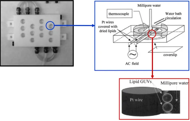
Due to the complexity of biological membrane peptide-membrane interaction studies are usually initiated with simple model membranes so that different properties such as lipid charge, membrane viscosity and the presence of sterols can be modulated. Each of the several model membranes that have been used to study membrane-active peptides has advantages and disadvantages, depending on the techniques to be employed. To start our studies, peptide/membrane interactions were followed using spectroscopic methodologies and liposomes.
Figure 2.4. Preparation of LUVs and
SUVs. A) Lipids are dissolved in chloroform and the solvent is removed to yield a lipid film. After addition of water/buffer, hydrated lipid sheets detach spontaneously. B) Upon agitation and freeze-thaw cycles, a MLVs suspension is obtained. C) Sonication may be used to produce SUVs while D) extrusion can be used to obtain LUVs. Image adapted from:
http://www.avantilipids.com.
.
When phospholipids are suspended in an excess of aqueous solution they spontaneously form multilamellar vesicles (MLVs). These aqueous dispersions produced by mechanical agitation of an aqueous medium in the presence of a dry lipid film were designated as liposome by Bangham et al. [133] (Figure 2.4). Generically the term liposome is applied for hydrated lipid dispersions which can be characterized based on size and number of lamellas [134]. MLVs are large vesicles with two or more lamellas; vesicles with one lamella can be: small unilamellar vesicles (SUVs) with a ∼30nm diameter; large unilamellar vesicles (LUVs) with a ∼100nm diameter; and giant unilamellar vesicles (GUVs) with a diameter higher than 10μm. The type of liposome
Dry lipid film Water Hydration of lipid Agitation MLVs Sonication homogenization SUVs LUVs Extrusion A B C D
 [pep-1] T a (µM) [pep-1] memb b(mM) I 0 /I c (0% fusion) I 0 /I d (100% fusion) I 0 /I (assay) fusion (%) e POPC 0.05 6.88 11.3 ( 3.9 1.30 ( 0.61 4.35 ( 0.07 1.45 ( 0.02 4.9 ( 0.07 POPC](https://thumb-eu.123doks.com/thumbv2/123dok_br/15570428.1047951/79.918.169.756.68.499/table-vesicle-fusion-induced-addition-fusion-fusion-fusion.webp)