Pathogenesis of
Salmonella
-induced
enteritis
1Departamento de Clínica e Cirurgia Veterinárias, Escola de Veterinária,
Universidade Federal de Minas Gerais, Belo Horizonte, MG, Brasil
2Department of Medical Microbiology and Immunology, College of Medicine,
Health Science Center, Texas A&M University System, College Station, TX, USA
3Department of Veterinary Pathobiology, College of Veterinary Medicine,
Texas A&M University, College Station, TX, USA R.L. Santos1,
R.M. Tsolis2,
A.J. Bäumler2
and L.G. Adams3
Abstract
Infections with Salmonella serotypes are a major cause of food-borne
diseases worldwide. Animal models other than the mouse have been employed for the study of nontyphoidal Salmonella infections
be-cause the murine model is not suitable for the study of Salmonella
-induced diarrhea. The microbe has developed mechanisms to exploit the host cell machinery to its own purpose. Bacterial proteins deliv-ered directly into the host cell cytosol cause cytoskeletal changes and interfere with host cell signaling pathways, which ultimately enhance disease manifestation. Recently, marked advances have been made in our understanding of the molecular interactions between Salmonella
serotypes and their hosts. Here, we discuss the molecular basis of the pathogenesis of Salmonella-induced enteritis.
Correspondence
R.L. Santos
Departamento de Clínica e Cirurgia Veterinárias
Escola de Veterinária, UFMG Av. Antônio Carlos, 6627 30161-970 Belo Horizonte, MG Brasil
Fax: +55-31-3499-2230 E-mail: [email protected]
Research supported by grant DHHS/PHS/NIH-1 RO1 A144170 from the National Institutes of Health and the Texas Agricultural Experiment Station Project (No. 8409). R.L. Santos was supported by CAPES.
Received February 14, 2002 Accepted September 17, 2002
Key words
·Salmonella typhimurium ·Enteritis
·Diarrhea ·Salmonellosis
Introduction
Salmonella serotypes have a broad host range and clinical manifestations that result from the combination between serotype and host species involved. Recently, a large amount of information on the molecular level of interactions between bacteria and host cells has allowed us to propose molecular mechanisms determining the pathologic and
clinical manifestations of nontyphoidal
Sal-monella infections. For simplification, Sal-monella enterica subsp. enterica ser. Typhi-murium will be referred to hereafter as either
S. typhimurium or serotype Typhimurium.
Salmonella infection is one of the most common food-borne infections worldwide. In the United States an estimated 1.41
mil-lion cases and more than 500 human deaths occur annually (1). Approximately 95% of
the human Salmonella infections are
food-borne, corresponding to approximately 30% of deaths caused by food-borne infections in
the United States (1). Salmonella infection is
even more detrimental in the developing world.
Experimental models for Salmonella infection
The mouse, which has been used for most of the studies addressing the various aspects
of host-pathogen interaction in S.
typhimu-rium infection, develops a systemic disease
when infected with S. typhimurium, but no
typhimu-rium results in a disease that is similar to human typhoid fever caused by infection with serotypes Typhi, Paratyphi A, B, and C, which are host-restricted serotypes that in-fect only man and few other primate species.
Thus, murine infection with S. typhimurium
has been employed as a model for human
typhoid fever, but not Salmonella-induced
diarrhea. In contrast, calves infected with S.
typhimurium develop a diarrheic disease with clinical manifestations similar to those ob-served in human infections, which also re-sult in diarrhea with a low mortality rate (reviewed in Refs. 2 and 3).
An important aspect of the pathology of
Salmonella-induced enteritis is the same pat-tern of inflammatory reaction developed by
calves after S. typhimurium infection, which
is characterized by a marked infiltration of neutrophils, also observed in non-human pri-mates in experimental infections, and in hu-man infections. In sharp contrast, mice
in-fected with S. typhimurium develop an
in-flammatory response with predominance of mononuclear leukocytes, which is not asso-ciated with diarrhea (reviewed in Ref. 3).
Interestingly, although several bacterial genes are required for disease progression and expression in both calves and mice, some genes that play a role in the murine typhoid model are not required for entero-pathogenesis in cattle and vice versa (4).
Many of the Salmonella virulence genes are
clustered in certain areas of the chromosome
known as Salmonella pathogenicity islands
(SPI). To date, five SPI have been described and two of them, SPI-1 and SPI-2, encode type III secretion systems. The SPI-1-en-coded type III secretion system translocates effector proteins into the cytosol of host cells. This system is required for invasion of nonphagocytic host cells (5) and enteropatho-genesis (6), while the SPI-2-encoded type III secretion system is required for intracellular survival in murine macrophages (7). In the
murine typhoid model, S. typhimurium strains
having mutations in SPI-1 and SPI-2 are
50-fold and >10,000-50-fold attenuated, respec-tively, after oral infection (5,7). In contrast, SPI-2 does not play a major role in entero-pathogenesis, whereas SPI-1 mutants are non-pathogenic for calves (6) as opposed to just a mild attenuation of these SPI-1 mutants in the mouse.
Invasion of epithelial cells by S. typhimurium
A remarkable aspect of Salmonella
patho-genesis is its ability to invade nonphagocytic cells in a process that morphologically re-sembles phagocytosis. M cells located in the follicle-associated epithelium in the Peyers patches are the primary intestinal epithelial
cell type targeted for invasion by Salmonella
in the mouse (8). In cattle, S. typhimurium is
able to invade both M cells and enterocytes with no predilection for a particular cell type (9).
Upon contact with intestinal epithelial
cells, S. typhimurium translocates bacterial
effector proteins into the host cell cytosol via the SPI-1-encoded type III secretion system previously discussed. Some of these pro-teins have kinase, phosphatase, or actin-bind-ing activity, and once in the epithelial cell cytosol, they alter host cell signaling path-ways that promote changes in the cytoskel-eton, with consequent bacterial internaliza-tion and changes in host gene expression
(reviewed in Ref. 10). Mutant strains of S.
typhimurium lacking structural components of the SPI-1-encoded type III secretion sys-tem, secreted proteins, or SPI-1 transcrip-tional regulators are unable to invade epithe-lial cells (11).
regulator HilA (12). HilA-dependent regula-tion of expression is not restricted to SPI-1, since SPI-4- and SPI-5-encoded genes were found to be regulated by HilA, whose ex-pression is regulated by SirA (13).
Studies in the early 1990s determined the morphologic features and dynamics of
the interaction between S. typhimurium and
intestinal epithelial cell monolayers. Shortly
after Salmonella enters in contact with the
apical surface of the epithelial monolayer, the epithelia develop cytoplasmic projec-tions with disruption of the underlying cyto-skeleton, and intracellular bacteria are de-tected 30 min after infection. Two hours post-infection, free bacteria are detected on the basolateral side of the monolayer (14).
Additional studies indicated that S.
typhimu-rium grown under conditions that favor
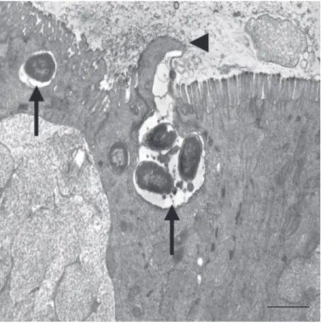
ex-pression of invasin induce morphologic changes in epithelial cells as quickly as 40 s after contact (15), which are associated with recruitment of cytoskeletal components (16). These morphological and cytoskeletal changes, characterized by formation of ruffle-like structures, mediate bacterial internaliza-tion into epithelial cells (17). As shown in Figure 1, similar morphologic changes occur
in calves infected with S. typhimurium in
vivo. SipC is an SPI-1-encoded protein that
acts as a translocase and is translocated itself into the host cytosol via the SPI-1-encoded type III secretion system. This protein bundles actin filaments and nucleates actin
polymer-ization in vitro, which results in cytoskeletal
rearrangements in vivo (18). SipA, which is
not required for invasion, binds to F-actin inhibiting depolymerization (19). Thus, SipC is essential for actin nucleation and bundling of actin filaments whereas SipA acts by enhancing the efficiency of this pro-cess (20).
Although sopE is absent in many S.
ty-phimurium strains, a homologue, sopE2, is
present in all strains of S. typhimurium (21).
Like SopE, SopE2 is also a guanine nucleo-tide exchange factor for Cdc42 and plays a
role recruiting the actin-nucleating complex Arp2/3 to the membrane ruffles (21). There-fore, SopE2 is required for optimal invasion
of cultured epithelial cells by S.
typhimu-rium (21). SptP, a Salmonella protein that
acts as a GTPase-activating factor for Rac-1 and Cdc42 and is also delivered into the epithelial cell cytosol via the SPI-1-encoded type III secretion system, has been shown to disrupt the actin cytoskeleton. Therefore it acts by reversing the cytoskeletal changes induced by the bacteria during invasion, re-storing the normal cytoskeletal structure (22). The effect of SptP antagonizing the action of other bacterial effector proteins clearly
indi-cates that S. typhimurium is able to finely
regulate cellular pathways in favor of its own purposes.
Inflammatory response during S. typhimurium infection
Although the complete mechanism of
Salmonella-induced diarrhea is still not clear, some previous reports indicate that it is dis-tinct from secretory diarrheas such as those caused by cholera toxin (23,24). As
dis-cussed above, infection of calves with S.
typhimurium results in enteritis in which neutrophils are the primary inflammatory cells involved. On the other hand, the mouse, which does not develop diarrhea, responds to the infection mostly with a mononuclear infiltrate in the intestine (3). Furthermore, experimental depletion of the polymorpho-nuclear leukocyte pool by administration of nitrogen mustard to rabbits results in a sig-nificant decrease in intestinal fluid secretion
induced by S. typhimurium infection (23). In
addition, administration of indomethacin, an anti-inflammatory agent, completely abol-ishes fluid secretion in rabbit intestinal loops
inoculated with S. typhimurium (24).
There-fore, neutrophils are proposed to play a very
important role in the pathogenesis of
Salmo-nella-induced diarrhea. Some of our recent experimental findings corroborate this hy-pothesis since the inflammatory response, characterized by neutrophil infiltration, pre-cedes intestinal fluid secretion after
infec-tion with S. typhimurium in calves (25).
Several Salmonella virulence factors required
for enteropathogenicity in calves, which are involved in eliciting neutrophil influx, are also required for fluid secretion (26,27), re-inforcing the significance of neutrophils in this process.
Following oral infection, intestinal epi-thelial cells are the first barrier to be crossed by S. typhimurium in order to invade and colonize the intestinal tissues and other or-gans. Current data indicate that epithelial cells play an important role in the outcome of infection by influencing the host inflam-matory response. Supporting this notion, a
study demonstrated that Salmonella strains
and serotypes that elicit enteritis and diar-rhea are also able to induce transepithelial signaling for neutrophil migration across epithelial cell layers, whereas strains that do not cause diarrhea failed to trigger neutro-phil transepithelial migration (28). Invasion of cultured epithelial cells by bacteria,
in-cluding Salmonella, results in expression
and secretion of interleukin 8 (IL-8), a
chemo-attractant for neutrophils (29). Salmonella
-induced IL-8 secretion by epithelial cells is dependent on the mitogen-activated protein kinase pathway and activation of the
trans-cription factor NF-kB and requires a
func-tional SPI-1-encoded type III secretion sys-tem (30). Cultured epithelial cells also
re-spond to S. typhimurium invasion with an
increase in the cytosolic concentration of calcium, which is absolutely required for
NF-kB activation and IL-8 expression (31).
Infiltration of neutrophils into the lamina
propria occurs shortly after infection with S.
typhimurium, which is followed by a mas-sive migration of neutrophils through the epithelium into the intestinal lumen (9,25). Thus, one can expect that chemoattractants for neutrophils are secreted into the intesti-nal lumen at the early stages of infection. Interestingly, experiments with polarized in-testinal epithelial cell monolayers indicate that IL-8 is secreted at the basolateral aspect of the epithelium, which implies that the role of IL-8 is primarily recruitment of neutro-phils to the subepithelial space rather than transepithelial migration into the intestinal lumen (32). Further experiments have led to the identification of a pathogen-elicited epi-thelial chemoattractant (PEEC) bioactivity, which is released in a polarized fashion to-wards the apical aspect of the epithelial mono-layer. Once secreted on the apical side of the epithelial cell, PEEC induces direct migra-tion of neutrophils across cultured intestinal epithelial cell monolayers (33). PEEC has a 1- to 3-kDa mass, stimulates neutrophils via a pertussis toxin-sensitive receptor and
still uncharacterized (33). Actual invasion of epithelial cells is not required for induction of epithelial promotion of neutrophil trans-epithelial migration since treatment of the monolayers with cytochalasin D, which
blocks S. typhimurium invasion, does not
reduce the promotion of neutrophil migra-tion (34). Importantly, although IL-8 and PEEC act in concert to promote neutrophil migration through the lamina propria and epithelia, respectively, secretion of these two chemotactic factors is mediated by distinct signaling pathways. In contrast to IL-8 ex-pression and secretion, PEEC activity is not
dependent on NF-kB activation (34). It has
been recently demonstrated that the bacte-rial protein SipA, an SPI-1-encoded effector protein translocated into the cytosol of the host cell via the SPI-1 type III secretion system, is sufficient to trigger neutrophil transepithelial migration in cultured
intesti-nal epithelial monolayers (35). Another
Sal-monella protein secreted by the SPI-1-en-coded type III secretion system, SopA, is also involved in the induction of neutrophil transepithelial migration (36).
Although some recent studies have
ad-dressed in vivo cytokine production in
re-sponse to Salmonella infection, most of these
studies were performed on mice. We have demonstrated that there is a marked increase in expression of CXC chemokines such as
IL-8, GROa/g and GCP2, and the
proinflam-matory cytokine IL-1ß in bovine Peyers patches as early as 1 h post-infection, in-creasing continuously until at least 5 h post-infection (9). Interestingly, anti-inflamma-tory cytokines such as IL-4 and the IL-1 receptor antagonist (IL-1Ra) are also
up-regulated in bovine Peyers patches in vivo
after infection with S. typhimurium (9).
Salmonella-induced host cell death
Although apoptosis has been defined clas-sically as a form of cell death that does not elicit an inflammatory reaction, under
spe-cific conditions this process may ultimately act as a proinflammatory signal. Several groups have reported that murine macro-phages and macrophage-like cell lines
un-dergo cell death when infected with S.
typhi-murium (37-39). A previous report indicated
that Salmonella-induced macrophage
apop-tosis is associated with marked IL-1 release (40). Thus, since IL-1 is a potent pro-inflam-matory cytokine, this was the first indication
of a possible link between Salmonella
-in-duced cell death and inflammation.
Salmo-nella-induced macrophage cell death is largely due to expression of genes associated with invasion, since mutant strains lacking functional SPI-1 or grown under conditions that prevent SPI-1 expression do not cause rapid cell death after infection of macro-phages (37,39,41,42), although SPI-1-inde-pendent cell death has also been described (38,42,43). Further investigation has led to the identification of the SipB protein as the bacterial effector responsible for induction of apoptosis (44). SipB is translocated into the host cell cytosol via the SPI-1-encoded type III secretion system, where it binds to and activates caspase-1, an intracellular cys-teine protease also known as IL-1ß convert-ing enzyme. Once activated, caspase-1 cleaves and activates IL-1ß (44). Caspase-1 is also responsible for triggering apoptosis in
Salmonella-infected macrophages, since a specific caspase-1 inhibitor blocks this mech-anism of cell death (44). Infection of
macro-phages with S. typhimurium also results in
degradation of the host protein Raf-1 in a SipB- and caspase-1-dependent manner, which favors the cytotoxic effect of SipB since Raf-1 acts by antagonizing the
cas-pase-1-mediated cell death (45). Thus,
Sal-monella-induced macrophage apoptosis re-sults in release of active IL-1ß, which is
thought to play a significant role in
mac-rophages infected with S. typhimurium (42). Although all the initial papers described
Salmonella-induced cell death as apoptotic in nature, more recent publications argue that it is a necrotic rather that an apoptotic mechanism of cell death. The conclusions of these reports are based on either failure to detect DNA fragmentation and morphologic features of apoptosis (46) or on the effect of
glycine blocking Salmonella-induced
cyto-toxicity (47). However, while the definition of a proper classification and terminology for this mechanism is still debatable, the requirement for caspase-1 activation and the proinflammatory nature of this mechanism is a consensus among different laboratories and has lead to the proposition of pyroptosis as a new term to describe proinflammatory programmed cell death (48).
In spite of the increasing amount of
infor-mation on the interaction between
Salmo-nella and macrophage in cell culture sys-tems, there are few data available regarding the significance of macrophage cell death and its putative proinflammatory effect on
the outcome of Salmonella infections in vivo.
The ability of S. typhimurium to induce
mac-rophage cell death has been demonstrated in
vivo in mice intravenously inoculated with small infectious doses (49). In addition, cas-pase-1 is required for colonization of Peyers patches and induction of systemic infection
in mice orally inoculated with S.
typhimu-rium (50). Although quite valuable, these
data are not applicable to the pathogenesis of
Salmonella-induced diarrhea. Indeed, we
have demonstrated that Salmonella-induced
host cell death is not sufficient to trigger the
inflammatory response after S. typhimurium
infection in calves (25).
Salmonella virulence factors involved in enteropathogenesis
There is clearly a difference between
Salmonella genes required for virulence in
mice, where the Salmonella virulence
plas-mid and SPI-2-encoded genes are essential for virulence, and those involved in eliciting diarrhea, in which SPI-1-encoded genes are
essential but SPI-2 and the Salmonella
viru-lence plasmid play only minor roles (6,13,26, 51,52). Disruption of the SPI-1 type III se-cretion decreases or abolishes the ability of
S. typhimurium to invade the intestinal epi-thelium, which correlates with its ability to elicit an inflammatory response and thereby induce diarrhea (6,13,26,51).
A fifth SPI has been identified and linked to the pathogenesis of diarrhea (27,53). An
SPI-5-encoded gene, sopB, has been
exten-sively studied. SopB, also known as SigD, is secreted via the SPI-1-encoded type III se-cretion system and its expression is
depend-ent on the regulator sirA, which is also an
activator of the SPI-1 regulator hilA (54). A
sopB mutant of S. dublin has a significantly
reduced ability to elicit inflammation and fluid secretion in bovine ligated ileal loops in spite of displaying wild-type levels of invasion in the Peyers patches (27). Similar
results were observed with a sopB mutant of
S. typhimurium (25). SopB is an inositol phosphate phosphatase that hydrolyzes phos-phatidylinositol 3,4,5-triphosphate, which is an inhibitor of chloride secretion. In addi-tion, SopB hydrolyzes inositol 1,3,4,5,6 pentakisphosphate, generating inositol 1,4,5,6 tetrakisphosphate (55), which may be involved in increasing chloride secretion (56). Thus, SopB is thought to mediate fluid secretion by increasing chloride secretion. However, changes in chloride secretion alone are not compatible with the pathologic
fea-tures of Salmonella-induced diarrhea, which
is associated with a severe acute neutro-philic infiltration. The most significant events
in the pathogenesis of Salmonella-induced
enteritis are illustrated in Figure 2. Interest-ingly, SopB also affects host cell signaling pathways that may be involved in regulation of cytokine expression such as activation of the serine-threonine kinase Akt (57). The
se-creted in an SPI-1-dependent manner, has an additive effect to SopB in the induction of enteritis (58), whereas SopA influences the inflammatory response by a mechanism dis-tinct from SopB and SopD. SopA is involved in induction of transepithelial migration of neutrophils, a phenomenon that is not influ-enced by SopB or SopD (36). Recent exper-imental findings from our laboratory indi-cate that the secreted effectors SipA, SopA, SopB, SopD, and SopE2 act in concert to
induce diarrhea, since a strain lacking all of
these genes (DsipAsopABDE) had additive
attenuation when compared to the single gene mutants. The quintuplet mutant (DsipAsopABDE) was as attenuated as a mu-tant with a defective SPI-1-encoded type III secretion system in the bovine ileal loop
model (59). The role of Salmonella
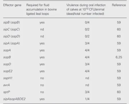
viru-lence genes in enteropathogenesis is sum-marized in Table 1.
Salmonella-induced diarrhea is an
effi-Figure 2. Schematic representation of the pathogenesis of Salmonella-induced enteritis, with the most significant events described from A through H.
E. At least in cell culture systems,
Salmonella induces macrophage (MO) cell death, which is triggered by SipB binding and activation of caspase-1. IL-1b is then released and may enhance the inflamma-tory reaction. In sharp contrast, neutrophils do not undergo cell death due to infection with Sal-monella.
F. As the inflammatory reaction progresses, neutrophils migrate through the epithelial layer, with accumulation of inflammatory cells and protein-rich fluid into the intestinal lumen. These events develop from 1 to 3 h post-inoculation.
G. The overwhelming inflamma-tory reaction results in massive transepithelial migration of neu-trophils, which causes epithelial detachment from the basal mem-brane, favoring fluid secretion into the intestinal lumen and diarrhea.
H. Due to the release of proteases and other mediators from inflam-matory cells, extensive necrosis of the superficial mucosa takes place at 24 to 48 h post-infection. The resulting necrotic debris pro-vide an adequate substrate for bacterial growth facilitating shed-ding and environmental contami-nation.
MO A. Salmonella (S) attaches to the apical surface of the intestinal epi-thelia (EP) and injects the effector proteins through the SPI-1-en-coded type III secretion system (TTSS) into the host cell cytosol. Resident macrophages (MO), a blood vessel (BV), and an intravas-cular neutrophil (PMN) are located in the lamina propria.
B. Salmonella effector proteins, particularly SipA and SipC, modu-late actin polymerization resulting in ruffling formation at the apical surface of the epithelial layer. This event takes place in both M cells and enterocytes as early as 15 min post-inoculation.
C. In addition to cytoskeletal re-modeling, some of the Salmonella
effector proteins trigger nuclear responses that ultimately result in increased expression of chemo-tactic factors. Interestingly, patho-gen-elicited epithelial chemoat-tractant (PEEC) is secreted toward the apical side whereas IL-8 is se-creted towards the basolateral side of the epithelial layer.
D. In response to chemotactic stimuli, there is infiltration of neutrophils in the lamina propria. At 1 h post-infection Salmonella
reaches the basal portion of the epithelial layer and undergoes phagocytosis by neutrophils and macrophages.
MO BV
PMN
EP
S PEEC
IL-8 GRO GCP2 TTSS-secreted
References
1. Mead PS, Slutsker L, Dietz V, McCaig LF, Bresee JS, Shapiro C, Griffin PM & Tauxe RV (1999). Food-related illnesses and death in the United States. Emerging Infectious Diseases, 5: 607-625. 2. Tsolis RM, Kingsley RA, Townsend SM, Ficht TA, Adams LG &
Bäumler AJ (1999). Of mice, calves, and men. Comparison of the mouse typhoid model with other Salmonella infections. Advances in Experimental Medicine and Biology, 473: 261-274.
3. Santos RL, Zhang S, Tsolis RM, Kingsley RA, Adams LG & Bäumler AJ (2001). Animal models of Salmonella infections: gastroenteritis
vs typhoid fever. Microbes and Infection, 3: 1335-1344.
4. Tsolis RM, Townsend SM, Miao EA, Miller SI, Ficht TA, Adams LG & Bäumler AJ (1999). Identification of a putative Salmonella enterica
serotype typhimurium host range factor with homology to IpaH and YopM by signature-tagged mutagenesis. Infection and Immunity, 67: 6385-6393.
5. Galán JE & Curtiss III R (1989). Cloning and molecular characteriza-tion of genes whose products allow Salmonella typhimurium to penetrate tissue culture cells. Proceedings of the National Academy of Sciences, USA, 86: 6383-6387.
6. Tsolis RM, Adams LG, Ficht TA & Bäumler AJ (1999). Contribution of Salmonella typhimurium virulence factors to diarrheal disease in calves. Infection and Immunity, 67: 4879-4885.
7. Ochman H, Soncini FC, Solomon F & Groisman EA (1996). Identifica-tion of a pathogenicity island for Salmonella survival in host cells.
Proceedings of the National Academy of Sciences, USA, 93: 7800-7804.
8. Jones BD, Ghori N & Falkow S (1994). Salmonella typhimurium
initiates murine infection by penetrating and destroying the
special-cient way of spreading the organism in the
environment. Recently, a gene, named shdA,
found only in strains of Salmonella adapted
to warm blooded-organisms (subspecies I) has been demonstrated to be involved in
prolonged shedding of S. typhimurium in
mice. Mutation of shdA causes a decrease in
the number of organisms shed in feces and the duration of shedding (61). Subsequent
studies indicated that the shdA gene product
binds to extracellular matrix proteins, par-ticularly fibronectin (62).
From these discussions of the
mechan-isms of Salmonella-induced diarrhea, it is
exceedingly clear that there is an intensive and intricate series of highly regulated adap-tive gene expression events by both the host
and the Salmonella microbe. Unraveling the
intricacies of the molecular basis and the regu-lation of these interactions holds great promise for developing new vaccination strategies as well as improved therapeutic rationales.
Table 1. Role of virulence genes in enteropathogenesis.
Effector gene Required for fluid Virulence during oral infection Reference accumulation in bovine of calves at 1010 CFU/animal
ligated ileal loops (dead/total number infected)
sipB (sspB) yes 0/4 59
sipC (sspC) nd 0/2 60
sipD (sspD) nd 0/2 60
sipA (sspA) yes 3/4 59
sopA yes 4/4 59
sopB yes 4/4 6,25
sopD yes 3/4 59
sopE2 yes 4/4 59
sspH1 no nd 59
avrA no nd 59
sptP no 3/4 60
sipAsopABDE2 yes 1/4 59
nd = not determined, CFU = colony-forming units.
ized epithelial M cells of the Peyer’s patches. Journal of Experimen-tal Medicine, 180: 15-23.
9. Santos RL, Zhang S, Tsolis RM, Bäumler AJ & Adams LG (2002). Morphologic and molecular characterization of Salmonella typhimu-rium infection in neonatal calves. Veterinary Pathology, 39: 200-215. 10. Galán JE & Zhou D (2000). Striking a balance: modulation of the actin cytoskeleton by Salmonella. Proceedings of the National Academy of Sciences, USA, 97: 8754-8761.
11. Penheiter KL, Mathur N, Giles D, Fahlen T & Jones BD (1997). Non-invasive Salmonella typhimurium mutants are avirulent because of an inability to enter and destroy M cells of ileal Peyer’s patches.
Molecular Microbiology, 24: 697-709.
12. Bajaj V, Lucas RL, Hwang C & Lee CA (1996). Co-ordinate regulation of Salmonella typhimurium invasion genes by environmental and regulatory factors is mediated by control of hilA expression. Molecu-lar Microbiology, 22: 703-714.
13. Ahmer BMM, Van Reeuwijk J, Watson PR, Wallis TS & Heffron F (1999). Salmonella SirA is a global regulator of genes mediating enteropathogenesis. Molecular Microbiology, 31: 971-982. 14. Finlay BB & Falkow S (1990). Salmonella interactions with polarized
human intestinal Caco-2 epithelial cells. Journal of Infectious Dis-eases, 162: 1096-1106.
15. Francis CL, Starnbach MN & Falkow S (1992). Morphological and cytoskeletal changes in epithelial cells occur immediately upon in-teraction with Salmonella typhimurium grown under low-oxygen conditions. Molecular Microbiology, 6: 3077-3087.
Journal of Cell Science, 99: 283-296.
17. Francis CL, Ryan TA, Jones BD, Smith SJ & Falkow S (1993). Ruffles induced by Salmonella and other stimuli direct macropinocytosis of bacteria. Nature, 364: 639-642.
18. Hayward RD & Koronakis V (1999). Direct nucleation and bundling of actin by the SipC protein of invasive Salmonella. EMBO Journal, 18: 4926-4934.
19. Zhou D, Mooseker MS & Galán JE (1999). Role of the S. typhimu-rium actin-binding protein SipA in bacterial internalization. Science, 283: 2092-2095.
20. McGhie EJ, Hayward RD & Koronakis V (2001). Cooperation be-tween actin-binding proteins of invasive Salmonella: SipA potenti-ates SipC nucleation and bundling of actin. EMBO Journal, 20: 2131-2139.
21. Stender S, Friebel A, Linder S, Rohde M, Mirold S & Hardt WD (2000). Identification of SopE2 from Salmonella typhimurium, a con-served guanine nucleotide exchange factor for Cdc42 of the host cell. Molecular Microbiology, 36: 1206-1221.
22. Fu Y & Galán JE (1999). A Salmonella protein antagonizes Rac-1 and Cdc42 to mediate host-cell recovery after bacterial invasion. Nature, 401: 293-297.
23. Giannella RA (1979). Importance of the intestinal inflammatory reac-tion in Salmonella-mediated intestinal secretion. Infection and Im-munity, 23: 140-145.
24. Giannella RA, Gots RE, Charney AN, Greenough SB & Formal SB (1975). Pathogenesis of Salmonella-mediated intestinal fluid secre-tion. Gastroenterology, 69: 1238-1245.
25. Santos RL, Tsolis RM, Zhang S, Ficht TA, Bäumler AJ & Adams LG (2001). Salmonella-induced cell death is not required for enteritis in calves. Infection and Immunity, 69: 4610-4617.
26. Watson PR, Galyov EE, Paulin SM, Jones PW & Wallis TS (1998). Mutation of invH, but not stn, reduces Salmonella-induced enteritis in cattle. Infection and Immunity, 66: 1432-1438.
27. Galyov EE, Wood MW, Rosqvist R, Mullan PB, Watson PR, Hedges S & Wallis TS (1997). A secreted effector protein of Salmonella dublin is translocated into eukaryotic cells and mediates inflamma-tion and fluid secreinflamma-tion in infected ileal mucosa. Molecular Microbi-ology, 25: 903-912.
28. McCormick BA, Miller SI, Carnes D & Madara JL (1995). Transepi-thelial signaling to neutrophils by Salmonellae: a novel virulence mechanism for gastroenteritis. Infection and Immunity, 63: 2302-2309.
29. Eckmann L, Kagnoff MF & Fierer J (1993). Epithelial cells secrete the chemokine interleukin-8 in response to bacterial entry. Infection and Immunity, 61: 4569-4574.
30. Hobbie S, Chen LM, Davis RJ & Galán JE (1997). Involvement of mitogen-activated protein kinase pathways in the nuclear responses and cytokine production induced by Salmonella typhimurium in cul-tured intestinal epithelial cells. Journal of Immunology, 159: 5550-5559.
31. Gewirtz AT, Rao AS, Simon Jr PO, Merlin D, Carnes D, Madara JL & Neish AS (2000). Salmonella typhimurium induces epithelial IL-8 expression via Ca2+-mediated activation of NF-kB pathway. Journal of Clinical Investigation, 105: 79-92.
32. McCormick BA, Hofman PM, Kim J, Carnes DK, Miller SI & Madara JL (1995). Surface attachment of Salmonella typhimurium to intesti-nal epithelia imprints the subepithelial matrix with gradients chemo-tactic for neutrophils. Journal of Cell Biology, 131: 1599-1608. 33. McCormick BA, Parkos CA, Colgan SP, Carnes DK & Madara JL
(1998). Apical secretion of a pathogen-elicited epithelial chemoat-tractant activity in response to surface colonization of intestinal
epithelia by Salmonella typhimurium. Journal of Immunology, 160: 455-466.
34. Gewirtz AT, Siber AM, Madara JL & McCormick BA (1999). Orches-tration of neutrophil movement by intestinal epithelial cells in re-sponse to Salmonella typhimurium can be uncoupled from bacterial internalization. Infection and Immunity, 67: 608-617.
35. Lee CA, Silva M, Siber AM, Kelly AJ, Galyov E & McCormick BA (2000). A secreted Salmonella protein induces a proinflammatory response in epithelial cells, which promotes neutrophil migration.
Proceedings of the National Academy of Sciences, USA, 97: 12283-12288.
36. Wood MW, Jones MA, Watson PR, Siber AM, McCormick BA, Hedges S, Rosqvist R, Wallis TS & Galyov EE (2000). The secreted effector protein of Salmonella dublin, SopA, is translocated into eukaryotic cells and influences the induction of enteritis. Cellular Microbiology, 2: 293-303.
37. Chen LM, Kaniga K & Galán JE (1996). Salmonella spp. are cytotoxic for cultured macrophages. Molecular Microbiology, 21: 1101-1115. 38. Lindgren SW, Stojiljkovic I & Heffron F (1996). Macrophage killing is
an essential virulence mechanism of Salmonella typhimurium. Pro-ceedings of the National Academy of Sciences, USA, 93: 4197-4201. 39. Monack DM, Raupach B, Hromockyj AE & Falkow S (1996). Salmo-nella typhimurium invasion induces apoptosis in infected macro-phages. Proceedings of the National Academy of Sciences, USA, 93: 9833-9838.
40. Arai T, Hiromatsu K, Nishimura H, Kimura Y, Kobayashi N, Ishida H, Nimura Y & Yoshikai Y (1995). Endogenous interleukin 10 prevents apoptosis in macrophages during Salmonella infection. Biochemical and Biophysical Research Communications, 213: 600-607. 41. Lundberg U, Vinatzer U, Berdnik D, Gabain A & Baccarini M (1999).
Growth phase-regulated induction of Salmonella-induced macro-phage apoptosis correlates with transient expression of SPI-1 genes.
Journal of Bacteriology, 181: 3433-3437.
42. Santos RL, Tsolis RM, Bäumler AJ, Smith III R & Adams LG (2001).
Salmonella enterica serovar typhimurium induces cell death in bo-vine monocyte-derived macrophages by early sipB-dependent and delayed sipB-independent mechanisms. Infection and Immunity, 69: 2293-2301.
43. Van der Velden AWM, Lindgren SW, Worley MJ & Heffron F (2000).
Salmonella pathogenicity island 1-independent induction of apopto-sis in infected macrophages by Salmonella enterica serotype typhi-murium. Infection and Immunity, 68: 5702-5709.
44. Hersh D, Monack DM, Smith MR, Ghori N, Falkow S & Zychlinsky A (1999). The Salmonella invasin SipB induces macrophage apoptosis by binding to caspase-1. Proceedings of the National Academy of Sciences, USA, 96: 2396-2401.
45. Jesenberger V, Procyk KJ, Rüth J, Schreiber M, Theussl HC, Wagner EF & Baccarini M (2001). Protective role of Raf-1 in Salmonella -induced macrophage apoptosis. Journal of Experimental Medicine, 193: 353-364.
46. Watson PR, Gautier AV, Paulin SM, Bland AP, Jones PW & Wallis TS (2000). Salmonella enterica serovars Typhimurium and Dublin can lyse macrophages by a mechanism distinct from apoptosis. Infec-tion and Immunity, 68: 3744-3747.
47. Brennan MA & Cookson BT (2000). Salmonella induces macrophage death by caspase-1-dependent necrosis. Molecular Microbiology, 38: 31-40.
48. Cookson BT & Brennan MA (2001). Pro-inflammatory programmed cell death. Trends in Microbiology, 9: 113-114.
resides intracellularly inside macrophages and exerts a cytotoxic effect on phagocytes in vivo. Journal of Experimental Medicine, 186: 569-580.
50. Monack DM, Hersh D, Ghori N, Bouley D, Zychlinsky A & Falkow S (2000). Salmonella exploits caspase-1 to colonize Peyer’s patches in a murine typhoid model. Journal of Experimental Medicine, 192: 249-258.
51. Watson PR, Paulin SM, Bland AP, Libby SJ, Jones PW & Wallis TS (1995). Characterization of intestinal invasion by Salmonella typhi-murium and Salmonella dublin and effect of a mutation in the invH
gene. Infection and Immunity, 63: 2743-2754.
52. Wallis TS, Paulin SM, Plested JS, Watson PR & Jones PW (1995). The Salmonella dublin virulence plasmid mediates systemic but not enteric phases of salmonellosis in cattle. Infection and Immunity, 63: 2755-2761.
53. Wood MW, Jones MA, Watson PR, Hedges S, Wallis TS & Galyov EE (1998). Identification of a pathogenicity island required for Salmo-nella enteropathogenicity. Molecular Microbiology, 29: 883-891. 54. Hong KH & Miller VL (1998). Identification of a novel Salmonella
invasion locus homologous to ShigellaipgDE. Journal of Bacteriol-ogy, 180: 1793-1802.
55. Norris FA, Wilson MP, Wallis TS, Galyov EE & Majerus PW (1998). SopB, a protein required for virulence of Salmonella dublin, is an inositol phosphate phosphatase. Proceedings of the National Acade-my of Sciences, USA, 95: 14057-14059.
56. Eckmann L, Rudolf MT, Ptasznik A, Schultz C, Jiang T, Wolfson N, Tsien R, Fierer J, Shears SB, Kagnoff MF & Traynor-Kaplan AE (1997). D-myo-inositol 1,4,5,6-tetrakisphosphate produced in human
intestinal epithelial cells in response to Salmonella invasion inhibits phosphoinositide 3-kinase signaling pathways. Proceedings of the National Academy of Sciences, USA, 94: 14456-14460.
57. Steele-Mortimer O, Knodler LA, Marcus SL, Scheid MP, Goh BG, Pfeifer C, Duronio V & Finlay BB (2000). Activation of Akt/protein kinase B in epithelial cells by the Salmonella typhimurium effector SigD. Journal of Biological Chemistry, 275: 37718-37724.
58. Jones MA, Wood MW, Mullan PB, Watson PR, Wallis TS & Galyov EE (1998). Secreted proteins of Salmonella dublin act in concert to induce enteritis. Infection and Immunity, 66: 5799-5804.
59. Zhang S, Santos RL, Tsolis RM, Stender S, Hardt WD, Bäumler AJ & Adams LG (2002). The Salmonella enterica serotype typhimurium effector proteins SipA, SopA, SopB, SopD and SopE2 act in concert to induce diarrhea in calves. Infection and Immunity, 70: 3843-3855. 60. Tsolis RM, Adams LG, Hantman MJ, Scherer CA, Kimbrough T, Kingsley RA, Ficht TA, Miller SI & Baumler AJ (2000). SspA is required for lethal Salmonella enterica serovar typhimurium infec-tions in calves but is not essential for diarrhea. Infection and Immu-nity, 68: 3158-3163.
61. Kingsley RA, Van Amsterdam K, Kramer N & Bäumler AJ (2000). The
shdA gene is restricted to serotypes of Salmonella enterica subspe-cies I and contributes to efficient and prolonged fecal shedding.
Infection and Immunity, 68: 2720-2727.