III
Mestrado em Medicina e Oncologia Molecular
Rafaela Isabel Magalhães Leal Silva
Clinical and functional impact of a CDH1 gene mutation:
From the definition of a common ancestor to the functional
characterization of a causative mutation
Dissertação de Candidatura ao grau de Mestre em Medicina e Oncologia Molecular submetida à Faculdade de Medicina da Universidade do Porto. Orientador: Doutora Carla Oliveira
Afiliação: Expression Regulation in cancer, i3S.
Co-orientador: Professor Doutor Sérgio Castedo. Afiliaçao: FMUP/Centro Hospitalar de São João.
V Acknowledgements
Em primeiro lugar dirijo umas breves palavras à direcção do programa de Mestrado em Medicina e Oncologia Molecular. Quero parabenizar a excelente qualidade de ensino e agradecer esta oportunidade de formação.
Uma boa orientação é o primeiro fator para o sucesso de um aluno. Por esse motivo, o meu primeiro agradecimento é dirigido à Doutora Carla Oliveira. Agradeço-lhe a receptividade para realização da tese de mestrado, o envolvimento neste interessante projecto e por toda a sua disponibilidade ao longo deste percurso. A Carla é uma inspiração. Agradeço ainda aos restantes membros do grupo de investigação Expression
Regulation in Cancer, i3S, pela boa receção e por me terem ajudado nos momentos em que
precisei.
O trabalho de equipa e a multidisciplinariedade marcam a investigação, sendo as parcerias uma mais valia para qualquer tipo de trabalho. Neste âmbito quero agradecer ao Professor Doutor Sérgio Castedo, do serviço de Genética Médica e Centro de Mama (Centro Hospitalar São João do Porto), e co-orientador deste projecto. Ainda deste serviço, quero agradecer à Enfermeira Luzia Garrido, pela dedicação e apoio que me deu na realização deste trabalho. A Luzia acompanha, de coração, os doentes que aqui foram alvo de estudo. Admiro imenso a sua dedicação e empenho. É inspirador poder trabalhar com alguém assim.
Quero deixar o meu agradecimento à Doutora Susana Seixas, do grupo Genetic
Diversity, i3S. Foi uma mais valia neste trabalho e mostrou-se sempre disponível para nos
ajudar. Aprendi muito com a sua experiência.
A todos os membros do Ipatimup Diagnósticos, onde será sempre a “minha bancada”. Foi aqui que comecei a minha formação e o meu caminho profissional. Um especial obrigada “às meninas do LDG” e ao coordenador Luís Cirnes. Obrigada por todos os momentos, por todo o apoio e amizade. Equipa como esta jamais encontrarei em qualquer parte do Mundo!
Ao Miguel Silva, o meu melhor amigo e companheiro. Obrigada pela paciência e motivação. Que seja uma de muitas etapas que possamos partilhar.
Aos meus pais. Por todos os sacrificios a que se dispuseram para me possibilitarem a formação que tive. Por todo o amor incondicional, apoio e motivação ao longo destes anos.
1
Table of Contents
Figures Index... 3 Tables Index ... 4 Abbreviations ... 5 Resumo ... 6 Abstract ... 7 Introduction ... 9 Gastric Cancer ... 9Epidemiology and general features ... 9
Gastric Cancer histology and staging ... 10
Hereditary Diffuse Gastric Cancer ... 11
Penetrance ... 14
Macroscopy and Histology of HDGC ... 14
Surveillance and treatment options ... 16
CDH1 – gene and protein overview ... 18
CDH1 as a cause of HDGC ... 18
Rational and Aims ... 24
Methodology ... 25
Samples ... 25
Methods ... 31
Identification of the recombination point in 16q22.1 chromosome’s region: haplotyping of different polymorphic markers. ... 31
Characterize and estimate the age of the c.1901C>T CDH1 ... 33
In Silico predictions to access the c.1901C>T pathogenicity ... 35
CDH1 expression at RNA level in c.1901C>T CDH1 mutation carriers ... 36
Results ... 40
Identification of the recombination point in 16q22.1 chromosome’s region: through haplotyping to the age estimation of c.1901C>T CDH1, a founder mutation with 490 years old ... 40
Frequency of DGC and LBC in c.1901C>T CDH1 carriers ... 41
In Silico predictions: c.1901C>T CDH1 pathogenicity ... 42
The c.1901C>T CDH1 mutation generates a cryptic splice-site that leads to premature truncation and decrease CDH1 RNA levels ... 43
Discussion ... 48
Conclusion ... 56
3
Figures Index
Figure 1 - Gastric cancer: estimated incidence and mortality rates in both sexes, worldwide in
2012. (Adapted from Globocan 2012, WHO, IARC, 2012 (2)……….……….………9
Figure 2 - Algorithm for testing criteria and management of HDGC patients. (Adapted from van
der Post, RS. et al. 2015) (7) ……….……….……….13
Figure 3 - Proposed model for early development of diffuse gastric cancer in CDH1 mutation
carriers. (Adapted from Carneiro, F. et al. 2004) (29) …….………..………15
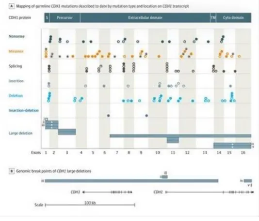
Figure 4 - Mapping of germline CDH1 mutations. Plots of the mutations according to exon location. A and B panels represent known break points for deletions in the transcript/protein view (A) and in the genome view (B). Roman numerals i to v show the large deletions with known genomic coordinates. Cyto stands for cytoplasmic; S, signal peptide; and TM, transmembrane
domain (20) …….……….………..…….……….19
Figure 5 - Pedigree of family A. In this pedigree information has been added regarding: presence of mutation in the germline; histological type of clinical disease presentation; risk reduction surgery; presence of foci in the risk reduction surgical specimen; age of onset (followed by y)..25 Figure 6 - Pedigree of family B. In this pedigree information has been added regarding: presence of mutation in the germline; histological type of clinical disease presentation; risk reduction surgery; presence of foci in the risk reduction surgical specimen; information about lack of surveillance; age of onset (followed by y). ………...…………26 Figure 7 - Pedigree of family C. In this pedigree information has been added regarding: presence of mutation in the germline; histological type of clinical disease presentation; risk reduction surgery; presence of foci in the risk reduction surgical specimen; age of onset (followed by y). Image in Annex I. Figure 8 is a close-up of this figure and contain the respective legend……...27 Figure 8 - Pedigree of family C. Close-up of the C.1 and C.2 branches, members used in the haplotyping study. In this pedigree information has been added regarding: presence of mutation in the germline; histological type of clinical disease presentation; risk reduction surgery; presence of foci in the risk reduction surgical specimen; information about lack of surveillance; age of onset
(followed by y). ……….…………28
Figure 9 - Pedigree of family D. In this pedigree information has been added regarding: presence of mutation in the germline; histological type of clinical disease presentation; risk reduction surgery; presence of foci in the risk reduction surgical specimen; age of onset (followed by y). Image in annexes: Annex II. ………..…………29 Figure 10 - Pedigree of family E. Information in the close-up of Figure 11. ………..……29 Figure 11 - Close-up of the family E studied branch. In this pedigree information has been added regarding: presence of mutation in the germline; histological type of clinical disease presentation; risk reduction surgery; presence of foci in the risk reduction surgical specimen; presence of other features related with HDGC; age of onset (followed by y). Image in annexes: Annex III………...30 Figure 12 – Chromosome 16 and 16q22.1 region. Depiction of the polymorphic markers analyzed and their location within this region. Black box: CDH1 gene from the transcription Start Site (TSS) to the end of the 3-UTR. Polymorphic markers and their distance to 5’-UTR highlighted in blue. Polymorphic markers and their distance from 3’-UTR highlighted in red. ………..32 Figure 13 – Frequency of DGC and LBC in the c.1901C>T CDH1 families..………..41 Figure 14- Colony PCR for 30 colonies from the BD1 sample. Electrophoresis in 2% agarose gel of the M13 PCR products. Small products highlighted with SP. No amplification occurred for colony 5. ………....………44
4
Figure 15 - Colony PCR for 50 colonies from the BD2 sample. Electrophoresis in a 2% agarose gel of the M13 PCR products. Small products highlighted with SP. Wells number 23 and 31 are the result of bacteria that incorporated the empty vector (without the cloned product)………….44 Figure 16- cDNA sequence of LP product. Sequencing of the colony 35 in sample BD2. c.1901C and c.2076C alleles highlited with an arrow. ………..….45 Figure 17 - cDNA sequence of SP product. Sequencing of the SP colony 43 in sample BD2. The same allele carries c.1901C and c.2076T, which have been highlighted with an arrow. ………46 Figure 18 – Snapshot result on the rs1801552 in sample BD2 for genomic DNA (left) and cDNA (right). Black peaks reflect the Cytosine (C) allele in the position c.2076 (rs1801552), while red peaks represent the Thymine (T) allele in the same position. At the RNA level, the c.2076T allele is less represented in the PBMCs carriers. ……….…47 Figure 19 – c.1901C>T CDH1 mutation. Representative scheme of the effect of the mutation in CDH1 mRNA and protein. The mutant c.1901T allele showed a deletion of 37 bp, from c.1900 to c.1937, with frameshift in the reading frame, leading to a premature stop codon. The c.1901T allele encodes a truncated form of CDH1 that lose the cytoplasmic domain, and not to a protein bearing a missense change. ……….……52
Tables Index
Table 1- TNM staging system for gastric cancer. Tumour (T), regional lymph nodes (N) metastases and distant metastases (M). (Adaptaded from AJCC, 2017 (13) ………. 11 Table 2 – List of dinucleotide markers in the vicinity of the CDH1 locus. Information regarding: Location of the marker, distance of each marker to the TSS (coordinate 16:68737225) or to the end of the 3-UTR (coordinate 16:68835548) of CDH1, Primers used and PCR product size. ……… 32 Table 3- PCR conditions used in the different experiments. ………...…... 33 Table 4 - Haplotypes from the 18 controls and from 4 c.1901C>T chromosomes found in each HDGC family. B, III-1 and D, IV-6 are the 2 combinations of descendent affected haplotypes..34 Table 5 - Primers of the different PCR reactions performed……….……37 Table 6 – Estimation of the c.1901C>T CDH1 mutation and it 95% CI. A and B are the two different analyses performed……… .…40 Table 7 – Overview of the c.1901C>T families studied. Frequency of DGC and LBC, number of risk reduction surgery, age, number of specimens with foci of carcinoma and number of carriers without surveillance………..……42 Table 8 – Bioinformatics prediction results… ………..………...………..……43
5
Abbreviations
AJCC American Joint Committee on Cancer
BD Blood donor
bp base pairs
cM Centimorgan
CRC Colorectal cancer
CDH1 cadherin 1 gene
DHS Decay of Haplotype Sharing
DHSMAP Decay of Haplotype Sharing MAPping
DGC Diffuse gastric cancer
EMT Epithelial to mesenchymal transition
ERAD Reticulum Endoplasmaticum Associated Degradation
FIGC Familial Intestinal gastric cancer
GC Gastric cancer
GAPPS Gastric adenocarcinoma and proximal polyposis of the stomach
HDGC Hereditary diffuse gastric cancer
IGC Intestinal gastric cancer
LP large product
LBC Lobular breast cancer
Mbp mega base pairs
MLPA Multiplex ligation dependent probe amplification
MRI Magnetic resonance imaging
NMD Nonsense-mediated mRNA decay
PBMCs Peripheral Blood Mononuclear Cells
PCR Polymerase Chain Reaction
PG Prophylatic gastrectomy
RT Room temperature
RRM Risk reduction mastectomy
RT-PCR Reverse Transcriptase Polymerase Chain Reaction
RTKs Receptor tyrosine kinases
SBE Single-base extension
SRCC Single ring cell carcinoma
SP small product
SDGC Sporadic diffuse gastric cancer
SGC Sporadic gastric cancer
TCGA The Cancer Genome Atlas
TMRCA The most recent common ancestor
TSG Tumour supressor gene
6
Resumo
Cancro Gástrico (GC) é o quinto cancro mais incidente e a terceira maior causa de morte relacionada com cancro a nível mundial, em ambos os sexos. O cancro gástrico difuso hereditário (HDGC) é o tipo mais comum de GC hereditário, correspondendo a <1% de todos os casos de GC. O HDGC é caracterizado por cancro gástrico difuso (DGC) multigeracional, de início precoce, que pode ser acompanhado por outras doenças, como o carcinoma lobular da mama (LBC). Mutações germinativas do gene CDH1 justificam 10-40% dos casos de HDGC. No Norte de Portugal existe um número elevado de famílias (não relacionadas) com HDGC e a mesma variante de mutação no CDH1: c.1901C> T (A634V). Este fato levanta a hipótese da existência de um um efeito fundador nesta região. O nosso objetivo é definir a idade desse efeito fundador, calcular a frequência do desenvolvimento de DGC e LBC nestes pacientes e perceber, ao nível do RNA, o efeito da mutação c.1901C> T no gene CDH1.
Para estimar a idade do efeito fundador, foram haplotipadas 4 famílias com HDGC para posterior aplicação do método Decay of Haplotype Sharing (DHS) desenvolvido por McPeek e Strahs. Para avaliar o efeito da mutação c.1901C>T no RNA, foi isolado e clonado RNA de 2 portadores (com e sem doença clínica) e estudámos o seu impacto funcional por estudos de desequilíbrio alélico usando o SnapShot.
De um total de 52 portadores da mutação, 8 (15.4%) desenvolveram DGC. Nas mulheres, de 26 portadoras, 2 (7.4%), com 49 anos, desenvolveram LBC. Dos 18 portadores que fizeram gastrectomia profilática (PG), 16 (88.9%) tinham focus de DGC. A única paciente que fez mastectomia profilática tinha focus de LBC. O RNA dos portadores da mutação c.1901C>T no
CDH1 apresentou uma deleção de 37bp a partir da posição c.1900 (exão 12) até ao início do
exão 13. A deleção cria um cryptic splice-site com alteração frameshift que leva à introdução de um codão stop e, consequentemente, a uma proteína truncada. Adicionalmente, verificámos que o alelo mutado estava menos representado que o alelo wild-type. Assim, propomos a
mudança de nomenclatura desta mutação para c.1901C>T; r.1900_1936del; p.Ala634ProfsTer7.
Estimamos que a idade da mutação c.1901C>T no gene CDH1 é de 490 anos, embora o CI tenha um intervalo muito abrangente (445 - 10 900 anos; 449,75 – 1 492 anos; 449,75 – 1
475,5 anos). Uma vez que a mutação c.1901C>T no CDH1 nos pacientes com HDGC é
frequente no Norte de Portugal devido a este efeito fundador, é provável que se propague na população. Por este motivo, é importante continuar a estudar esta mutação e a identificar mais famílias com esta síndrome.
Este estudo possibilitou a identificação do efeito fundador da mutação c.1901C>T no
gene CDH1no Norte de Portugal e a datação da origem deste event. Além disso, o estudo propõe
uma nova classificação para esta mutação patogénica.
Palavras-chave: Cancro gástrico difuso hereditário (HDGC); cancro gástrico difuso (DGC);
carcinoma lobular da mama (LBC); mutação fundadora c.1901C>T no gene CDH1; activaçao de cryptic
7
Abstract
Gastric Cancer (GC) is the fifth most incident cancer worldwide and the third leading cause of cancer-related death in the world for both sexes. The hereditary diffuse gastric cancer (HDGC) is the most common type of hereditary GC, accounting for <1% of all GC cases. HDGC is characterized by early-onset multigenerational DGC which might be accompanied by other diseases such as lobular breast cancer (LBC). Germline mutations of the CDH1 gene, which occur in 10-40% of HDGC cases, are thought to be the underlying cause of disease in a subset of patients. In Northern Portugal, there is an unusually high number of HDGC unrelated families with the c.1901C>T variant in CDH1 (A634V), which leads to the hypothesis that a founder effect
might have happened in this region. Our aim is to define the age of this founder effect, calculate the frequency of DGC and LBC development, and understand the effect of the c.1901C>T mutation on the CDH1 gene at the RNA level.
To estimate the age of the founder effect, we haplotyped 4 HDGC families and applied the Decay of Haplotype Sharing (DHS) method for fine-scale linkage disequilibrium mapping developed by McPeek and Strahs. To assess the effect of the c.1901C>T CDH1 mutation on the RNA we isolated and cloned RNA from 2 carriers (with and without clinical disease) and studied its functional impact by allelic imbalance studies using SnapShot.
In our cohort, from a total of 52 mutation carriers, 8 (15.4%) developed DGC. In women, from 26 carriers, 2 (7.4%), aged 49, developed LBC. Clinical data from these families indicated that from the 18 patients who underwent prophylactic gastrectomy (PG), 16 (88,9%) had already developed DGC foci. The only patient who underwent risk reduction mastectomy (RRM) had LBC foci. The RNA from c.1901C>T CDH1 mutation carriers showed a deletion of 37 bp from the c.1900 position (in exon 12) till the starting point of exon 13. This deletion creates a cryptic splice-site with a shift in the reading frame that leads to a premature stop codon and thus to a truncated protein. Furthermore, we saw that the mutated allele was less represented than the wild-type (wt)
in the patients studied. Accordingly, we propose a different nomenclature for the c.1901C>T;
r.1900_1936del; p.Ala634ProfsTer7.
According to the analysis done we calculated the c.1901C>T CDH1 mutation to be 490 years old, although the CI are very broad (445 - 10 900 years; 449,75 – 1 492 years; 449,75 – 1
475,5 years). Since the c.1901C>T CDH1 mutation in HDGC patients is highly prevalent in the
Northern Portugal due to the founder effect, it is likely that it is spread in the population. Therefore, it is of utmost importance to continue to study this mutation and identify additional families with this syndrome.
Taken together, this study allowed characterizing the clinical in phenotypes in c.1901C>T
mutation carriers, identifying the first CDH1-related founder effect in HDGC families from Portugal, date the origin of the founder event and gather evidence towards the reclassification of this pathogenic mutation.
Keywords: Hereditary Diffuse Gastric Cancer (HDGC); Diffuse Gastric Cancer (DGC); Lobular
Breast Cancer (LBC); CDH1 c.1901C>T founder mutation; cryptic splice-site activation; patients’
9
Introduction
Gastric Cancer
Epidemiology and general features
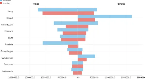
Gastric Cancer (GC) is the fifth most incident cancer with 951,000 new cases in 2012 and 723,000 deaths, making it the third leading cause of cancer-related death in the world for both sexes (1, 2). The reported 5-year survival of GC ranges between 68.1% (early disease stages) to 5,2% (advanced stages) depending if the carcinoma is localized or metastatic (3). GC is the 4th most common cancer in men and the 3th most mortal. On the other hand, in women, GC is the 4th most incident and the 4th cause of cancer related death (Figure 1) (2).
Figure 1- Gastric cancer: estimated incidence and mortality rates in both sexes, worldwide in 2012. (Adapted from Globocan 2012, WHO, IARC, 2012 (2))
Specifically in Portugal, 1834 men were diagnosed with GC, and 1387 died of this disease, whereas in women there were 1184 newly diagnosed cases and 898 reported deaths making GC the 4th and 3th cause of cancer associated deaths in men and women respectively, in 2012 (2, 4). Although Portugal has the highest rates of GC among Western European countries, its incidence has been steadily decreasing since 1970 likely due to the improvements in diagnosis and screening of the disease, as well as changes in dietary habits and hygiene conditions (4). The high mortality associated with GC is a result of late diagnosis and limited therapeutic options.
The majority of GCs are sporadic (approximately 90%), however roughly 10% (~100.000/year) of them occur in a familial setting (5). This means that the incidence of
10
a specific type of cancer(s) in a family is higher than what one would expected by chance. This familial clustering might be associated with shared genetic susceptibility, life-style and environmental factors (5). One to three percent of GC cases are the result of hereditary cancer predisposition syndromes (10.000 to 30.000/year), which is expected to arise as a result of a causal mutation in the germline of these patients. The three main GC-associated syndromes are gastric adenocarcinoma and proximal polyposis of the stomach (GAPPS), familial intestinal gastric cancer (FIGC) and hereditary diffuse gastric cancer (HDGC), the latter being the most common type of hereditary GC, accounting for <1% of all GC cases, where Diffuse Gastric Cancer (DGC) is the most prevalent cancer type, and/or Lobular Breast Cancer (LBC) the second (5, 6). Per year, it is predicted that ~10.000 patients diagnosed worldwide with DGC belong to HDGC families.
GC has been also identified, although with less prevalence, as part of the tumor spectrum of other syndromes, namely the Li-Fraumeni syndrome, Lynch syndrome, Peutz-Jeghers syndrome, hereditary breast and ovarian cancer, MUTYH-associated adenomatous polyposis, familial adenomatous polyposis, juvenile polyposis syndrome and PTEN hamartoma tumour syndrome (Cowden syndrome) (5, 7).
The present Master Thesis work will mainly focus on GC occurring in the context of HDGC.
Gastric Cancer histology and staging
There are two major subtypes of GC, the DGC and intestinal gastric cancer (IGC), which are distinct in epidemiology, morphology, and molecular features (8, 9).
The IGC usually develops under the sequence chronic atrophic gastritis, intestinal metaplasia, and dysplasia. This type is more frequent in males and older patients and is more often linked to environmental factors, as Helicobacter pylori infection (10, 11). DGC is more common in younger females and is believed to be more related to genetic predisposition (8, 12) .
Histologically, the IGC is characterized by recognizable glands that arise on an intestinal metaplasia’s background (8). In contrast, the DGC presents poor cohesiveness and round small cells, that infiltrate the gastric wall (8).
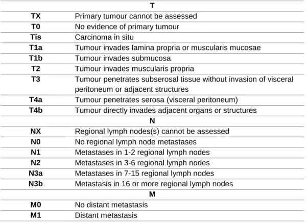
GC is staged following the TNM classification system. This system takes in consideration the extension of the tumor (T), the extent of lymph nodes affected (N), and the presence of distant metastasis (M). The TNM system is described in Table 1,
11
Table 2- TNM staging system for gastric cancer. Tumour (T), regional lymph nodes (N) metastases and distant metastases (M). (Adaptaded from AJCC, 2017 (13)).
T
TX Primary tumour cannot be assessed
T0 No evidence of primary tumour
Tis Carcinoma in situ
T1a Tumour invades lamina propria or muscularis mucosae
T1b Tumour invades submucosa
T2 Tumour invades muscularis propria
T3 Tumour penetrates subserosal tissue without invasion of visceral
peritoneum or adjacent structures
T4a Tumour penetrates serosa (visceral peritoneum)
T4b Tumour directly invades adjacent organs or structures
N
NX Regional lymph nodes(s) cannot be assessed
N0 No regional lymph node metastases
N1 Metastases in 1-2 regional lymph nodes
N2 Metastases in 3-6 regional lymph nodes
N3a Metastases in 7-15 regional lymph nodes
N3b Metastasis in 16 or more regional lymph nodes
M
M0 No distant metastasis
M1 Distant metastasis
Most of the patients are diagnosed with GC in an advanced stage due to the lack of symptoms. At an advanced stage, IGC is characterized by expansive growth and liver metastasis, whereas DGC disseminating spreads on the peritoneum and, also frequent, could metastasize to the bone (14).
This work will mainly focus on DGC, as this is the only histotype so far associated with the established GC-associated HDGC syndrome.
Hereditary Diffuse Gastric Cancer
HDGC is characterized by early-onset, multigenerational DGC which might be accompanied by other diseases such as LBC and less frequently colorectal cancer (CRC) (15). The onset of the disease is, on average, 38 years (range: 14-69 years old), but it is variable within and between families (16, 17). Around 10-40% of families affected by HDGC have a germline mutation in the E-cadherin gene (CDH1), depending on their geographical origin, leading to loss of function and reduced expression of the protein (5,
12
7), and therefore regarded as the most likely molecular basis for HDGC (17). In CDH1-mutant HDGC patients, E-cadherin expression is absent, reduced or aberrant in all precursor lesions, early and invasive GC. In normal adjacent mucosa, CDH1 expression should be normal (18). In CDH1 carriers mutation, many cancers occur before age 40 years (16, 17).
Given that a large proportion of HDGC families seem to fail a link to CDH1 coding mutations and deletions, and therefore remain genetically unexplained, other genes and also full exomes have been explored. Mutations in multiple other genes have been advanced as a potential genetic cause for HDGC in families from several ethnicities, such as in CTNNA1, BRCA2, PRSS1, ATM, PALB2, SDHB, STK11, MSR1, MSH2, ATR, NBN, and RECQL5, however at very low frequencies (19-21). From these, only germline mutations in CTNNA1 are believed to mimic the role of CDH1 as a cause of HDGC (22). For instance, biallelic MYD88 germline mutations were found in a single patient, being a
rare event that increased GC susceptibility. In another study, Gaston D. et al, associated germline mutations in MAP3K6 with familial GC, however, the high number of MAP3K6 variants found in controls shows that MAP3K6 variants are unlikely to be involved in high-penetrant GC predisposition (22-24).
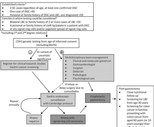
The identification of the inherited alteration among individuals with family history of GC is of the utmost importance for early diagnosis and proper disease management. A genetic test in the proband (person serving as the starting point for genetic study of a given family) pinpoints which mutation is causing the disease and could be transmitted to the offspring within the family. Since approximately 10-40% of HDGC families have been described with germline mutations in CDH1, in order to manage these patients and their families, clinicians should ask for a genetic screening on CDH1 gene (5, 7, 15, 20, 25). For a better assessment of these patients’ disease status, guidelines for genetic and clinical management of HDGC families were created in 1999 and later revised and updated in 2010 and 2015 (Figure 2) (7, 15, 26).
13
Figure 2 - Algorithm for testing criteria and management of HDGC patients. (Adapted from van der Post, RS. et al. 2015 (7)).
Patients who fulfill the criteria for the development of HDGC should be followed by a multidisciplinary team so that they can be informed about their condition and the impact the disease could have in their lives, as well as in their relatives. CDH1 pathogenic mutation carriers should undergo prophylactic gastrectomy (PG), the unique treatment option available in HDGC (7). Relatives of carriers who refuse to be tested, and families that fulfill the criteria for HDGC with no CDH1 mutations, should undergo yearly surveillance by endoscopic screening with random biopsy (7). Carriers that refuse or are not possible to be submitted to PG are also included in this group (7).
For women carrying CDH1 mutations, it is recommended to undergo yearly breast magnetic resonance imaging (MRI) and mammography from age 30 years onwards as they have a high lifetime risk of developing LBC. Risk reduction mastectomy (RRM) is also an option for these patients, that should be properly discussed in a multidisciplinary environment (7).
14
Penetrance
Mutations in CDH1 are the best known HDGC related-cause. The spectrum of the disease includes, mainly, DGC and LBC. To better understand the risk of carriers, penetrance estimation should be useful for genetic counseling. Pharoah, PD., et al. were the firsts to report the incidence of DGC and LBC in 11 HDGC kindred with CDH1 mutations, showing its incomplete and relatively high penetrance (27). The cumulative risk of gastric cancer by age 80 years was 67% in men (95% confidence interval [95% CI], 39-99) and 83% in women (95% CI, 58-99) (27). For women, the cumulative risk of breast cancer was 39% (95% CI, 12-84), while the combined risk of gastric cancer and breast cancer was 90% by age 80 years (27).
Kaurah, P., et al. estimated the cumulative risk of DGC and LBC in 4 families with the founder 2398delC mutation (28). By age 75 years, the cumulative risk of GC was estimated to be 40% (95% confidence interval [CI], 12%-91%) for males and 63% (95% CI, 19%-99%) for females (28). The cumulative risk for LBC for females by the age of 75 years was found to be 52% (95% CI, 29%-94%) (28).
Hansford, S. et al. studied the largest series of CDH1 mutant patients by grouping analysis of cohorts that included: i) previously published families; ii) newly identified families with mutation, and; iii) previously unpublished families. The study concluded that the cumulative risk of GC development for carriers by age 80 years is 70% for men (95% CI 59% to 80%) and 56% for women (95% CI 44% to 69%). Additionally, the risk of developing LBC for female is 42% (95%CI, 23%-68%) by 80 years (20).
Although being very high, the penetrance is different among the different studies. The studies exposed included different CDH1 mutations, revealing the importance of estimating DGC and LBC penetrance in each family/mutation context.
Macroscopy and Histology of HDGC
Cancers arising in patients who develop DGC in the context of HDGC are macroscopically indistinguishable from the sporadic diffuse gastric cancer (SDGC) patients. Both present features of linitis plastica, and can implicate all topographical regions of the stomach which makes it difficult to differentiate the two disease settings (29). In asymptomatic CDH1 mutation carriers submitted to prophylactic gastrectomy, the macroscopic features and mucosal thickness (when slicing) seem normal (30, 31). In some cases, upon standard white light endoscopy pale areas can be seen, which also
15
appears as white patches after fixation, a typical event in intramucosal diffuse carcinoma or single ring cell carcinoma (SRCC) (32).
Microscopically, DGC in advanced HDGC is poorly differentiated and can have undifferentiated or mixed subtypes with mucinous and occasionally tubular components. DGC in the HDGC context is characterized by the presence of individual and small foci, ranging from 0-1 mm to 10 mm, that extends to all topographical region of the stomach (29, 33). The two intraepithelial hallmark lesions regarded as precursors of invasive cancers in CDH1 mutation carriers are in-situ SRCC (signet ring carcinoma cells generally with hyperchromatic and depolarized nuclei, within the basal membrane) and
pagetoid spread of SRCC (structures with an inner layer composed of benign mucous
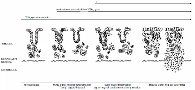
cells and an outer layer of SRCC) below the normal epithelium of gastric glands and/or foveolae (29). In advanced stages of the disease, SRCC invades most of the gastric wall, which becomes rigid (linitis plastica), without forming a distinct mass. Patients with HDGC normally have mild chronic gastritis, but rarely have Helicobacter pylori and intestinal metaplasia (29). Carneiro, F. et al., proposed a model for the early development DGC in CDH1 mutation carriers (Figure 3) which depicts the morphologic evolution of the disease. Inactivation of the second allele of CDH1 leads to tumour initiation, however the other molecular effectors which fuel tumour progression and drive the development of a full blown carcinoma are not yet defined (29).
Figure 3 - Proposed model for early development of diffuse gastric cancer in CDH1 mutation carriers. (Adapted from Carneiro, F. et al. 2004 (29)).
16
Surveillance and treatment options
The high penetrance of HDGC justifies PG as a treatment in both symptomatic and asymptomatic CDH1 mutation carriers. This approach is currently the unique option for preventing DGC in the context of HDGC. Although this is highly recommended for asymptomatic carriers of CDH1 mutation, not all patients are eligible for surgery, nor all are ready for this procedure (through patient choice or existence of physical or psychological comorbidity) due to the big impact it has on their lives (34). This treatment brings 100% morbidity to the patients as this surgery affect the quality of life due to short-term and long-short-term adverse events, reflected in long-short-term complications including rapid intestinal transit, dumping syndrome, diarrhea, eating habit alterations, weight loss and an increased risk for malabsorption, incidence of osteoporosis, osteomalacia, and malnutrition (26, 35-38). These comorbidities coupled with a 2% mortality rate associated with this procedure hampers patients’ decision and sometimes the clinical window in which clinicians can act may close (39). In these cases, patients are followed by regular endoscopic surveillance with biopsy sampling to early detect precursor lesions, in-situ SRCC and pagetoid spread of SRCC, both characteristic of early HDGC(32, 38).
Most of the tumors found in PG from asymptomatic CDH1 mutation carriers are intramucosal SRCC (29, 31, 33, 40). By definition these are invasive carcinomas, although they appear to remain indolent for a long time and are not accompanied by distant metastasis in this clinical setting (18). The majority of SRCC foci limited to the gastric mucosa (T1) in CDH1 carriers PG samples are <1 mm diameter and contain mitotically inactive SRCCs. Progression to advanced HDGC is associated with development of larger SRCC foci (>3 mm) containing increased numbers of poorly differentiated cancer cells, which are highly proliferative and invade the muscularis
mucosae (T1a) and muscularis propria (T2) (41).
According to Humar, B. et al, tissue dedifferentiation and c-Src kinase activation are crucial events required for the progression of early HDGC and SDGC. In this study, they showed, in 13 paraffin embedded tissues samples from CDH1 mutation carriers and 10 sporadic early DGC cases, c-Src kinase activation (overexpression) in small signet ring cells located in the upper neck region which increased with the depth of invasion, correlating it with tumour progression. In light with these findings, the authors also proposed that c-Src antagonists may be suitable for the treatment of early stage cancer (42).
Additionally, van der Post, RS. and Gullo, I. et al. showed that all early stage neoplasic lesions (Tis and T1) in HDGCs patients had “indolent” morphologic features. Also, these lesions were negative for p53 and Ki-67 protein expression which greatly
17
contrasted with a more advanced setting of the disease (pT>1) where lesions showed an “aggressive” phenotype due to high Ki-67 labelling index and p53 overexpression in pleomorphic tumor cells (18).
Lee, HE., et al. showed that most HDGCs from PGs have two groups of cells, corresponding to a poorly differentiated and well-differentiated phenotype (43). The poorly differentiated cells are strongly positive for p16 by immunohistochemistry, lack mucin in pleomorphic tumor cells and present large atypical nuclei and dense pink cytoplasm. These are occasionally accompanied by subtle desmoplastic reaction as well. On the other hand, in the well-differentiated phenotype cells include the large SRCCs (mucin-rich) in the superficial mucosa and small SRCCs (mucin-poor) around the neck region. Both of these cells are negative for p16. Still in this study, 15/17 advanced SDGCs (control samples) were positive for p16, and given the morphological similarities between the poorly-differentiated cells in HDGC and the tumor cells in SDGC with more aggressive behavior, an aberrant p16 expression may predict disease progression of early HDGC in asymptomatic patients (43).
These are crucial findings that underlie the importance in understanding the mechanisms and the definition of pathognomonic features of this disease that can help understand how does the tumor progresses in order to aid clinical decision-making.
Mi, EZ., et al. assessed the value of endoscopic surveillance for HDGC patients in patients with CDH1 germline mutations, in which gastrectomy was delayed and in patients with no pathogenic variants in CDH1 (41). The yield of SRCC foci on endoscopy and the change in yield with time on the endoscopic surveillance context, were the evaluated parameters in this study. The sensitivity of the detection of SRCC by random and targeted biopsy sampling were 75% and 41.7%, respectively (41). This study showed better results than others (9-16%), in a larger cohort, which can be explained by the use of an optimized and standardized endoscopic protocol performed by an experienced team of endoscopists (41). The success of endoscopic screening for detection of SRCC in HDGC patients without CDH1 mutations is low, (9.7% ), which could be explained by the fact that half of the individuals in these families are probably wild-type and therefore, not at risk, or the mechanism/pathway that leads to DGC in these patients could be different, thus in this setting, SRCC may not be the precursor lesions (7, 41).
Patients with symptomatic invasive HDGC are usually treated with the same ineffective chemotherapies as patients with sporadic gastric cancer (SGC) (5). However, the therapeutic response of the HDGC patients is lower when compared to patients with
18
SDGC or DGC with non-pathogenic CDH1 mutations (44, 45). Only 10% of these patients have potentially curable disease, and the 5-year survival rate still does not exceed 30% (7).
CDH1 – gene and protein overview
CDH1 is located on 16q22.1 chromosome’s region and has 16 exons that encode
for the CDH1 or E-cadherin, a 120 kD transmembrane glycoprotein. CDH1 has: i) five extracellular cadherin repeats, encoded by exons 4 to 13, which mediates homophilic cellular interaction, ii) a transmembrane region encoded by exons 13 and 14, and; iii) a highly conserved cytoplasmic tail encoded by exons 14 and 16 that bind to the actin-myosin cytoskeleton by association with various catenins (β, p120- and α-catenins), mediating structural integrity, cellular polarity, morphogenesis and epithelium function. CDH1 is localized at the junctions of epithelial cells and functions as a mediator of hemophilic calcium-dependent cell-cell adhesion (46-48).
In the context of cancer, E-cadherin has tumor suppressor activity, and it is crucial to prevent cell invasion (49-52). Somatic mutations in CDH1 lead to a loss of function of E-cadherin by reducing the expression of the functional protein, contributing to cancer progression by increasing proliferation, invasion, and/or metastasis, since there is a loss of cell-cell adhesion in the cells of the tissue (46, 53, 54). CDH1 is commonly somatically mutated in many cancer types including gastric, breast, colorectal, thyroid and ovarian cancer (46, 55).
CDH1 as a cause of HDGC
Since 1998, when Guilford, P., et al. reported CDH1 mutations in Maori families and correlated them with HDGC, others have reported similar findings, confirming the autosomal-dominant pattern of inheritance associated with germline mutations of the
CDH1 gene, which occurs in approximately 10-40% of HDGC cases (7, 17, 28, 56-59).
As a tumour suppressor gene (TSG), CDH1 needs biallelic inactivation (of the remaining functional allele) by a second molecular hit, to cause aberrant E-cadherin expression and development of HDGC in germline mutation carriers. This is usually achieved by hypermethylation of the promoter in the normal allele. A second mutation or deletion may also arise as a second hit although not so frequently (40, 60-63). Strikingly, Oliveira, C. et al., showed that the same patient can present distinct second hits in distinct neoplasic foci, and that the same neoplastic lesion can present different second-hit
19
mechanisms (63). In contrast to SDGC in which there are some known hot spots in
CDH1, in HDGC the genetic alterations are not site-specific and affect the entire coding
sequence and all the functional domains of the protein (Figure 4) (64).
There are 60-90% of HDGC families that lack CDH1 germline mutations. Surprisingly, in 70% of these CDH1 mutation negative HDGC families, allele-specific expression analysis revealed a germline phenotype of monoallelic CDH1 downregulation (65, 66). More than 90% of these patients showed loss or low expression of E-cadherin in their tumors (45, 63). This likely indicates alternative genetic mechanism targeting non-exonic regions of the CDH1 gene.
Figure 4 – Mapping of germline CDH1 mutations. Plots of the mutations according to exon location. A and B panels represent known break points for deletions in the transcript/protein view (A) and in the genome view (B). Roman numerals i to v show the large deletions with known genomic coordinates. Cyto stands for cytoplasmic; S, signal peptide; and TM, transmembrane domain. (Adapted from Hansford, S. et al. 2015 (20))
Up until now, more than 155 different CDH1 mutations were identified, of which 126 are pathogenic and 29 are unclassified variants (20). Eighty percent of CDH1 mutations are truncating, leading to the production of premature stop codons and therefore to the loss of E-cadherin expression and function (20). Five percent of these mutations are large deletions (from 357 bp to 275 kb in length), and this justifies the necessity to add multiplex ligation dependent probe amplification (MLPA) to the molecular diagnosis in HDGC families who test negative for CDH1 point mutations (7,
20
17, 25, 67). The genotype–phenotype correlation is straightforward for truncating mutations, but the same is not true for the approximately 20% of missense mutations described to date (5, 68).
Functional significance of missense mutations: facts and tools
An additional layer of complexity exists when dealing with missense mutations, as some of these mutations can trigger or activate cryptic splice-sites (5), which are dormant splice-sites that when activated through mutations, are used extremely efficiently, resulting in a wide range of genetic diseases (69, 70).
Missense mutations are expected to result in full-length E-cadherin molecules with a single amino acid substitution. In some instances, these single substitutions are sufficient to impair protein function to an extent that it becomes disease causative. So far, 18 missense mutations have been classified as potentially pathogenic, based on published data (5).
The pathogenicity of missense variants should take into account their recurrence in unrelated HDGC families and the allelic frequency in healthy controls, as well as co-segregation within families (56, 71). However, it is difficult to predict the functional impact of missense variants because of the low number of affected individuals and small pedigrees that prevent segregation analysis, thus representing a major drawback in genetic counselling and clinical surveillance (5, 67, 71). It is therefore mandatory to perform additional studies to assess if and how the variant could affect the expression and function of E-cadherin, as well as related signaling and cellular mechanisms (5, 7, 72).
Kaurah P. et al, reported the effect of both 715G>A (G239R) and 2195G>A (R732Q) CDH1 missense mutations in RNA isolated from white blood cells and gastric tissue, respectively. They showed that the 715G>A (G239R) mutation caused deletion of the first 29 bp from exon 6 and the 2195G>A (R732Q) mutation resulted in a deletion of 32 bp at the start of exon 14 (28). Through minigene analysis, the authors showed that the 715G>A (G239R) mutation creates a preferential splice-site in exon 6 and that the 2195G>A (R732Q) mutation activates a cryptic acceptor splice-site in exon 14 (28).
The c.1901C>T (A634V) missense mutation was also studied in colon cancer cell lines (73). The RNA from these cells had a deletion of the last 37 nucleotides of exon 12 and
subsequent frameshift (73). These results reflect the importance of studying missense
mutations since it may improve classification of a mutation towards pathogenicity of non-pathogenicity. Additionally, the studies using RNA from patients, in the diagnostic field, are few probably due to the lack of implemented routine techniques. Therefore, the use
21
of in silico tools to predict the effect of variants in cryptic splicing is likely the most widely used tool for this purpose.
The aforementioned limitations when studying HDGC families and human gastric samples, in the clinical and diagnostic context, led to the development of in-silico and in
vitro approaches to predict the pathogenicity of CDH1 missense mutations (68, 74).
The in silico approach use algorithms that take into account the degree of conservation of a particular nucleotide among species, the location and context within the protein sequence, the biochemical properties of the amino acid substitution, the presumed impact of the variant in protein native-state, and the possible effect in splice-sites (68, 74). Because different bioinformatics tools are designed under different algorithms and might give different outputs/results, it is recommend to use multiple software tools (68, 75).
Functional biochemical comparison between wild-type CDH1 and pathogenic mutated cell lines (with missense variants) showed that the latter had disruption of cell adhesion, incorrect binding of cadherin to adhesion-complex regulators, damage of E-cadherin stability at the plasma membrane, and induction of cell migration or invasion (56, 74, 76).
Melo, S., et al, established a pipeline that correlate familial data with in silico studies, expression analysis, and functional characterization of CDH1 missense mutants
in vitro and in vivo in order to classify germline CDH1 missense variants as neutral or
deleterious (pathogenic variants). However, the pipeline is not sufficient to ensure a confident result in 17% of the missense variants (68).
Discriminating between pure missense mutations and single nucleotide changes inducing cryptic splicing is very important, as it will allow a better classification of the variant and its potential deleterious effect, however none of the instruments developed so far are perfect.
If a germline missense CDH1 variant has been classified as deleterious, the carriers should enter a surveillance program and clinicians should discuss the therapeutic options, similarly to carriers of truncating CDH1 mutations. When it is not possible to access the pathogenicity of a variant, carriers should be closely followed and submitted to intensive endoscopic surveillance. The relevance of these mutations and their classification is crucial because in some missense variant carriers, microscopic foci of invasive cancer were detected in PG specimens (62).
22 Recurrence of CDH1 germline mutations in HDGC families
In the HDGC context, there are no mutational hot spots in CDH1 and the variations affect the entire coding sequence (20). However, approximately one third of CDH1 pathogenic mutations found in HDGC, are present in apparently unrelated families (5), which may be due to both independent mutational events or common ancestry (28). Kaurah, P. et al. were the first to report recurrence of 8 mutations in different families (28). To understand if these mutations occurred in different families due to independent mutational events or founder effects, authors haplotyped 5 of these mutations in different families. Four of which (1901C>T, 1137G>A, 2064-2065delTG, and 2398delC) were found to be associated with similar haplotypes in more than one HDGC family. Four families within a genetically homogeneous, ethnically or geographically defined population were shown to harbour the 2398delC mutation, therefore it is a likely founder mutation. The 1137G>A and the 1901C>T mutations occurred on common haplotypes in 2 families, but in a third family they showed different haplotypes (independent event). This data has implications on the genetic testing and counseling of families with HDGC, since the genetic test could be directed to specific mutations, in areas where founder effects where previous detected. Also, knowing about the recurrence of these events, doctors could easily order the screening of a specific mutation when a new case appear in a potentially relevant population.
From the 3 families with the pathogenic missense variant c.1901C>T [p.A634V]
studied by Kaurah, P. et al, the two who shared common haplotypes were Portuguese. These families shared 6 of the 7 polymorphic markers studied, suggesting that they could
share a common ancestor. This mutation is expected to interfere with E-cadherin
function due to a change in a single amminoacid. However, this alteration may also lead to the disruption of the full length CDH1 protein, due to the induction of cryptic splicing in exon 12 of the gene sequence, as demonstrated in a colorectal cancer cell line (28, 56, 58, 73).
In Northern Portugal, there is an unusually high number of HDGC unrelated families with the c.1901C>T variant in CDH1, which leads to the hypothesis of a founder effect that happened in this region (77). By definition, a founder effect is a phenomenon that leads to the colonization by a different genetic group. In this case, this founder effect may be due to a founder pathogenic mutation leading the fixation of the mutation (78).
To address this question, Ana Aguiar, in the scope of her Master Thesis, haplotyped 4 unrelated families from this region and reported that they shared approximately 2,7Mb of genetic material. In that work, it was shown that this could be a founder mutation in this geographic area, however, the length of the shared haplotype was not disclosed,
23
preventing the dating of the causing ancestral allele, and the formal proof of a common ancestry.
Understanding the etiology and penetrance of this mutation is of the utmost importance for the clinical management of both the patients and their relatives in order to also determine whether unaffected carrier relatives are in risk for DGC and LBC. The CDH1 genetic testing presents itself as a valuable assessment of risk, however given the incomplete penetrance of this disease and the highly invasive prophylactic options (total PG or surveillance through gastroscopy with multiple random biopsies, which showed poor test sensitivity when performed by non-expert professionals) make this a challenge when considering therapeutic options, as a non-neglectable proportion of carriers never develop clinical disease (20).
24
Rational and Aims
In this work, we aim to characterize and trace the origins of the founder effect in HDGC patients with c.1901C>T CDH1 mutation from Northern Portugal, and collect all data for future penetrance analysis of the disease in families with this mutation. Although previous studies with cancer cell lines showed that this mutation has an impact on splicing, in has never been proved that this was the case in the germline of HDGC patients, therefore this mutation remains classified as a missense mutation (A634V). To formally demonstrate whether the c.1901C>T CDH1 mutation is a missense, a splice-site or a mixed mutation, we will analyze RNA from carriers.
There are still many challenges regarding the proper clinical management of families with HDGC, for instance to whom prophylatic gastrectomy should be directed or even how would one predict which individuals will develop disease. This would allow a more accurate diagnosis and management in this and other HDGC families. Moreover, understanding the effect of this specific mutation at the RNA level could help defining new strategies to prevent disease development and even think about new treatment options.
In order to understand better the molecular and clinical impact of this mutation in HDGC patients and families, we designed the following aims:
- Date the origin of this founder effect by identifying point of recombination in 16q22.1 chromosome’s region in different HDGC patients, thus allowing the comparison between different families from Northern Portugal, through haplotyping of different polymorphic markers.
- Characterize and estimate the age of the c.1901C>T CDH1 mutation in the population to identify the point in time when these families started to share the common ancestor.
- Determine the frequency of DGC and LBC in carriers with this variant based on the clinical information from the patients.
- Understand the functional impact of the c.1901C>T mutation by analyzing RNA expression levels in c.1901C>T CDH1 mutation carriers with and without clinical disease.
25
Methodology
Samples
Twenty-seven DNA samples and clinical information of all HDGC probands and their relatives were provided by Centro Hospitalar São João, Porto.
Blood was collected from 2 selected patients for RNA studies. The 2 donors (BD1, BD2), were selected based on the following criteria: one (BD1) younger than 20 years with foci development assessed through PG and another (BD2) carrier, with more than 35 years, from a different family, with LBC, besides SRCC foci found in the PG. BD1 and BD2 belongs to the family A and D, respectively.
All of the patients studied are currently enrolled in genetic counselling and follow-up consultation/treatment at the Centro Hospitalar São João, and signed informed consents to be part of the study. This project had the approval from the Centro Hospitalar
São João Ethics Committee (307-17) (Annex VII).
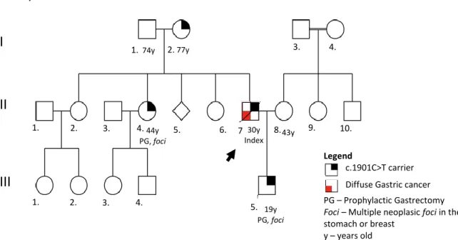
Family A: The index case is a patient with DGC diagnosed at 30 years of age (II-7), and died at 31. His son (III - 5; BD1) was tested when he was 19 years old for CDH1 mutations and is carrier of c.1901C>T CDH1 mutation. At 19 years old, he was submitted to PG. In this family, another two members were positive for the mutation, the index’s mother (I-2) and the index’s sister (II-4), the latter also underwent PG (Figure 5).
Figure 5 - Pedigree of family A. In this pedigree information has been added regarding: presence of mutation in the germline; histological type of clinical disease presentation; risk reduction surgery; presence of foci in the risk reduction surgical specimen; age of onset (followed by y).
77y 74y 30y Index 19y PG, foci 43y 44y PG, foci
I
II
III
1. 2. 3. 4. 1. 2. 3. 4. 5. 6. 7 . 8. 9. 10. 1. 2. 3. 4. 5.Family A
Legend c.1901C>T carrierDiffuse Gastric cancer PG – Prophylactic Gastrectomy
Foci – Multiple neoplasic foci in the
stomach or breast y – years old
26
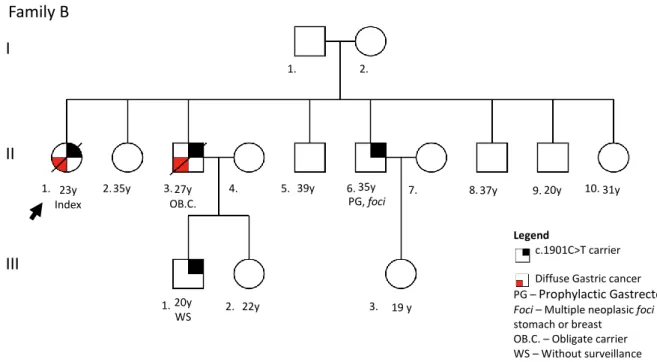
Family B: The index case (II-1), female, was diagnosed with DGC at 23 years old and died in the same year. Six of her 7 brothers were tested for this CDH1 mutation, but only one was positive (II-6). This brother underwent PG at the age of 35 and his stomach already presented multiple foci of diffuse carcinoma. Currently he is disease free. The brother who did not perform the genetic test (II-3) died at 27 years with DGC. His 2 children were tested and one of them (III-1) is c.1901C>T carrier, but refuses surveillance. From the 10 individuals tested for CDH1 mutation in this family, 3 tested positive. Of these 1 deceased (the index case), and 1 underwent PG. One extra individual (II-3) is an obligate carrier since his son (III-1) carries the c.1901C>T. (Figure 6).
Figure 6 - Pedigree of family B. In this pedigree information has been added regarding: presence of mutation in the germline; histological type of clinical disease presentation; risk reduction surgery; presence of foci in the risk reduction surgical specimen; information about lack of surveillance; age of onset (followed by y).
I
II
III
1. 2. 1. 2. 3. 4. 5. 6. 7. 8. 9. 10. 1. 2. 3. Legend c.1901C>T carrier Diffuse Gastric cancer PG – Prophylactic Gastrectomy Foci – Multiple neoplasic foci in the stomach or breastOB.C. – Obligate carrier WS – Without surveillance y – years old 27y OB.C. 20y WS 35y PG, foci 19 y 23y Index
Family B
22y 39y 37y27
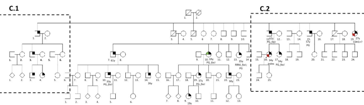
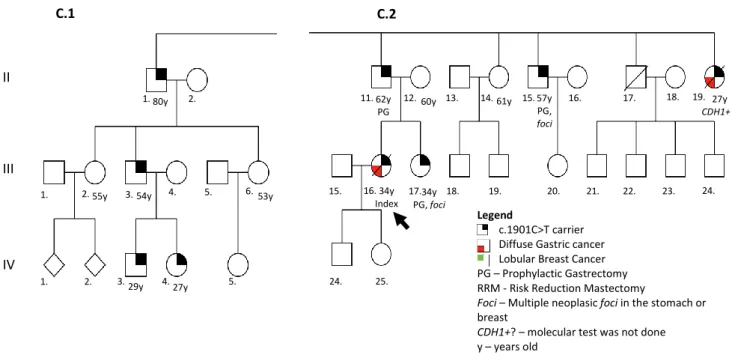
Family C: The index case is a female (III-16) who was diagnosed with DGC at the age of 34 and died at 35. As her father (II-11) was positive for the c.1901C>T mutation, this family was also studied. From the 33 members studied, 16 were proven to be carriers and from those, 1 was identified with disease at the initial steps of surveillance: a female cousin who developed LBC at 59 (III-10). She was submitted to a bilateral mastectomy with curative approach on the affected breast and as risk reduction approach on the healthy breast, as well as PG. Currently, at the age of 64, she is disease free. The II-19 individual, index’s aunt, died at 27 years with DGC, but she was not tested. The remaining carriers are under surveillance with regular endoscopy and multiple biopsies (for both genders) and also breast MRI (for females). In this family, 1 of the 16 proven carriers are deceased (the index), 1 individual deceased with DGC but lacking information regarding the CDH1 mutation status, 7 performed PG and of these, 1 female performed also bilateral RRM. The remaining carriers are under surveillance program (Figure 7 and 8).
Figure 7 - Pedigree of family C. In this pedigree information has been added regarding: presence of mutation in the germline; histological type of clinical disease presentation; risk reduction surgery; presence of foci in the risk reduction surgical specimen; age of onset (followed by y). Image in Annex I. Figure 8 is a close-up of this figure and contain the respective legend. I II III IV V 1. 2. 1. 2. 3. 4. 5. 6 7. 8. 9. 10. . 11. 12. . 13.. 14. 15. 16. 17. 18. 19. . 1. 2. 3. 4. 5. 6. 7. 8. 9. 10. 11. 12. 13. 14 . 15. 16. 17. 18. 19. 20. 21. 22. 23. 24. 1. 2. 3. 4. 5. 6. 7. 8. 9. 10. 11. 12. 13. 14. 15. 16.. 17.. 18. 19. 20. 21. 22. 23. 24. 25. 1. 2. 3. 4. 5. 6. 61y 33y 26y .59y PG, foci 37y PG, foci 19y 30y 7. 8. 9. 10. 11. 12. 13. 35y RRM, foci; PG
Family C
C.1 C.2 PG, foci 27y CDH1+? 62y PG, foci 34y PG, foci 34y Index 57y PG28
Figure 8 - Pedigree of family C. Close-up of the C.1 and C.2 branches, members used in the haplotyping study. In this pedigree information has been added regarding: presence of mutation in the germline; histological type of clinical disease presentation; risk reduction surgery; presence of foci in the risk reduction surgical specimen; information about lack of surveillance; age of onset (followed by y).
Family D: The index case (III-8, BD2) is a female with 53 years that developed LBC at the age of 49, performed genetic test at the age of 50 and underwent curative mastectomy to the affected breast and RRM to the healthy one. Because she is a carrier, she also underwent PG. The index’s sister (III-2) also developed LBC at the initial steps of surveillance and her brother (III-15) was diagnosed and died with DGC when he was 33 years old (not confirmed to be carrier). Overall, from the 21 family members tested, 12 were positive: 1 is deceased, 2 underwent PG and unilateral RRM since, they were both diagnosed with LBC on the other breast and 9 are in the surveillance program. Two other family members deceased with DGC but we don’t have information about CDH1 status (Figure 9). 1. 2. 1. 2. 3. 4. 5. 6. 1. 2. 3. 4. 5. II III IV 80y 54y 29y 27y 11. 12. 13. 14. 15. 16. 17. 15. 16. 17. 18. 19. 20. 21. 22. 23. 24. 24. 25. 62y PG 34y Index 34y PG, foci 57y PG, foci 27y CDH1+ ? 18. 19. C.1 C.2 61y 60y 53y 55y Legend c.1901C>T carrier Diffuse Gastric cancer Lobular Breast Cancer PG – Prophylactic Gastrectomy RRM - Risk Reduction Mastectomy
Foci – Multiple neoplasic foci in the stomach or breast
CDH1+? – molecular test was not done y – years old
29
Figure 9 - Pedigree of family D. In this pedigree information has been added regarding: presence of mutation in the germline; histological type of clinical disease presentation; risk reduction surgery; presence of foci in the risk reduction surgical specimen; age of onset (followed by y). Image in annexes: Annex II.
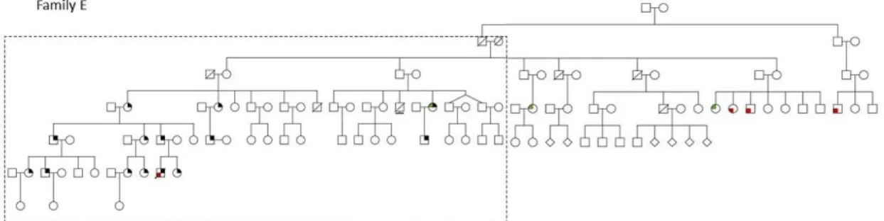
Family E: the index case is a male patient who died at the age of 18 years with DGC (VI-10). Sixteen of the 34 relatives who performed genetic testing were positive for c.1901C>T. The individual IV-17 developed LBC at 49 years of age. Six of them underwent PG. One of them was the proband’s sister who was tested at the age of 13 and underwent PG at the age of 14. Her pathology report revealed multiple foci of diffuse carcinoma. The individual V-18 has cleft lip and cleft palate (Figure 10 and 11).
Figure 10 - Pedigree of family E. Information in the close-up of Figure 11.
I II III IV 1. 2. 3. 4. 5. 6. 7. 8. 9. 10. 11. 12. 13. 14. 15. 16. 17. 18. 19. 20. 21. 22. 23. 24. 25. 26. 27. 28. 1. 2. 3. 4. 5. 6. 7. 8. 9. 10. 11. 1. 2. 3. 4. 5. 6. 7. 8. 9 . 10. 1. 2. 61y PG, foci 50y PG, foci; Index 29y 48y 23y 21y 33y CDH1+? 39y 19y 45y Family D 41y 55y 30y CDH1+? Legend c.1901C>T carrier Diffuse Gastric cancer Lobular Breast Cancer PG – Prophylactic Gastrectomy RRM - Risk Reduction Mastectomy
Foci – Multiple neoplasic foci in the stomach or breast
CDH1+? – molecular test was not done y – years old
30
Figure 11 - Close-up of the family E studied branch. In this pedigree information has been added regarding: presence of mutation in the germline; histological type of clinical disease presentation; risk reduction surgery; presence of foci in the risk reduction surgical specimen; presence of other features related with HDGC; age of onset (followed by y). Image in annexes: Annex III.
III IV V VI VII 1. 2. 3. 41y PG, foci 38y PG, foci 18y Index 21y 19y 22y 16y
PG, foci 13y PG, foci 24y 58y PG, foci 59y 83y WS 49y 23y CL, CP 80y WS 42y PG, foci Family E 36y 1. 2. 3. 4. 5. 6. 7. 8. 9. 10. 11. 1. 2. 3. 4. 5. 6. 7. 8. 9. 10. 11. 12. 13. 14. 15. 16. 17. 18 19. 20 21. 1. 2. 3. 4. . 1. 2. 3. 4. 5. 6. 7. 8 9. 10 . 11. 12. 13. 14.. 15. 16. 17. 18.. 19. 20. 21. 22. Legend c.1901C>T carrier Diffuse Gastric cancer Lobular Breast Cancer PG – prophylactic Gastrectomy RRM - Risk Reduction Mastectomy
Foci – Multiple neoplasic foci in the stomach or breast
CDH1+? – molecular test was not done CL,CP – Cleft Lift, Cleft Palate
y – years old
31
Methods
Identification of the recombination point in 16q22.1 chromosome’s region: haplotyping
of different polymorphic markers.
Twenty-seven DNA samples provided by Centro Hospitalar São João, Porto were used to amplify, by Polymerase Chain Reaction (PCR), the polymorphic markers (upstream and downstream of CDH1) described in Table 2 and Figure 12. To this purpose, we used the Qiagen® Multiplex PCR Kit (Qiagen, Hilden) and the primers described in Table 2, under the conditions described in Table 3 on the T100™ Thermal Cycler (Bio-Rad, Foster City, CA, USA). The PCR for the markers D16S318, D16S3025, rs16260, rs1801552, D16S496, D16S496, D16S3067, D16S3095 were previously performed in the scope of Ana Aguiar’s Master Thesis. For each PCR reaction negative controls were also included. PCR products were then run on a 2% agarose gel for confirmation of the amplified sequences.
Regarding the microsatellites analyses, 1 µL PCR products were mixed with 9,5 µL Hi-Di™Formamide (Applied Biosystems, Foster City, CA, USA) and 0,5 µL of molecular size standard dye GeneScan™ 120 LIZ™ or GeneScan™ 500 ROX™ (Applied Biosystems, Foster City, CA, USA) depending on the molecular size of the amplified fragment. The mixture was run in BI PRISM® 3130xl Genetic Analyzer (Applied Biosystems, Foster City, CA, USA) and Peak ScannerTM software (ThermoFisher, Massachusetts, EUA) was used to the bioinformatic analysis of the results.
For SNPs analysis the PCR products were purified by FastAP™ Thermosensitive Alkaline Phosphatase and Exonuclease I (Thermo Scientific, Wilmington), following manufacturer’s instructions, and then sequenced by Sanger sequencing approach. For this we used 2.5x BigDye® Terminator Cycle v3.1 (Applied Biosystems, Foster City, CA, USA), 1x BigDye® Terminator Buffer and 10 µM primer (Table 2), to a final volume of 5μl. The reactions were submitted to an initial denaturation step of 95ºC for 2 minutes, followed by 35 cycles of 20 seconds denaturation at 95ºC, 20 seconds of annealing at 55ºC and an extension of 60ºC for 10 minutes. The final extension was done at 60ºC for 10 minutes. The reactions were done in a T100™ Thermal Cycler (Bio-Rad, Foster City, CA, USA). Sequencing products were purified with 7% Sephadex® G-50 Fine (GE Healthcare, Uppsala) through a 4 minutes centrifugation at 3800 rpm (Eppendorf 5810R, Hamburg, Germany). After adding 10 µl of Hi-Di™Formamide (Applied Biosystems, Foster City, CA, USA), the purified products were run in ABI PRISM® 3130xl Genetic Analyzer (Applied Biosystems, Foster City) and the analysis was done using Mutation Surveyor® software (SoftGenetics, State College, PA, USA).