S
TUDY OF THE
Legionella-
DEPENDENT INDUCTION OF
APOPTOSIS IN THE NATURAL HOST
Acanthamoeba
castellanii
J
OANAC
ATARINAP
ENAD
ANTASD
ISSERTATION SUBMITTED FOR THE DEGREE OFM
ASTER OFS
CIENCE INM
EDICALM
ICROBIOLOGYS
TUDY OF THELegionella-
DEPENDENT INDUCTION OF APOPTOSIS IN THE NATURAL HOSTAcanthamoeba castellanii
Joana Catarina Pena Dantas
Dissertation submitted for the degree of Master of Science in Medical
Microbiology
Advisor: Professor Maria Teresa Marques MD, PhD
Co-advisors: Maria de Jesus Chasqueira MSc
Professor Guadalupe Cabral PhD
NOVA Medical School | Faculdade de Ciências Médicas (NMS|FCM)
J. Dantas i
Bibliographic Elements Resulting From This Dissertation
Poster
Dantas J., Chasqueira M.J., Rodrigues L., Cabral M.G., Marques M.T. Estudo do processo de morte celular induzido por Legionella pneumophila em Acanthamoeba castellanii. 14º Encontro Nacional de Actualização em Infecciologia. October 2015. Porto, Portugal (Appendix 1).
J. Dantas ii
Dedication
To my grandmother, Avó Vina, the most loving and caring woman I have ever known. I will always miss the unconditional love I felt every time I looked into your eyes.
J. Dantas iii
Acknowledgements
Though only my name appears on the cover of this dissertation, several people have contributed to its production. I owe my sincere gratitude:
To my advisor, Professor Maria Teresa Marques, whose expertise, understanding and patience were crucial for the completion of this dissertation.
To my co-advisor, Master Maria de Jesus Chasqueira, for all her encouragement, insightful comments and motivation; for teaching me that an investigation project can be made of sadness before it reaches happiness.
To my co-advisor, Professor Guadalupe Cabral, who was always available to share her knowledge with me and to sort out several details of my work, including countless revisions that were fundamental for the scientific accuracy of this dissertation.
To everyone at the “Infection: Etiology, Pathogenesis and Therapeutic Bases” unit, mainly to Lúcia Rodrigues for all her encouragement and practical advice.
To the members of the Flow Cytometry Lab at Instituto Gulbenkian de Ciência, who so kindly received me when I had my major setback during this journey. Particularly, I would like to acknowledge Master Cláudia Bispo for sharing her knowledge with me and for the many valuable conversations that helped me understand my research area better.
J. Dantas iv
To my family: my father Jorge, my mother Maria and my brother David, for their constant love and support. Each one of you, in your own way, is essential to me and I know that I would never be able to come this far in my life without you guys. You inspire me, every day, with your resilience dad, your self-confidence mum and your pure kindness little brother, to be a better person. I will always love you, with all my heart.
To Aquiles, my canine buddy, for bringing joy to my life, every day, for ten years now.
To my girls, Cate and Marilu, for being the best friends I could have ever asked for in this graduate experience. I will forever cherish the memories we made together for the past two years. “Este é para a vida”.
To my lab buddies, Tita and Fábio, for all their support when I most needed it and all the good times we spent together. For all the laughs, the dinners, the ice creams, the YouTube videos... It really was a pleasure.
To all my friends that have been patiently waiting for me to finish this demanding step in my life. I knew I could count on you, no matter what.
Last, but not least, to my partner in so many ways, Francisco Esteves. I know we do not say “thank you” to each other but… Thank you so much my love. For helping me find my strength when I am not able to, for your amazing hugs, for our laughs, for all the singing, for making me smile so easily, for all the dreams we share, for knowing me so well by now, for the simple gestures you know I treasure… For making me fall in love with you every day.
J. Dantas v
Abstract
Legionella pneumophila, a Gram-negative bacillus first identified in 1977, was the etiologic agent responsible for an outbreak of pneumonia that occurred in the American Legion Convention in 1976, Philadelphia. Although several other species of the genus Legionella were subsequently identified, this species is the most frequent cause of two distinct clinical syndromes: Legionnaires’ disease and Pontiac fever.
The life cycle of Legionella pneumophila is divided in two distinct phases: a replicative phase followed by an infectious or transmissive phase. In the replicative phase the bacterium shows a nonmotile stage with a low or nonexistent toxicity. In the infectious phase the bacterium develops a flagellum becoming motile and highly toxic.
The main hosts of Legionella pneumophila are amoeba and human macrophages. The interaction of the bacteria with these hosts shows several similarities, except for the death mechanism that it induces in order to evade before beginning a new infection cycle. In macrophages, this process has already been studied in great detail but in amoeba it has rarely been addressed and so far, the published data was based in only a few conditions of the infection.
For a better understanding of this process we performed a study that considered several conditions of the infection. The results obtained in our study show a significant difference in the percentage of apoptotic cells between the Acanthamoeba castellanii infected by Legionella pneumophila and the uninfected Acanthamoeba castellanii. This data suggests that Legionella pneumophila induces apoptosis in amoeba and that the process of killing and exiting the macrophages by apoptosis may have evolved from the interaction with amoeba in the environment.
J. Dantas vi
Resumo
Legionella pneumophila, um bacilo Gram-negativo identificado pela primeira vez em 1977, foi o agente etiológico responsável pelo surto de pneumonia que ocorreu na Convenção da Legião Americana em 1976, em Filadélfia. Apesar de muitas outras espécies do género Legionella terem já sido identificadas, esta espécie é a causa mais frequente de duas síndromes clínicas distintas no Homem: a Doença dos Legionários e a Febre de Pontiac.
O ciclo de vida da Legionella pneumophila divide-se em duas fases: a fase replicativa seguida por uma fase infecciosa ou transmissiva. Na fase replicativa a bactéria apresenta-se numa fase não-móvel e com uma toxicidade baixa ou quase inexistente. Na fase infecciosa a bactéria desenvolve um flagelo tornando-se móvel e altamente tóxica. Os principais hospedeiros de Legionella pneumophila são as amibas e os macrófagos. A interacção da bactéria com estes hospedeiros mostra várias semelhanças, à excepção do mecanismo de morte que induz para se evadir antes de iniciar um novo ciclo de infecção. Nos macrófagos este processo já foi estudado em grande detalhe mas nas amibas raramente tem sido explorado e os dados reportados até agora são baseados em poucos parâmetros da infecção.
De modo a esclarecer melhor este processo, procedemos a um estudo mais abrangente considerando várias condições de infecção. Os resultados obtidos no nosso estudo mostraram que existe uma diferença significativa na percentagem de células apoptóticas entre Acanthamoeba castellanii infectadas por Legionella pneumophila e Acanthamoeba castellanii não-infectadas. Estes dados sugerem que Legionella pneumophila induz apoptose nas amibas e que o processo de morte e evasão dos macrófagos por apoptose pode ter evoluído da interacção com as amibas no ambiente.
J. Dantas vii
Table of Contents
Bibliographic Elements Resulting From This Dissertation ... iDedication ... ii
Acknowledgements ... iii
Abstract ... v
Resumo ... vi
Table of Contents ... vii
List of Figures ... x
List of Tables ... xiii
Symbols and Abbreviations... xiv
Chapter 1. Introduction 1.1. Legionella pneumophila: Physiology and Ecology ... 2
1.2. Epidemiology ... 3
1.3. Transmission ... 4
1.4. Clinical Presentation ... 6
1.5. Diagnosis ... 6
1.5.1. Culture ... 8
1.5.2. Urinary Antigen Test ... 9
1.5.3. Serological assays ... 10
1.5.4. Polymerase Chain Reaction ... 10
1.6. Management and Risk Factors ... 11
1.7. Legionella pneumophila natural host: Acanthamoeba spp. ... 12
1.7.1. Life cycle in Acanthamoeba spp. ... 13
1.7.2. From Amoebae to Macrophage... 14
1.7.3. Evasion of the host for new infection cycle ... 15
1.8. Apoptosis ... 16
1.8.1. Morphological features of apoptosis ... 16
1.8.2. Mechanisms of apoptosis ... 18
1.8.3. Biochemical features ... 19
J. Dantas viii
1.9. Research objectives ... 20
Chapter 2. Material and Methods 2.1. Microorganisms, culture media and growth conditions ... 22
2.1.1. Growth of Legionella pneumophila strain Paris ... 22
2.1.2. Growth of Acanthamoeba castellanii ... 22
2.2. Infection Protocol of Acanthamoeba castellanii with Legionella pneumophila strain Paris ... 23
2.2.1. Preparation of Legionella pneumophila strain Paris liquid culture for infection ... 23
2.2.2. Preparation of the Acanthamoeba castellanii confluent layer for infection ... 24
2.2.3. Infection of Acanthamoeba castellanii with Legionella pneumophila strain Paris ... 25
2.3. Apoptosis analysis ... 25
2.3.1. Flow cytometry ... 26
2.3.1.1. Staining of Acanthamoeba castellanii with Annexin V-FITC and 7- -AAD ... 26
2.3.1.2. Strategy to analyze flow cytometry results ... 27
2.3.1.3. Statistical analysis ... 28
2.3.2. Agarose gel electrophoresis to assess DNA fragmentation ... 28
2.3.2.1. Agarose gel electrophoresis ... 29
2.4. Analysis of the intracellular Legionella pneumophila strain Paris replication 29 2.4.1. Spread and drop plate method ... 29
2.4.2. Counting and CFU calculation ... 30
Chapter 3. Results 3.1. Study of the death mechanisms induced by Legionella pneumophila in Acanthamoeba castellanii ... 31
3.1.1. Legionella pneumophila strain Paris culture for infection ... 31
3.1.2. Optimization of the positive control for apoptosis ... 32
J. Dantas ix
3.1.4. Detection of apoptosis using agarose gel electrophoresis ... 43
3.2. Intracellular replication of Legionella pneumophila strain Paris ... 44
3.2.1. Validation of the drop plate method for bacteria enumeration ... 44
3.2.2. Determination of the intracellular replication rate of Legionella pneumophila strain Paris ... 45
Chapter 4. Discussion and Conclusions ... 47
Bibliographic References ... 53 Appendix 1 ... 64 Appendix 2 ... 65 Appendix 3 ... 66 Appendix 4 ... 67 Appendix 5 ... 68 Appendix 6 ... 69
J. Dantas x
List of Figures
Figure 1 - The life cycle of Lp.. ... 3 Figure 2 - Route of Legionella dissemination from natural waters to human exposure.. 5 Figure 3 - Anatomical locations of each specimen type used for Legionella detection
and the specific diagnostic tests... ... 7
Figure 4 - Culture of Lp in BCYE-α agar with 4 days of incubation (photo obtained in
the present study). ... 8
Figure 5 - Rapid diagnostic test for detection of Lp antigen in urine ... 9 Figure 6 - The environmental life cycle of Lp within protozoa. ... 14 Figure 7 - Morphological and biochemical changes that occur during apoptosis and
necrosis... ... 17
Figure 8 - The two main apoptotic signalling pathways... ... 18 Figure 9 - Phosphatidylserine exposure during apoptosis.. ... 19 Figure 10 - Lp Paris was inoculated, in duplicate, on AYE (A) and incubated at 37ºC,
170 rpm (B). The Lp Paris growth was monitored by measure of OD600 until the culture reached the stationary phase (C). ... 24
Figure 11 - The Lp Paris liquid culture in the stationary phase was collected (A) and
centrifuged for 15 minutes at 3095 x g (B). For the infection, the concentration of Lp Paris was measured by spectrophotometry (C). ... 24
Figure 12 - The Ac concentration for infection was determined by counting on a Thoma
Chamber with a transmitted light microscope (A) at 10x (B). ... 25
Figure 13 - Definition of the gates according to the emitted fluorescence. (A) Positive
control - Ac treated with heatshock, single stained with 7-AAD; (B) Positive control - Ac treated with heatshock, single stained with Annexin V-FITC.. ... 28
Figure 14 - Lp Paris growth curves in AYE. ... .32 Figure 15 - Agarose gel electrophoresis (1.5%). (1) 1 kb DNA Ladder; (2) Ac treated
J. Dantas xi
for 3.5 minutes; (3) Ac treated for 4 minutes; (4) Ac treated for 4.5 minutes; (5) Ac treated for 5 minutes; (6) Ac treated for 5.5 minutes; (7) Negative control (untreated Ac). ... 33
Figure 16 - Fluorescent microscopy analysis of Ac double stained with Annexin
V-FITC and 7-AAD. (A) Normal Ac; (B) Ac submitted to heat shock at 56ºC for 5 minutes.. ... 34
Figure 17 - Means of the obtained percentages from each stage (live/encysted, early
apoptotic, late apoptotic and necrotic) of the uninfected Ac (negative control), from all the infection assays. ... 35
Figure 18 - Flow cytometry analysis of the Ac death mechanism inducted by Lp Paris at
20 hours post-infection.. ... 35
Figure 19 - Flow cytometry analysis of the Ac death mechanism inducted by Lp Paris:
(A) at 18h post-infection; (B) 20h and (C) 23h. ... 36
Figure 20 - Flow cytometry analysis of the Ac death mechanism inducted by Lp Paris:
(A) at 18.5 h post-infection, (B) 20h and (C) 21.5 h. ... 36
Figure 21 - Flow cytometry analysis of the Ac death mechanism inducted by Lp Paris at
12h post-infection, with a MOI of 100. ... 37
Figure 22 - Light microscopic images of the infected Ac, 3 hours post-infection, with
three different MOI values (40x). (A) MOI 100; (B) MOI 50; (C) MOI 25.. ... 38
Figure 23 - Flow cytometry analysis of the Ac death mechanism inducted by Lp Paris at
8h (total population). (A) We can observe by the graphic (FSC vs SSC) that the total population has two subpopulations. (B) Percentages of the total Ac population ... 38
Figure 24 - Flow cytometry analysis of the two sub-populations, separately. A and B
represent the first subpopulation; C and D represent the second subpopulation.. ... 39
Figure 25 - Flow cytometry analysis of the suspended Ac population at 8h
post-infection. ... 39
Figure 26 - Graphic where the Ac from the negative control are compared with the total
population of infected Ac.. ... 42
J. Dantas xii suspended subpopulation of infected Ac.. ... 42
Figure 28 - Agarose gel electrophoresis (1.5%). (1) 1 kb DNA Ladder; (2) Lp Paris; (3)
Untreated Ac; (4) Positive control (heat shock); (5) Uninfected Ac; (6) Infected Ac (8h, MOI 100)... ... 43
Figure 29 - Example of plates with the drop plate method. Two BCYE agar plates with
different dilutions of the Lp Paris liquid culture by the drop plate method with drops of 30µl (photo obtained in the present study). ... 45
Figure 30 - Example of a plate with the spread method. A BCYE agar plate with a
specific dilution of the Lp Paris liquid culture by the spread method (photo obtained in the present study).. ... 45
J. Dantas xiii
List of Tables
Table 1 - Distinguishing apoptosis from necrosis using Annexin V-FITC and 7-AAD
staining. ... 27
Table 2 - OD600nm measures of Lp Paris in AYE ... 31
Table 3 - Apoptosis detection by flow cytometry after heat shock with different times at
56ºC. The results are presented in percentages. ... 34
Table 4 - First flow cytometry analysis of the infected Ac with MOI 100 at 8h
post-infection, in the CyAn™ ADP Analyzer. ... 40
Table 5 - Flow cytometry analysis of the triplicates with MOI 100 at 8h post-infection,
in the CyAn™ ADP Analyzer. ... 41
Table 6 - Calculation of CFU/ml of spread and drop plate methods. ... 44 Table 7 - Drop plate of initial suspension (0h) and 1 and 24 hours post-infection ... 46
J. Dantas xiv
Symbols and Abbreviations
°C - Degree Centigrade
7-AAD - 7-Aminoactinomycin D Ac – Acanthamoeba castellanii
ACES - N-2-acetamido-2-aminoethansulfonic acid Ca - Calcium
CFU - Colony-forming unit DMSO - Dimethyl Sulfoxide DNA - Deoxyribonucleic acid
ELISA - Enzyme-Linked Immunosorbent Assay FCS - Fetal Calf Serum
HCl - Hydrochloric acid
IFA - Indirect Fluorescent-antibody Assay LD - Legionnaires’ Disease
Lp - Legionella pneumophila ml - Milliliter
mM - Millimolar
MOI - Multiplicity Of Infection PBS - Phosphate-Buffered Saline PS - Phosphatidylserine TBE - Tris-Borate–EDTA UV - Ultraviolet V - Volt x g - Acceleration of gravity μl - Microliter
J. Dantas 1
Chapter 1. Introduction
In late July 1976, the Centers for Disease Control and Prevention (CDC) in Atlanta, Georgia, responded to an epidemic of respiratory disease that affected the attendees of the 58th annual convention of the American Legion, Department of Pennsylvania, held in Philadelphia. Although the cause and etiologic agent were unknown, the disease appeared to be characterized by flu-like symptoms [1].
By early August, the first deaths from pneumonia among men who had attended the convention began to be reported by news organizations from all over the country. Since by that time the cause of the illness was yet to be determined, the new threat was named “Legionnaires’ Disease” (LD) [2].
To investigate the cause of the outbreak, the CDC sent to the field the largest team in the center’s history to that date (32 people). The team was led by David Fraser and included 20 epidemiologists. The initial findings revealed that in total, 182 people met the criteria of a case, from which 29 died. Additionally, the team identified 39 people that showed a similar illness but had not attended the convention. These people had only been within a block of the hotel and the cases were called “Broad Street pneumonias” [3].
In order to identify the etiologic agent of LD, the team began to examine patients' serum and tissue specimens in a search for toxins, bacteria, fungi, chlamydiae, rickettsiae and viruses. With samples from the lungs of four patients (from a total of six) they were able to isolate a Gram-negative, nonacid-fast bacillus in guinea pigs. However, the classification of the organism was still incomplete [4].
Later on, using deoxyribonucleic acid (DNA) relatedness, Joseph McDade and colleagues showed that all strains of the LD bacterium were members of the same species and that the DNA from the LD bacterium was not significantly related to DNA from any other group of bacteria already known. Considering these data, in April 1979, they proposed that the LD bacterium should be named Legionella pneumophila and the type strain Philadelphia 1 [5].
Chapter 1. Introduction
J. Dantas 2
1.1. Legionella pneumophila: Physiology and Ecology
The most well-known species, Legionella pneumophila (Lp), has a biphasic life cycle that features two phenotypically distinct phases: a replicative phase, where Lp shows a nonmotile shape; and a transmissive phase where Lp is highly virulent and flagellated [6, 7].
Legionella spp. is highly pleomorphic since it can alternate between coccoid, bacillary (~0.3 to 0.6µm by ~3µm) and long filamentous (~8 to 50µm) forms [8]. These pleomorphic changes are influenced by factors like temperature, nutrient availability, growth environment (e.g., as intracellular parasite) and the medium type [9, 10, 11, 12]. The bacterium are ubiquitous in aquatic environments and can be found in several freshwater habitats worldwide (e.g. rivers, lakes, streams, ponds, hot spring) [13, 14, 15, 16] and some species have also been found in potting mixes [17, 18, 19] and soil [20, 21, 22].
At the time of writing, the genus Legionella comprised approximately 56 distinct species (and many unnamed species) including at least 70 serogroups. Despite only half of them were isolated from, or identified in, clinical samples, all species are considered potential human pathogens [8].
Over 90% of the isolates associated with LD are Lp, and serogroup 1 (sg1) was identified in 83% of the cases [23].
In natural aquatic environments, Lp thrives at 25ºC to 37ºC [9], and is able to establish within biofilm communities or persist as facultative intracellular parasite that can invade and replicate inside amoebae [6]. Once ingested by the amoeba, Lp is able to avoid ingestion and begins to highly replicate inside the protozoan host. Once nutrients become limited, Lp induces the host death in order to evade and begin a new infection cycle [7].
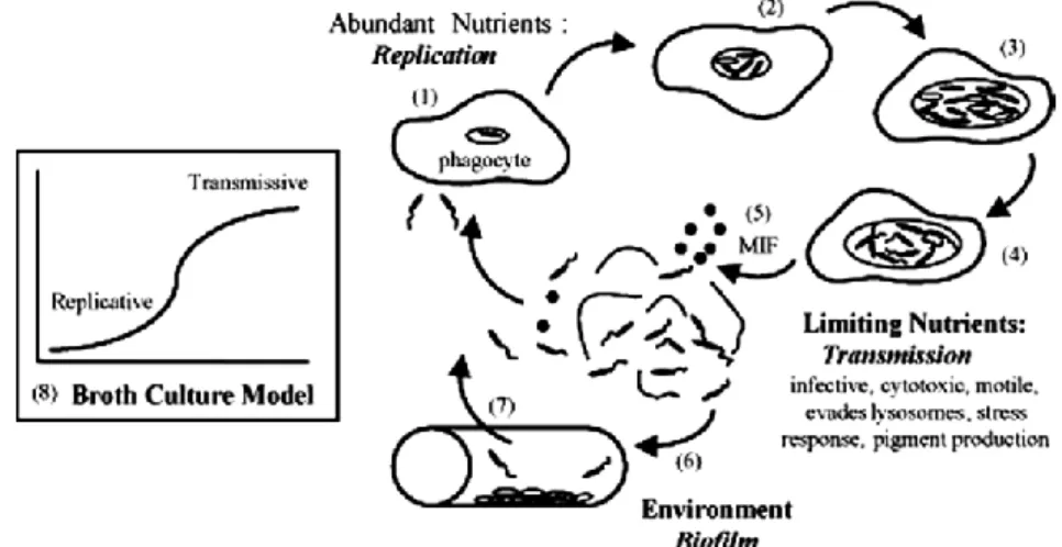
Inside host cells Lp differentiates into a replicative form and when nutrients become limited into a transmissive form (Figure 1) [7]. Garduño et al. have shown that Lp can differentiate in vivo in Tetrahymena tropicalis directly from the transmissive form into a highly infectious mature intracellular form (MIF), indicating that transmissive form and MIFs constitute a differentiation continuum. In addition, MIFs are thought to be the infectious forms implicated in the transmission of LD [24].
Chapter 1. Introduction
J. Dantas 3
1.2. Epidemiology
Much has been learnt about the epidemiology of LD and Legionella since the organism was first identified in 1976. In the meantime, surveillance schemes for LD have been applied in several countries including the USA, Canada, New Zealand, Australia, Japan, Singapore, and Europe, where LD became a notifiable disease and coordinated European surveillance has been established since 1995 [25].
In Portugal, LD was included in the Portuguese system of mandatory notifications of infectious diseases in 1999 and in 2004 an Integrated Programme of Epidemiological Surveillance was implemented in order to improve reporting (clinical and laboratory notification) [26].
Despite being a notifiable disease, LD is likely to be underestimated in many countries mainly because of the lack of common definitions, diagnostics and surveillance Figure 1 - The life cycle of Lp.
1. Free-swimming transmissive Lp that are engulfed by the phagocyte host (amoeba) establish vacuoles that provide protection from lysosomal digestion.
2. When nutrients and other conditions are favorable, intracellular bacteria repress transmission traits and activate pathways that promote replication.
3. As conditions in the replication compartment become unfavourable, the progeny stop dividing and coordinately express traits that promote survival in the environment and transmission to a new phagocytic host.
4. After a long period, the microbes may continue to develop into a mature intracellular form (MIF), a cell type that is highly resilient and infectious.
5. The amoeba is lysed, and the microbes are released into the aqueous environment.
6. Lp that do not immediately encounter a new phagocyte probably establish biofilms in both water
systems and ponds, where they are resistant to biocidal agents.
Chapter 1. Introduction
J. Dantas 4
systems. This fact makes it impossible for LD to be directly comparable between countries [25].
The distribution of LD cases by age and gender is similar between countries: the disease is rare in children and more frequent in older people (74-91% of patients are ≥50 years) and males account for the majority of the notifications (1.4-4.3 male patients for every female patient) [27-31]. In Europe, the age-standardized notification rate of LD was 10.7 per million people in 2013 and the number of notifications per million inhabitants was 11.4 [23].
Several studies have shown that LD has a seasonal pattern that is associated with warmer and wetter weather conditions [32-35]. There is also evidence that suggests that the persistence of Lp is increased in aerosols at high relative humidity [36, 37].
There is also an association between LD cases and a history of recent travel, particularly involving overnight stays in hotel accommodations. Rooms that have not been used for a long time and the presence of large numbers of water outlets with long pipe runs can contribute to water stagnation, which will promote Legionella spp. growth, unless adequate control measures are applied [38, 39]. Cruise ships can also be sources of Legionella for similar reasons, and have already been associated with several outbreaks [40].
In view of the association with travel, specific surveillance systems for travel-related cases of LD have been implemented in order to improve source identification and public health action [40].
In Europe, the surveillance of LD is carried out by the European Legionnaires’ disease Surveillance Network (ELDSNet) since 2010 and is coordinated by the European Centre for Disease Prevention and Control (ECDC). ELDSNet works in partnership with several entities like the World Health Organization (WHO), public health authorities of non-EU countries and tour operators [41].
1.3. Transmission
Transmission of LD is usually by inhalation of aerosols or aspiration of water containing Legionella spp. and until the present year no evidence of person-to-person transmission existed [25]. However, there has been documented recently (February,
Chapter 1. Introduction
J. Dantas 5
2016) by Correia et al [42] a probable case of Lp transmission between two individuals after a cluster of cases of LD occurred in Vila Franca de Xira, Portugal, in 2014. The second patient is believed to have been infected by the first patient since the second patient was not geographically linked to the cluster epicenter and strains of both patients showed the novel ST1905 profile (identified as the causative strain in the cluster) [43]. In cases of LD where the etiologic agent is Legionella longbeachae, the transmission route is not well known to date, but it is believed to have a different source. In these cases, activities like exposure to potting compost or soil or poor hand-washing practices after gardening are regarded as risk factors of infection by this species [44, 45, 46].
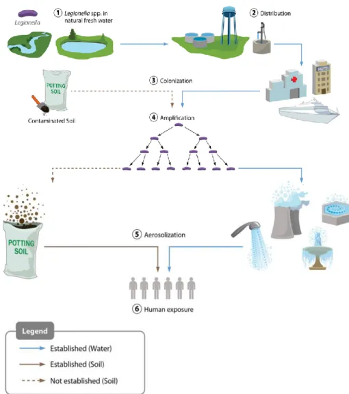
Figure 2 - Route of Legionella dissemination from natural waters to human exposure. Legionella from
freshwater sources (1) is distributed at low concentrations from points of water purification (2) to colonize downstream local plumbing networks and cooling systems (among other sites) (3) and amplifies under permissive environmental conditions (4). Subsequent aerosolization (5) exposes a human population (6), leading to a potential disease spectrum. The route of LD caused by contaminated soil is less well understood but also appears to involve aerosol exposure (Adapted from [8]).
Chapter 1. Introduction
J. Dantas 6
Contaminated cooling towers have been associated with the largest outbreaks of LD (Murcia, Spain, in 2001 with 449 confirmed cases and Vila Franca de Xira, Portugal, in 2014 with 334 confirmed cases) [43, 47-49]. Hot and cold water systems and whirlpool spas have also been shown to be sources of transmission [50, 51].
In fact, a great number of LD outbreaks have been shown to be linked to aerosol-producing devices as the source of Legionella spp. transmission and a major variety of mechanisms and settings (e.g. evaporative air conditioning units, decorative fountains and showers) have been described [52, 53].
1.4. Clinical Presentation
In humans, Legionella can cause two distinct clinical syndromes: Pontiac Fever (PF) and LD [54]. PF is an acute, self-limited form of legionellosis characterized by flu-like symptoms and was named after an explosive outbreak that occurred in the city of Pontiac, in Michigan, USA [55].
LD, the most severe form of legionellosis, is characterized by pneumonia that can be associated with systemic infection [25]. LD does not have specific, defining clinical features because it presents as a variety of clinical manifestations and symptoms (e.g., fever, myalgia, cough, and pneumonia) [56-58]. The incubation period of LD is thought to be 2–10 days (median 6–7 days); however, longer incubation periods have been reported [48, 51]. In the last years, the mortality from LD has decreased, probably due to earlier diagnosis associated with better treatment [59].
1.5. Diagnosis
The diagnosis of LD can be a challenging process, considering that the clinical symptoms are often indistinguishable from other causes of pneumonia.Considering this fact, laboratory confirmation becomes essential for an accurate diagnosis [60]. There are several laboratory-based methods for the LD diagnosis, which can be divided in two major groups: phenotypic and genotypic methods. The phenotypic methods currently used are urinary antigen test (UAT), serum antibody titration and culture; the genotypic methods are based on the Polymerase Chain Reaction (PCR) [61].
Chapter 1. Introduction
J. Dantas 7
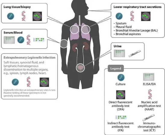
Figure 3 shows the anatomical locations of each specimen type used for the LD diagnosis and the possible diagnostic tests for each one.
Current LD case classification is divided in probable and confirmed case. Probable case is defined when any person has pneumonia and at least one of the following four laboratory criterion: detection of Lp antigen in respiratory secretions or lung tissue, for example by Direct Fluorescent Antibody (DFA) staining using monoclonal-antibody derived reagents; detection of Legionella spp. nucleic acid in respiratory secretions, lung tissue or any normally sterile site; significant rise in specific antibody level to Lp other than sg 1 or other Legionella spp. in paired serum samples; or single high level of specific antibody to Lp sg 1 in serum. Confirmed case is defined when any person has pneumonia and at least one of the following three laboratory criterion: isolation of Legionella spp. from respiratory secretions or any normally sterile site; detection of Lp sg1 antigen in urine or significant rise in specific antibody level to Lp sg 1 in paired Figure 3 - Anatomical locations of each specimen type used for Legionella detection and the specific diagnostic tests. The clinical identification of Legionella can be made by several assays and
different specimens. Some assays can be applied to multiple specimen types, such as culture and nucleic acid amplification. Although it is not recurrent, Legionella infection at extrapulmonary sites can also be observed (Adapted from [8]).
Chapter 1. Introduction
J. Dantas 8 serum samples [62].
In the following sections, four diagnostic tests will be described in greater detail: culture, UAT, serological assays and PCR.
1.5.1. Culture
Despite the improvements in other diagnostic tests, isolation by culture remains the gold standard for Legionella detection and LD diagnosis. Is the most specific test and requires special media, adequate processing of specimens, and technical expertise [63]. Legionella can be cultured from a number of specimens, including respiratory secretions (sputum, bronchial alveolar lavage (BAL), and bronchial aspirates), lung tissue/biopsy, serum, blood and stool [64, 65]. However, the preferable specimens are the lower respiratory tract secretions (e.g. sputum), which should be promptly processed since Legionella may survive poorly in these secretions [64].
Reports of Legionella infection at extrapulmonary sites are rare, but have already been verified. In those cases, the samples are collected from soft tissues, joint fluids, and blood and culture should be attempted only when other etiologies have been ruled out since the recovery of an isolate is extremely difficult [8].
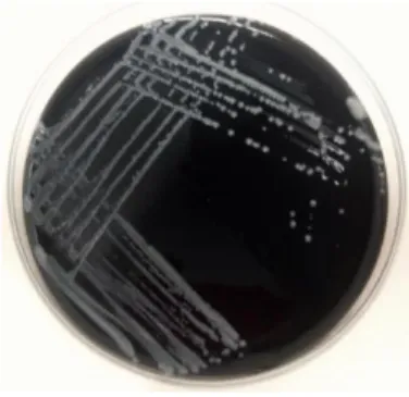
The standard medium used to culture Legionella spp. is the Buffered Charcoal Yeast Extract (BCYE-α) agar, and contains yeast extract, activated charcoal, α-ketoglutarate, L-cysteine and soluble ferric pyrophosphate. The pH of the agar must be adjusted to 6.9 with the addition of ACES (N-2-acetamido-2-aminoethansulfonic acid) buffer in order to enhance bacterial growth [66] and the optimum growth temperature is 37ºC.
The main disadvantage of this method is the fact that it requires several days to obtain a positive result since Legionella, being a fastidious organism, takes at least 5 days to grow [60].
Figure 4 – Culture of Lp in BCYE-α agar with 4 days of incubation (photo obtained in the present
Chapter 1. Introduction
J. Dantas 9
1.5.2. Urinary Antigen Test
The UAT for the diagnosis of LD is available since 1980 [59] and it has become the first-line diagnostic test [67]. Its use has been increasing over the years, surpassing other diagnostic methods, mainly because it is fast, easy to perform and provides timely, accurate results, with high sensitivity (70%-80%) and specificity (>95%) [68].
The urinary antigen in question is a component of the cell wall LPS of Lp and is detected by an enzyme immunoassay (EIA), early in the course of the illness [67, 61, 69]. The test becomes positive after 48-72h of the beginning of the symptoms [67], and the antigen may continue to be excreted in detectable quantities in urine for several months, even after appropriate treatment and apparent recovery from infection, which becomes a disadvantage for diagnosis [69]. However, it has been shown that in the majority of cases antigenuria became negative within 2 months after the first diagnosis [70].
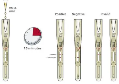
At present, the Legionella UAT is available in two main formats: a 96-well plate-based EIA or ELISA, and a rapid immunochromatographic test, in a card/strip-based format, similar to a home pregnancy test (Figure 5) [8].
However, despite being a valuable tool, the UAT only detects the most prevalent species and serogroup, Lp sg1 (the causative agent in 70%-80% of LD cases), which fails the diagnosis of LD infections caused by non-Lp sg1 strains [63, 59, 71].
Figure 5 – Rapid diagnostic test for detection of Lp antigen in urine.
Chapter 1. Introduction
J. Dantas 10
1.5.3. Serological assays
Serological testing was one of the main methods used for LD diagnosis in the early 1980s [59]. Nowadays, this method is useful for cases where the patients have already started antibiotherapy or when it is difficult to obtain respiratory samples. It is considered a late diagnostic because it requires a paired serum samples with 2 weeks apart.
The main disadvantage of the serological assays is the possibility of cross-reactivity between different serogroups or between different species of Legionella spp., which may interfere with the interpretation of the results and therefore influence the diagnosis, leading to incorrect and ineffective treatment [72, 73].
Nevertheless, serology remains important for LD confirmation in the cases where the etiologic agent cannot be isolated. Furthermore, this method may be of interest for retrospective epidemiological studies, such as general seroprevalence, to identify patterns of disease or potential ongoing outbreaks [8].
1.5.4. Polymerase Chain Reaction
The first report of PCR as a method to detect Legionella was in 1989 [74] and since then it has become frequently used to identify Legionella in clinical samples [75]. PCR enables specific amplification of small amounts of Legionella DNA, provides rapid diagnosis (with outcome in a day) and is one of the few diagnostic tests with the potential to detect infections caused by all of the known species and serogroups of Legionella spp. [63, 67, 75].
It has been successfully used to detect Legionella DNA in a range of clinical samples, such as sputum, urine and serum. However, the reliableness of the results obtained from nonrespiratory specimens is not fully known [63].
Previous tests with samples from the lower respiratory tract have shown that PCR is highly specific and has sensitivity greater than culture, suggesting that it is suitable for use in the routine microbiological diagnostic laboratory [76].
Additionally, this method can be used when the patient have already started the antibiotherapy [77].
Chapter 1. Introduction
J. Dantas 11
Despite all the advantages, the difficulty in assessing bacterial viability is the main disadvantage of all nucleic acid amplification methods, since they fail to discriminate between nucleic acids from dead or dying bacteria. Therefore, they cannot distinguish disease resolution from current disease [8].
1.6. Management and Risk Factors
When a case of LD is diagnosed, the physicians must be aware of the possible presence of other cases related in time and place, which can be crucial for identification of the potential source of infections. It is also important to have a detailed history of the recent activities of the patient, including any potential exposure to aerosolized water droplets (especially during the previous 10 days) in order to support the epidemiological follow-up, trace any other patients and identify the source of infection [25].
LD can occur in previously healthy individuals, but is more frequent in those who gather the major risk factors, such as age (≥50 years), sex (male), smoking habits, chronic cardiovascular/respiratory disease, diabetes, alcohol misuse, and immunosuppression (e.g. after solid organ transplantation or any other immunosuppressive therapy). Immunosuppressed patients might present with more severe clinical disease and frequently require intensive care, intravenous antibiotics, and a longer duration of therapy [78-81].
The empiric therapy for LD is antibiotic treatment of the infection and management of any complications [58]. The chance of recovery is higher if the appropriate antibiotics are given early [57, 82]. Since LD does not have any defining clinical features and that β-lactam antibiotics (usually used to treat bacterial community-acquired pneumonia) are unsuccessful for treatment of LD, it is wise to give effective antibiotic therapy against Legionella spp. in the early stage of all moderate-to-severe community-acquired and hospital-acquired pneumonias, until a specific microbiological diagnosis is made. The dose and route of administration of treatment (which can be either oral or intravenous) is guided by severity, underlying risk factors, consciousness level, and gastrointestinal disorders [25].
Because Lp is an intracellular pathogen residing within tissue and alveolar macrophages, successful treatment depends on use of antibiotics that achieve
Chapter 1. Introduction
J. Dantas 12
therapeutic intracellular concentrations within macrophages, such as the macrolides and fluoroquinolones [83-88]. Erythromycin had been the drug of choice for treatment of LD until the 1990s but its use have been decreasing since it is bacteriostatic and has side-effects, particularly when used intravenously [89, 90]. However, the newer macrolides such as azithromycin have fewer side-effects. Regarding fluoroquinolones, they are bactericidal and in vitro their activity against Legionella spp. in animal models has been shown to be higher the one from erythromycin [91, 92].
1.7. Legionella pneumophila natural host: Acanthamoeba spp.
Amoebae are ubiquitous organisms that can be found in humid soil and water reservoirs, being the most common genus Acanthamoeba spp. [93, 94]. They are unicellular protozoans that can display two different phases during their life cycle, according to the environmental conditions. When the conditions are unfavorable to their growth (e.g. limited nutrients, temperature) they remain as a dormant cyst, a process called encystment. Conversely, when the conditions are suitable for their growth, they change to an active vegetative trophozoite, a process called excystment [94].
Free-living amoebae are frequently isolated from several man-made reservoirs, such as tap water, air-conditioning units, and cooling towers, where they feed on the existing microbial biofilm. However, though amoebae are bacterial predators, several bacteria have developed mechanisms to survive phagocytosis, being able to use the amoebae as hosts [95].
Transient association with amoebae has been reported for a number of different bacteria including Lp, many Mycobacterium spp., Francisella tularensis, and Escherichia coli O157, among others [96-99].
Since most of these bacteria are pathogens of humans, it has been suggested that protozoa have an important role in the development of bacterial pathogenesis and that the interaction bacteria/protozoa is significant in terms of human disease [100].
To date, only the interaction of Lp with free-living amoebae has been studied in greater detail [95].
Chapter 1. Introduction
J. Dantas 13
1.7.1. Life cycle in Acanthamoeba spp.
Lp is ubiquitous in freshwater, often in close association with freshwater protozoa [95] and replicates at temperatures of 25–42°C with an optimal growth temperature of 35°C. Consistent with what Lp would encounter in the environment, motility and adherence to host cells are optimal at temperatures below 37°C [101].
Thermal conditions have been shown to affect the interaction between Lp and amoebae [102]. At temperatures over 25°C Lp is able to infect the trophozoite form of free-living amoebae and replicate intracellularly, increasing the number of bacteria in the water. Consequently, the chance of transmission to humans increases as well [102, 103]. In contrast, with temperatures below 20°C, the amoebae encyst and Lp is not able to replicate inside the host [63].
Intracellular Lp is capable of survival within this cysts that ensure their survival by protecting them from harsh environments. In this stage, the bacteria are not capable to proliferate and thrive to a dormant state [104].
Fourteen species of amoebae, with Hartmannellae and Acanthamoeba being the most prominent, and two species of ciliated protozoa have been shown to support intracellular replication of Lp [95]. When compared with bacteria grown in vitro, bacteria grown in amoebae have an increased resistance to harsh conditions [105, 106] and show changes in biochemistry, physiology, and virulence potential [103]. From those changes it has been verified: an enhanced resistance to chemical disinfectants, treatment with biocides and antibiotics [95]; shorter size and larger diameter; different protein expression [107]; enhanced intracellular survival and replication and increased ability to infect not only amoebae but also mammalian cells [108]. It has also been shown that amoeba are capable of resuscitate viable non-culturable Lp, for instance after disinfection [95, 109, 110]. Additionally, it is believed that the enhanced infectivity of Lp after growth within amoebae may compensate the low concentration of bacteria usually detected in the aquatic reservoirs from which the bacteria are transmitted to humans during LD outbreaks [111-114].
Besides enhancing the pathogenicity of Lp, amoebae are also responsible for their persistence in the environment. Since Legionella species cannot multiply extracellularly, the presence of amoebae as hosts in their life cycle is fundamental
Chapter 1. Introduction
J. Dantas 14 (Figure 6) [103].
1.7.2. From Amoebae to Macrophage
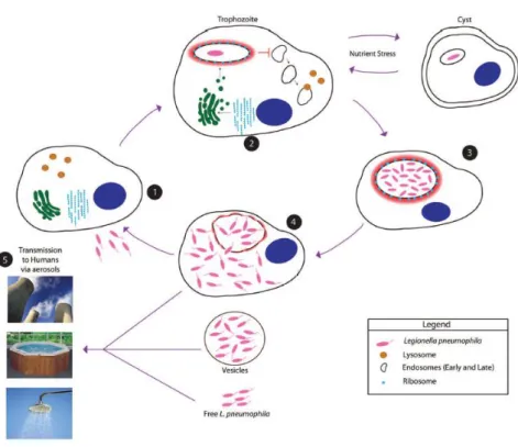
Lp are able to infect, multiply within, and kill human macrophages, as well as free-living amoebae [116]. This ability is thought to be a consequence of previous interaction with several protozoan hosts that allowed Lp to adapt to intracellular growth within macrophages [117]. This adaptation process is thought to include the acquisition of eukaryotic genes during its co-evolution with amoebae [118], which is supported by Figure 6 - The environmental life cycle of Lp within protozoa.
1. Flagellated Lp infects protozoa in the aquatic environment.
2. The Legionella-containing vacuole (LCV) evades the default endosomal–lysosomal degradation
pathway and becomes rapidly remodeled by the ER through intercepting ER-to-Golgi vesicle traffic.
3. Under unfavorable stress conditions, such as nutrient deprivation, amoeba encysts, and bacterial
proliferation will not occur due to nutrient limitation.
4. During late stages of infection, the LCV becomes disrupted leading to bacterial egress into the
cytosol where the last 1–2 rounds of proliferations are completed. Nutrient depletion triggers a phenotypic transition into a flagellated virulent phenotype followed by lysis of the amoeba and bacterial escape from the host cell. Excreted vesicles filled with bacteria are also released. The infectious particle is not known but may include excreted Legionella-filled vesicles, intact
Legionella-filled amoeba, or free Legionella that have been released from host cell.
5. Transmission to humans occurs via aerosols generated from man-made devices and installations,
Chapter 1. Introduction
J. Dantas 15
findings that show that all the genes that are required for intracellular growth in human macrophages are the same required for intracellular growth in Acanthamoeba castellanii (Ac) [116].
Additionally, the life cycle of Lp in macrophages strongly resembles the one observed in amoebae, mainly in the process by which Lp is able to avoid digestion. In both phagocytes vacuoles containing Lp neither acidify nor fuse with the lysosomal compartment, allowing the association between the phagosome and the endoplasmic reticulum which leads to the high intracellular replication of Lp [119].
Moreover, the similar cell biology between amoebae and macrophages can also be an indicator that the virulence of Lp for macrophages is in fact a consequence of its evolution as an intracellular parasite of protozoa [119].
Lp and amoebae have been isolated from the same source of infection during outbreaks of LD [95] which shows that amoeba are a natural reservoir for the opportunistic pathogens of macrophages [119].
1.7.3. Evasion of the host for new infection cycle
The ability to lyse and exit host cells after intracellular replication is an essential step in the life cycle of all intracellular pathogens [120]. After this step, the released pathogens are able to infect other cells within the same host or be transmitted to a new susceptible host [120].
It is known that Legionella has the ability to kill a wide variety of host cells (e.g. human phagocytic cells, amoeba and ciliated protozoa) and it has been suggested that the bacteria induce apoptosis, or programmed cell death, in the host [121].
Furthermore, it has been shown that Lp is able to induce major changes in the nuclear morphology of the host as well as increase the proportion of fragmented DNA, which correlates with the cell death process that occurs in macrophage-like HL-60 cells [122]. Even though this issue has already been addressed, apoptotic death of amoeba infected with Lp has not been observed yet [121].
Chapter 1. Introduction
J. Dantas 16
1.8. Apoptosis
The first studies dedicated to cell death date back to the nineteenth century and were generally related to the metamorphosis of tadpoles and insects, and later with transient embryonic structures [123, 124]. The subject of “programmed cell death” – in which “apoptosis” is included as one type of cell death - appeared later in the 1970s when scientists first began to observe a sequence of events in the cell that once established could lead to its death. These observations showed that cell death during development is not of accidental nature but instead follows a sequence of controlled steps that lead to self-destruction of the cell [124].
The term “apoptosis” was then proposed in 1972 to name one type of programmed cell death that plays an important role in the regulation of animal cell populations. Moreover, it was shown that the apoptotic process could be initiated or inhibited by a variety of environmental stimuli, both physiological and pathological [125].
Since then, the field of apoptosis research began to grow exponentially with a new set of mind in which cell death is not an incidental part of life, but instead a highly controlled and medically important element of existence [124, 126].
Although apoptosis is the most frequent form of programmed cell death, it is important to note that there are other non-apoptotic types of programmed cell death that have already been described and have biological significance [127, 126].
1.8.1. Morphological features of apoptosis
When a cell is triggered to suffer apoptosis it means that a cascade of molecular events has been activated, resulting in the total disintegration of the cell [128].
One of the first events of this type of “programmed cell death” is the loss of intracellular water, which leads to a smaller and denser cell. Later, one of the most characteristic features of apoptosis occurs: the condensation of the nuclear chromatin to heterochromatin (in one or more masses in the nucleus). The nuclear membrane of the cell begins to disintegrate and lamin proteins undergo proteolytic degradation, followed by nuclear fragmentation. Activation of endonuclease(s) results in a selective degradation of DNA, in fragments up to 50–300kb, and is followed (in many but not all
Chapter 1. Introduction
J. Dantas 17
cell systems) by internucleosomal DNA cleavage. As a result, many nuclear fragments (that resemble with DNA droplets of different sizes) are then scattered throughout the cytoplasm. Afterwards, those nuclear fragments, together with constituents of the cytoplasm (e.g. undamaged organelles), are packaged and enclosed by fragments of the plasma membrane. These structures, named ‘‘apoptotic bodies’’, are then shed from the dying cell (Figure 7) [124, 128].
During this process, there are two important characteristics that are exclusive of apoptosis: the activation of endonuclease(s) that preferentially cleave DNA at the internucleosomal sections and the preservation of the structural integrity of the plasma membrane as well as some cellular organelles, such as mitochondria and lysosomes [128].
Opposing to apoptosis, there is a different type of cell death that can be interpreted as an “accidental death”, named necrosis [126]. In this case, the cell death pathway begins with the mitochondrial swelling that eventually leads to the rupture of the plasma membrane, which causes the releasing of the cytoplasmic constituents. At the end of this process, nuclear chromatin shows irregular areas of condensation and the nucleus is slowly dissolute [128].
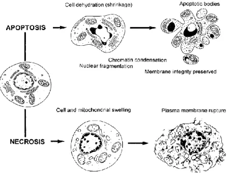
Figure 7 – Morphological and biochemical changes that occur during apoptosis and necrosis. The
apoptotic pathway begins with the loss of intracellular water and increase in the concentration of ionized calcium in the cytoplasm, resulting in the cell shrinkage. Subsequently, the chromatin condensation occurs followed by nuclear disintegration and formation of apoptotic bodies. The integrity of the plasma membrane is preserved to the late stages of apoptosis. In contrast, the necrotic pathway begins with the swelling of mitochondria as well as swelling of the whole cell, combined with marginal chromatin condensation. Finally, the rupture of the plasma membrane results in the releasing of the cytoplasmic content of the cell (Adapted from [128]).
Chapter 1. Introduction
J. Dantas 18
1.8.2. Mechanisms of apoptosis
The apoptotic machinery is tightly controlled and can be initiated by two main alternative pathways: the death-receptor pathway (usually named “extrinsic pathway”); or the mitochondrial pathway (usually named “intrinsic pathway”) [129, 126].
Both pathways have in common the presence of initiator cysteine aspartyl-specific proteases, called caspases (caspase-8 for the extrinsic pathway and caspase-9 for the intrinsic pathway) that once activated cleave and activate the ‘executioner’ caspases, such as caspase-3, which starts the execution pathway and is common to both extrinsic and intrinsic pathways. The active executioner caspases then cleave each other and an amplifying proteolytic cascade of caspase activation is started. Ultimately, the active executioner caspases cleave cellular substrates leading to the characteristic biochemical and morphological changes of apoptosis (Figure 8) [129, 126].
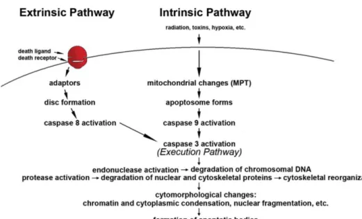
Figure 8 - The two main apoptotic signalling pathways. Apoptosis can be initiated by two alternative
pathways: either through death receptors on the cell surface (extrinsic pathway) or through mitochondria (intrinsic pathway). Both of them require specific triggering signals to begin an energy-dependent cascade of molecular events. Each pathway activates its own initiator caspase (8 and 9) which in turn will activate the executioner caspase-3. The execution pathway results in characteristic cytomorphological features including cell shrinkage, chromatin condensation, formation of cytoplasmic blebs and apoptotic bodies and finally phagocytosis of the apoptotic bodies by adjacent parenchymal cells, neoplastic cells or macrophages (Adapted from [126]).
Chapter 1. Introduction
J. Dantas 19
1.8.3. Biochemical features
There are several biochemical hallmarks of apoptosis (e.g. DNA degradation, protein cleavage, protein cross-linking, phagocytic recognition) that can be used to identify this mode of cell death [126].
One of those biochemical features is the expression of cell surface markers in the outer leaflet of the cell membrane. This membrane alteration allows the apoptotic cells to be recognized by adjacent cells, allowing a quick phagocytosis [126, 130].
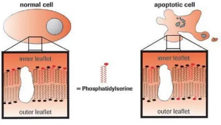
Phosphatidylserine (PS) is an anionic phospholipid of the cellular membrane that in normal cells is only present in the inner membrane. During apoptosis, the amount of phosphatidylserine (PS) on the outer surface of the membrane increases, becoming exposed outside the cell [130].
For the study of apoptotic cells, a recombinant phosphatidylserine-binding protein named Annexin V can be used to detect this type of cells. Annexin V has been shown to interact strongly and specifically with phosphatidylserine residues, which makes it a useful tool for apoptotic cell detection [131-133].
Figure 9 – Phosphatidylserine exposure during apoptosis. During apoptosis, the distribution of neutral
phospholipids (black symbols) and anionic phospholipids such as PS (red symbols) in the cell membrane changes. PS is present in the outer membrane of apoptotic cells, but not of normal cells. An exogenously added molecule specific for PS, such as Annexin V-FITC, will bind to PS on the outer membrane of apoptotic cells, but cannot react with the PS of normal cells (Adapted from [130]).
Chapter 1. Introduction
J. Dantas 20
1.8.4. Apoptosis measurement
Since apoptosis has so many characteristic changes that appeared this type of cell death, those changes have become markers that are used to identify this mode of cell death by several laboratory methods [128].
There are several commercial kits available to detect and count apoptotic cells and since many features of apoptosis and necrosis can overlap, it is crucial to employ two or more distinct assays to confirm that cell death is occurring via apoptosis. The assays should be based on a different principle in order to have a more complete approach. Other methodology for apoptosis detection that is becoming very popular is the multiplex, in which the same sample allows to gather more than one set of data [126].
Therefore, there are apoptosis assays based on several methodologies, such as cytomorphological alterations; DNA fragmentation; detection of caspases, cleaved substrates, regulators and inhibitors; membrane alterations; detection of apoptosis in whole mounts and mitochondrial assays [126].
All assays have advantages and disadvantages regarding the object of study. Consequently, it is crucial to understand the pros and cons of each assay and to define the best combination of assays to allow a more complete and accurate study [126].
1.9. Research objectives
The molecular mechanisms by which Lp kills protozoan host cells are largely unknown. In 1998, Hagele et al have demonstrated that Lp is able to induce apoptosis in human monocytes and that this process depends on the multiplicity of infection (MOI), i.e. the proportion between Lp and the host, and the time post-infection. However, by studying infection in Ac at 24h post-infection and a MOI of 50, they concluded that Lp was not able to induce apoptosis in Ac. This finding suggested that Lp induces different mechanisms of cell killing to evade the main hosts: protozoan and human cells [121]. More recently, Gao and Kwaik demonstrated that intracellular Lp kills and exits Acanthamoeba polyphaga preferentially by induction of necrosis. They have also confirmed that, as in the mammalian cells, PS is distributed in the inner leaflet of the plasma membrane in Acanthamoeba and that specifically Acanthamoeba polyphaga
Chapter 1. Introduction
J. Dantas 21 possesses a functional apoptotic pathway [133].
The main objective of this study was to clarify if Lp is able to induce apoptosis in the natural host Ac.
Since the molecular mechanisms by which Lp kills protozoan host cells remains uncertain and the published studies were based only in a few conditions of the infection, we proposed to clarify whether Lp induces apoptosis in Ac, using different MOIs and time post-infection.
The apoptosis detection in Ac was assessed by membrane alterations (PS externalization) using flow cytometry analysis and DNA fragmentation using electrophoresis.
J. Dantas 22
Chapter 2. Material and Methods
All the experimental procedures were performed in a tissue culture room, in a Class II Type A2 Biological Safety Cabinet that maintained the sterility of cell lines and protected both the user and the experiment. In the tissue culture room, an exclusive disposable lab coat was always used, proper aseptic techniques were assembled in order to eliminate possible contaminants and all the materials were disposable plastic.
2.1. Microorganisms, culture media and growth conditions
In this study, we used Lp strain Paris (Lp Paris), isolated from patients and the environment in the area of Paris, France, from 1987 to 1997 (courtesy of the Pasteur Institute) and a line of Acanthamoeba castellanii (Ac) strain Neff (ATCC® 30010™). A stock culture of both strains was preserved at -80ºC. Lp was stored in skim milk and Ac was stored in 10% DMSO + 90% FCS.
2.1.1. Growth of Legionella pneumophila strain Paris
After thawing, Lp Paris was cultured on Buffered Charcoal Yeast Extract (BCYE, Appendix 2) agar supplemented with ACES Buffer/potassium hydroxide, ferric pyrophosphate, L-cysteine HCl and α-ketoglutarate (Legionella BCYE Growth Supplement SR0110A, Oxoid), and incubated at 37°C, for 48h.
2.1.2. Growth of Acanthamoeba castellanii
Ac was grown in axenic static culture in culture T-flasks with a surface area of 25cm2, in 6ml of Peptone-Yeast Extract-Glucose medium (PYG, Appendix 3) at room temperature.
Chapter 2. Material and Methods
J. Dantas 23
Additionally, to ensure that the cultures remained viable for an extended period of time, we had to follow some procedures that will be described below:
Monitor the culture regularly, to confirm its viability and healthy morphology, using an inverted microscope (Meiji Techno, TC-5300).
When the culture reaches the peak density, i.e., confluent layer of Ac on the bottom surface of the flask, the culture was split to a new culture flask. In order to do this, first, the confluent layer of Ac was washed with 3ml of PYG, for three times, to assure that the wastes resulting from metabolism and unviable Ac were rejected. After this, the Ac were harvested in 3ml of PYG with a cell scrapper and approximately 0.25ml of the Ac suspension was transferred to a new flask. Repeat the procedure in 2-3 days intervals.
Prepare fresh PYG on a monthly basis in order to maintain this procedure.
2.2. Infection Protocol of Acanthamoeba castellanii with Legionella pneumophila strain Paris
For the infection protocol of Ac with Lp Paris, both microorganisms required previous growth conditions and specific procedures before the infection. The preparation of each microorganism for infection will be described below.
2.2.1. Preparation of Legionella pneumophila strain Paris liquid culture for infection
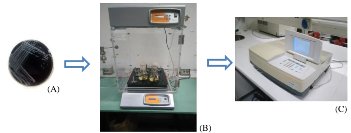
For each infection, three to four colonies from a new 48h passage culture in BCYE agar, were cultured to 100ml of ACES-buffered yeast extract broth (AYE, Appendix 4) and incubated at 37ºC with shaking at 170 rpm (incubator I10-C+ACOP. E, OVAN). To monitor the growth of the liquid culture we used a spectrophotometer (Shimadzu, UV-1700 PharmaSpec) to measure the optical density at 600nm (OD600), at hourly intervals. These measures were used to build the Lp Paris growth curve and determine when the culture reached the beginning of the stationary phase, i.e. when the difference between two consecutive measures was 0.05 (previously determined by the research group).
Chapter 2. Material and Methods
J. Dantas 24
Once the culture was in the stationary phase, 5ml of the suspension were collected to a 15ml sterile Falcon tube and centrifuged (centrifuge ROTINA 380R, Hettich) for 15 minutes at 3095 x g. The supernatant was discarded and the pellet was resuspended in 10ml of Minimum Medium (MM, Appendix 5). The OD600 of the suspension was measured and adjusted to 1.2, which represents a concentration of 109 CFU/ml (previously determined by the research group).
2.2.2. Preparation of the Acanthamoeba castellanii confluent layer for infection
For each infection, we used a confluent layer of Ac with 48h of growth. The Ac monolayer was washed with 3ml of minimum medium (MM, Appendix 5), for two
(C) (B) (A) (C) (B) (A)
Figure 11 – The Lp Paris liquid culture in the stationary phase was collected (A) and centrifuged for
15 minutes at 3095 x g (B). For the infection, the concentration of Lp Paris was measured by spectrophotometry (C).
Figure 10 – Lp Paris was inoculated, in duplicate, on AYE (A) and incubated at 37ºC, 170 rpm (B). The
Chapter 2. Material and Methods
J. Dantas 25 (B)
(A)
times. To detach the Ac from the bottom layer of the flask, we used a cell scrapper and 1,5ml of MM. A counting chamber (Thoma, 0.1mm) was used in order to count the Ac (Transmitted Light Microscope Standard 25 ICS, Carl Zeiss) and determine the concentration of the suspension.
2.2.3. Infection of Acanthamoeba castellanii with Legionella pneumophila strain Paris
For the infection protocol, we tested three different MOIs: 10, 50 and 100 (this values were previously established by the research group). In each infection, we prepared the required culture T-flasks with an Ac monolayer of 3x10⁶ Ac/ml. After this, we calculated the required volume of the Lp Paris suspension to infect the Ac (considering the intended MOI). Once the Lp Paris was pipetted into each flask, the flasks were incubated at 37ºC for one hour. After the incubation time, we washed each flask with 3ml of MM, for three times, to remove the extracellular Lp. After the washing process, we added a final volume of 6ml of MM and the flasks were incubated at 37ºC. The infection is considered to be started at this point.
Each infection assay had a negative control (flask with uninfected Ac in MM, incubated at 37ºC).
2.3. Apoptosis analysis
The apoptosis analysis was performed using two techniques: flow cytometry and Figure 12 – The Ac concentration for infection was determined by counting on a Thoma Chamber with a