Ivo Miguel Ferreira da Paixão Reis
Degree in Cell and Molecular Biology
Unveiling human Cardiac Stem Cells role in
myocardial ischemia-reperfusion injury
Dissertation to obtain a Master’s degree in Biotechnology
Supervisor: Patrícia Gomes Alves, PhD, Senior scientist, IBET/ITQB-NOVA
Jury:
President: Prof. Dr. Isabel Maria Godinho de Sá Nogueira
Arguer: Dr. Ana Fernandes-Platzgummer
Supervisor: Dr. Patrícia Gomes Alves
Un v eil ing hu man Ca rd iac S tem Ce lls r o le in m y o c ar d ial is chemia -r epe rf u sion inju ry Iv o M igu el F er rei ra da P ai xã o Rei s 2018
Ivo Miguel Ferreira da Paixão Reis
Degree in Cell and Molecular Biology
Unveiling human Cardiac Stem Cells role in
myocardial ischemia-reperfusion injury
Dissertation to obtain a Master’s degree in Biotechnology, by Universidade Nova de Lisboa, Faculdade de Ciências e Tecnologia
Supervisor: Patrícia Gomes-Alves, PhD, Senior scientist, IBET/ITQB-NOVA
Jury:
President: Prof. Dr. Isabel Maria Godinho de Sá Nogueira Arguer: Dr. Ana Fernandes-Platzgummer
Supervisor: Dr. Patrícia Gomes Alves
September 2018
Copyright
Unveiling human Cardiac Stem Cells role in myocardial
ischemia-reperfusion injury
Ivo Miguel Ferreira da Paixão Reis, Faculdade de Ciências e Tecnologias, Universidade Nova de Lisboa
A Faculdade de Ciências e Tecnologia e a Universidade Nova de Lisboa têm o direito, perpétuo e sem limites geográficos, de arquivar e publicar esta dissertação através de exemplares impressos reproduzidos em papel ou de forma digital, ou por qualquer outro meio conhecido ou que venha a ser inventado, e de a divulgar através de repositórios científicos e de admitir a sua cópia e distribuição com objectivos educacionais ou de investigação, não comerciais, desde que seja dado crédito ao autor e editor.
ACKNOWLEDGMENTS
Acknowledgments
“Science is far from a perfect instrument of knowledge. It's just the best we have.” Carl Sagan
I would like to acknowledge all the people directly or indirectly involved in this thesis. To Prof. Dr. Paula Alves, for giving me the opportunity to do my master thesis at Animal Cell Technology Unit at ITQB/IBET, for the good working conditions offered and for being a strong example of leadership.
To my supervisor Dr. Patricia Gomes-Alves and to Dr. Margarida Serra, for all the encouragement, motivation and precious support, thank you for the scientific discussions and advices. Thank you!
I would (particularly) like to thank to Dr. Maria João for her encouragement and never-ending support and guidance throughout this year. For always being there to help whenever I needed, for her confidence and friendship.
To all the Animal Cell Technology Unit colleagues, for the good working environment and help throughout this year, specially to Maria João, Marta Silva, Bernardo Abecasis, Henrique Almeida and Marta Paiva for all the good moments and support during this year. To my desk neighbour Pedro for the constant good mood and help. To Marcos Sousa for the availability to assist during the bioreactor runs. To Ricardo Gomes from UniMS for all the help regarding proteomic data.
Em especial, à Rita por todo o carinho, paciência e motivação e pelo apoio incondicional. À minha família, principalmente aos meus Pais, ao meu Irmão e Irmã, pelo apoio constante e por me terem proporcionado todas as condições para que fosse possível concluir esta etapa.
PREFACE
Preface
The following work was developed at the Animal Cell Technology Unit of IBET and ITQB-UNL within the scope of the funded projects CardioStem (ref. MITP-TB/ECE/0013/2013), NETDIAMOND (SAICTPAC/0047/2015) and iNOVA4Health (UID/Multi/04462/2013. Part of the work was developed in close collaboration with TiGenix (https://tigenix.com/).
Part of this work has been included in poster communications, a conference proceeding, and a manuscript is being prepared for publication:
Poster Presentations:
• Sebastião, M. J., Reis, I., Sousa, M., Palacios, I., Serra, M., Gomes-Alves, P., Alves, P. Applying proteomic tools to disclose human cardiac stem cells regenerative potential in ischemia/ reperfusion injury. 20-22/04/2018: Frontiers in Cardiovascular Biology, Vienna, Austria;
• Sebastião, M. J., Reis, I., Sousa, M., Palacios, I., Serra, M., Gomes-Alves, P., Alves, P. Decoding the role of human cardiac stem cells in acute myocardial infarction using proteomic tools. 28/02-02/03/2018: Proteomics in Cell Biology and Disease Mechanisms, Cambridge, UK;
Conference proceedings:
• Sebastião, M. J., Paiva, M., Reis, I., Palacios, I., Serra, M., Gomes-Alves P., & Alves, P. “P494 – Applying proteomic tools to disclose human cardiac stem cells regenerative potential in ischemia/ reperfusion injury.” Cardiovascular Research 114, S120, 2018. doi: 10.1093/cvr/cvy060.351
Manuscript in preparation:
• Sebastião, M.J., Reis I., Palacios, I., Serra, M., Gomes-Alves, P., Alves, P.M. Merging bioreactor technology with 3D culture: a novel myocardial I/R in vitro model.
ABSTRACT
Abstract
Acute myocardial infarction (AMI) is one of the leading causes of death worldwide. During AMI, Ischemia/Reperfusion (I/R) injury occurs: Ischemia, triggered by the complete artery blockage causes death to cardiomyocytes (CMs) while reperfusion, although mandatory for patient survival, further exacerbates the damage in the heart. Cell-based approaches are widely used in disease models, including myocardial I/R injury. However, most in vitro models rely on two-dimensional (2D) culture strategies and use murine cells, thus failing on the recapitulation of human AMI pathophysiology. To better mimic the physiology of the human myocardium regarding cell to cell contact as well as nutrient and gas gradients, a novel human in vitro model of CMs aggregates derived from human induced pluripotent stem cells (hiPSC-CMs) was developed in stirred-tank bioreactors (STBR). This strategy allowed the recapitulation of important features of AMI, including CMs death, mitochondria and sarcomere ultrastructural damage and increased release of pro-angiogenic, pro-migratory and pro-inflammatory cytokines.
Cell therapies using human Cardiac Stem/Progenitor Cells (hCSCs) in AMI context are now being explored. These heart-resident cells are usually quiescent and become activated after AMI, leading to improved cardiac function mainly due to paracrine signalling. Although CSCs have regenerative properties, their mechanisms of action are still not fully resolved. To further unveil the mechanism of action of hCSCs in AMI context, hCSCs were incubated with conditioned media of hiPSC-CMs subjected to the previously described I/R setup. Quantitative proteomics showed an activation of pathways and functions related to proliferation, migration, paracrine signalling and stress response in hCSCs exposed to I/R hiPSC-CMs conditioned media vs CTL conditioned media,which are features already described to be hallmarks of hCSC activation in AMI context.
This work provides a novel human-based I/R injury model as well as new insights regarding hCSCs response to AMI.
RESUMO
Resumo
O enfarte agudo do miocárdio (EAM) é uma das principais causas de morte. Durante o EAM, segue-se uma lesão designada por Isquemia/Reperfusão (I/R): A isquemia, provocada pelo bloqueio de uma artéria, provoca a morte dos cardiomiócitos (CMs), enquanto que a reperfusão, embora obrigatória para a sobrevivência, aumenta os danos no coração. Os ensaios in vitro são amplamente utilizados como modelos de doenças, incluindo na lesão do miocárdio causada por I/R. No entanto, a sua maioria baseia-se em cultura celular planar com células de ratinho, falhando assim na recapitulação da fisiopatologia humana. Para simular melhor a fisiologia do miocárdio humano em termos do contato célula-célula, bem como dos gradientes de nutrientes e gases, desenvolveu-se um novo modelo in vitro utilizando agregados de CMs derivados de células estaminais pluripotentes humanas induzidas (hiPSC-CMs) em biorreactores de tanque agitado. Nesta estratégia recapitularam-se aspectos do EAM, incluindo morte dos CMs, danos ultraestruturais nas mitocôndrias e sarcómeros e aumento da secreção de citocinas pró-angiogénicas, pró-migratórias e pró-inflamatórias.
As terapias celulares baseadas em células humanas estaminais cardíacas (hCSCs) são actualmente muito exploradas no contexto do EAM. Estas células residem no coração e encontram-se quiescentes. Ao serem activadas após o EAM, melhoram a função cardíaca, principalmente devido à comunicação parácrina. Embora as hCSCs tenham propriedades regenerativas, os mecanismos de acção ainda não são totalmente conhecidos. Para conhecer melhor o mecanismo de acção das hCSCs no contexto do EAM, incubaram-se hCSCs com os meios condicionados de hiPSC-CMs submetidos ao ensaio de I/R. Através de proteómica quantitativa, demonstrou-se que esta incubação activa nas hCSCs vias relacionadas com proliferação, migração, sinalização parácrina e resposta ao stress. Neste trabalho desenvolveu-se um novo modelo I/R com células humanas, que foi explorado para desenvolveu-se compreender melhor os mecanismos de resposta das hCSC ao EAM.
Palavras-chave: I/R, hiPSC-CMs, hCSC, análise proteómica quantitativa, biorreactor
LIST OF CONTENTS
List of Contents
1. Introduction ... 1
1.1. Acute Myocardial Infarction ... 1
1.1.1. Ischemia/Reperfusion Injury ... 2
1.1.1.1. The Ischemic phase ... 2
1.1.1.2. The Reperfusion phase ... 2
1.1.2. Strategies in AMI treatment ... 5
1.1.2.1. Current therapeutic strategies ... 5
1.1.2.2. Emerging strategies ... 5
1.2. Stem Cells ... 6
1.2.1. Cardiac Stem Cells ... 7
1.2.1.1. Cardiac Stem Cells in transplantation ... 7
1.2.1.2. Endogenous Cardiac Stem Cells Populations... 8
1.2.1.3. Endogenous Cardiac Stem Cells in cardiac regeneration ... 10
1.2.1.3.1. Mechanisms of action of Cardiac Stem Cells ... 10
1.3. I/R models ... 11
1.4. Bioreactor technology and 3D cell culture ... 13
1.4.1. 3D cell culture strategies ... 13
1.4.2. Bioreactors for culture of advanced cell models ... 14
1.5. Proteomics ... 15
1.5.1. SWATH-MS ... 17
2. Aim of the thesis ... 19
3. Materials and Methods ... 21
3.1. hiPSCs culture ... 21
3.1.1. hiPSCs cell source and maintenance ... 21
3.1.2. Differentiation, aggregation and maturation of hiPSCs-CMs ... 21
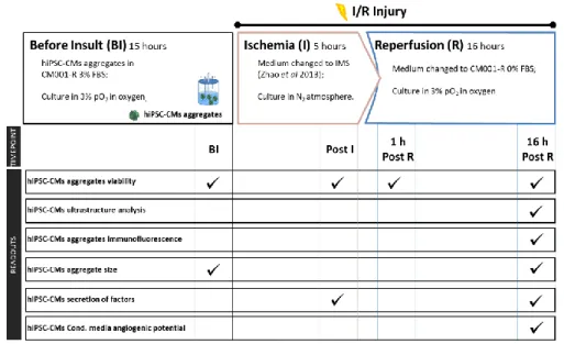
3.2. I/R setup in bioreactors ... 21
3.2.1. Generation of hiPSC-CM I/R Conditioned Media ... 22
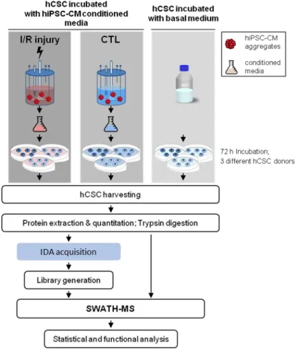
3.3. hCSCs culture ... 23
3.3.1. hCSCs source and maintenance ... 23
3.3.2. Incubation of hCSCs with I/R Conditioned Media ... 23
3.4. Cytokine/Chemokine quantification ... 23
3.4.1. Cytokine array ... 23
3.4.2. Growth factor quantification by ELISA ... 23
3.5. Scratch wound migration assay ... 24
3.6. HUVECs and tube formation assay ... 24
3.7. Determination of cell viability and concentration ... 24
Unveiling human Cardiac Stem Cells role in myocardial ischemia-reperfusion injury
3.7.2. Nuclei count ... 25
3.7.3. Fluorescent-based viability assay (FDA/Pi and NucView Staining) ... 25
3.7.4. Aggregate size determination ... 25
3.7.5. Immunofluorescence microscopy ... 25
3.7.6. Transmission electron microscopy ... 26
3.8. Mass Spectrometry... 26
3.8.1. Quantitative whole proteome analysis... 26
3.8.1.1. Generation of the spectral reference library ... 28
3.8.1.2. SWATH-MS analysis and targeted data extraction ... 28
3.8.1.3. Proteomic data analysis ... 29
3.9. Statistical analysis ... 29
4. Results and Discussion ... 31
4.1. Bioreactor I/R injury in 3D hiPSC-CMs aggregates ... 31
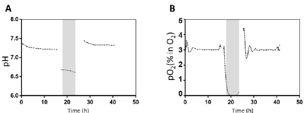
4.1.1. Bioreactor I/R injury setup ... 31
4.1.2. Effect of I/R injury on hiPSC-CMs ... 31
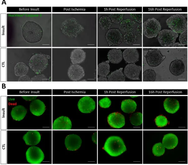
4.1.2.1. Effect of I/R injury on hiPSC-CMs aggregates’ viability ... 31
4.1.2.2. Effect of I/R injury on hiPSC-CMs aggregates’ phenotype and ultrastructure ... 33
4.1.2.2. Effect of I/R injury on hiPSC-CMs aggregates’ secretome profile ... 35
4.2. Human Cardiac Stem Cells’ response to hiPSC-CMs conditioned media ... 39
4.2.1. Effect on hCSCs migration ... 39
4.2.2. Effect on hCSCs proliferation ... 41
4.2.3. Effect on hCSCs’ secretion of IGF-1, HGF and CXCL6 ... 42
4.2.4. Effect on hCSC’s angiogenic potential ... 42
4.2.5. SWATH-MS of hCSCs after hiPSC-CM CondM incubation ... 43
4.2.5.1. Proteome profile of hCSCs incubated with hiPSC-CMs control conditioned media ... 44
4.2.5.2. Proteome profile of hCSCs incubated with hiPSC-CMs I/R injury paracrine factors ... 46
5. Conclusion ... 51
LIST OF FIGURES
List of Figures
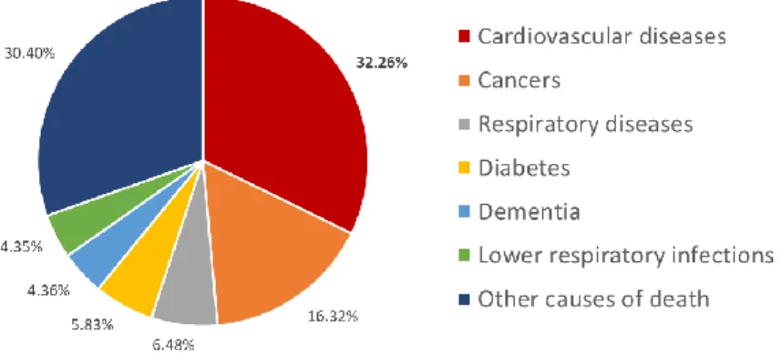
Figure 1.1 Share of deaths by cause in 2016. ... 1
Figure 1.2 Overall key mediators responsible for myocardial ischemia/reperfusion (I/R) injury. .. 4
Figure 1.3 Examples of different bioreactor systems used in cell culture ... 14
Figure 1.4 Generic workflow schematization of a mass spectrometry (MS) proteomics experiment ... 16
Figure 2.1 Thesis rational. Outlined strategy and major readouts.. ... 19
Figure 3.1 Schematic representation of Ischemia/Reperfusion injury (I/R) bioreactor-based experimental setup. ... 22
Figure 3.2 Experimental design and proteomic workflow. ... 27
Figure 4.1 pO2 and pH time-profiles throughout the I/R assay in bioreactors. ... 31
Figure 4.2 Viability of hiPSC-CMs aggregates subjected to the I/R injury. ... 32
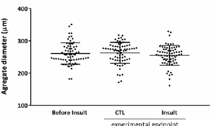
Figure 4.3 hiPSC-CMs aggregate size before and after I/R. ... 33
Figure 4.4 Characterization of cryosections of hiPSC-CMs aggregates. ... 34
Figure 4.5 Ultrastructural characterization of hiPSC-CM aggregates. ... 35
Figure 4.6 Secreted growth factors and cytokines by hiPSC-CMs aggregates during ischemia phase of I/R injury setup. ... 36
Figure 4.7 Secreted growth factors and cytokines by hiPSC-CMs aggregates during reperfusion phase of I/R injury setup. ... 37
Figure 4.8 Assessment of angiogenic potential of I/R bioreactors conditioned medium. ... 38
Figure 4.9 hiPSC-CMs conditioned media effect on hCSCs migration. ... 40
Figure 4.11 Secretion rate of IGF-1, HGF and CXCL6 factors by hCSCs after incubation with hiPSC-CMs conditioned media. ... 42
Figure 4.12 Assessment of angiogenic potential of hCSC supernatant after hiPSC-CMs conditioned media incubation. ... 43
Figure 4.13 Principal Component Analysis (PCA) biplot of hCSC samples, including technical and biological replicates. ... 44
Figure 4.14 Quantitative proteomic analysis and comparison between hCSCs incubated with basal medium and with hiPSC-CMs control conditioned medium. ... 45
Figure 4.15 Quantitative proteomic analysis between hCSCs incubated with Ischemia/Reperfusion injury (insult) and hiPSC-CMs control (CTL) conditioned media... 46
Figure 4.16 hCSCs activate pro-migratory and proliferation associated pathways. ... 48
Figure 4.17 hCSCs secrete factors that activate clathrin-mediated endocytosis mechanisms. . 49
LIST OF TABLES
List of Tables
Table 1.1 CSCs subtypes, molecular markers and transcription factors. ... 10 Table 4.1 Activated and inhibited canonical pathway and functions in hCSCs incubated with hiPSC-CMs Insult conditioned medium. ... 47
ABBREVIATIONS
Abbreviations
2D Two-dimensional
3D Three-dimensional
AMI Acute Myocardial Infarction
BMSCs Bone marrow mesenchymal stem cells CDCs Cardiosphere-derived CSCs
CMs Cardiomyocytes
CSCs Cardiac Stem Cells
CTL Control
CVD Cardiovascular diseases DIA Data-independent acquisition DMEM Dulbecco modified Eagle medium DNA Deoxyribonucleic acid
DPBS Dulbecco's phosphate-buffered saline ECGs Electrocardiogram
ECM Extracellular matrix
ECGM2 Endothelial Cell Growth medium 2 ELISA Enzyme-linked immunosorbent assay FBS Fetal bovine serum
FDA Fluorescein diacetate FDR False discovery rate FSG Fish skin gelatin
hCPC Human Cardiac Progenitor cells hCSCs Human Cardiac Stem cells hESC Human Embryonic Stem Cell
HF Heart failure
HGF Hepatocyte growth factor HIF-1 Hypoxia-inducible factor 1
hiPSC-CMs Human induced pluripotent stem cell derived cardiomyocytes HSP90 Heat shock protein 90
HUVECs Human umbilical vein endothelial cell iPSCs Induced pluripotent stem cells I/R Ischemia/Reperfusion
Unveiling human Cardiac Stem Cells role in myocardial ischemia-reperfusion injury
IDA Information-dependent acquisition IGF Insulin-like growth factor
IHD Ischemic Heart Disease IMS Ischemic mimetic solution IPA Ingenuity Pathway Analysis
LC-MS/MS Liquid chromatography-tandem mass spectrometry
miR microRNA
mPTP Mitochondrial permeability transition pore
MS Mass Spectrometry
NSTEMI non-ST segment elevation myocardial infarction PCA Principal component analysis
PCI Percutaneous coronary intervention
PI Propidium iodide
PSC Pluripotent stem cells ROS Reactive oxygen species
SP CSCs Side population cardiac stem cells STBR Stirred-tank bioreactor
STEMI ST-segment elevation myocardial infarction
SWATH-MS Sequential Window Acquisition of All Theoretical Mass Spectrometry Sca1 Stem cell antigen-1
TEM Transmission electron microscopy TOF Time-of-flight
INTRODUCTION
1. Introduction
1.1. Acute Myocardial Infarction
Cardiovascular diseases (CVD), a group of disorders of the heart and blood vessels, are currently the main cause of death worldwide, accounting for 17.3 million deaths per year (Figure 1.1) [1]. By the year 2030, this number is estimated to grow to more than 23.6 million deaths [1]. Within CVD, ischemic heart disease (IHD) is the largest contributor of mortality and includes diseases such as stable and unstable angina, sudden cardiac death and acute myocardial infarction (AMI) [2].
Figure 1.1 Share of deaths by cause in 2016. (adapted from Institute for Health Metrics and Evaluation, Global Burden of Disease, https://ourworldindata.org/causes-of-death, accessed on July 05th 2018)
AMI, commonly known as a heart attack, consists in the sudden decrease or cessation of blood flow to a part of the heart muscle, causing local ischemia. The most common cause of AMI is the complete blockage of a coronary artery triggered by the rupture of an atherosclerotic plaque (i.e. buildups of fatty deposits) with the formation of a vascular blood clot (i.e. a thrombus) [3]. This leads to changes in the balance between oxygen and nutrient supply of the affected myocardium causing death to the heart muscle cells - the cardiomyocytes (CMs) - and consequently myocardial tissue damage [4].
In order to help diagnose AMI correctly, a different number of tests can be performed, including coronary angiography to identify intra-coronary thrombus, blood tests to track specific markers in the serum such as cardiac troponins and/or creatine kinase MB and electrocardiograms (ECGs) [5]. Regarding the heart’s electrical activity visualized by ECG, an AMI can be generally classified in two different types [6]: a ST elevation Myocardial Infarction (STEMI) usually caused by a fully blocked artery and a non-ST segment elevation myocardial infarction (NSTEMI) which normally results from a partially clogged one [7].
After an AMI, rapid transportation to the hospital followed by initial assessment and treatment are crucial since the increasing extension of cell death (e.g. necrosis) and prehospital cardiac arrest play major roles in the patient’s prognostics. The initial management of acute cardiac
Unveiling human Cardiac Stem Cells role in myocardial ischemia-reperfusion injury
disorders comprises bed rest, patient’s monitoring by ECG and early administration of antithrombotic agents [8].
1.1.1. Ischemia/Reperfusion Injury
During AMI, the deficient blood flow is responsible for the ischemic condition. Therefore, the treatment of choice (later discussed in section 1.1.2.) is the restoration of coronary blood flow through reperfusion. However, reperfusion strategies, although needed, exacerbate the damage even further, thus the overall damage is usually referred as Ischemia/Reperfusion (I/R) injury.
Myocardial I/R injury was first observed in 1960 by Jennings et al. in canine hearts after observing that reperfusion enhanced the degree of necrosis [9]. The consequences of an I/R injury evolve in a time-dependent manner, starting with exacerbated oxidative stress, overload of intracellular Ca2+, inflammation and then quickly reaching to irreversible cell death by mechanisms such as necrosis and apoptosis [10, 11].
1.1.1.1. The Ischemic phase
Ischemia is a condition that is characterized by the decrease or restriction of the blood flow (and therefore of oxygen) in the tissues. Regarding cardiac ischemia, the deficiency in oxygen causes irreversible cell death (e.g. in CMs) within minutes of the ischemic event [12].
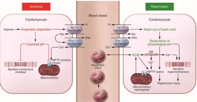
The deprivation of nutrients and oxygen supply leads to severe changes in terms of CMs metabolism and biochemical pathways within the myocardial tissue. Since no oxygen is available, CMs metabolism shifts from oxidative phosphorylation to anaerobic glycolysis, leading to energy depletion by the reduction of ATP (via degradation and defective synthesis), to the depolarization of the mitochondrial membrane and the inhibition of myocardial contractile function. The prevalence of anaerobic metabolism is also responsible for the reduction of intracellular pH caused by the production and accumulation of lactate, NAD+ and protons [12, 13]. The subsequently decrease in ATP deranges the activity and function of ion transporters (Na+-H+ and 2Na+-Ca2+ exchangers) within the cell, that overall result in an overload of intracellular Ca2+ which can contribute to cell death following I/R (Figure 1.2) [14].
1.1.1.2. The Reperfusion phase
Current reperfusion strategies, although crucial to improve the AMI patients’ health condition, are considered a “mixed blessing” [15]. Even though the reperfusion intervention restores the nutrients and oxygen required for the generation of ATP through aerobic metabolism and also normalizes the extracellular pH by washing out the accumulated protons, it comes with severe consequences [13]. During this phase, several mediators contribute to cell death, including oxidative stress, the overload of intracellular Ca2+, the opening of the mitochondrial permeability transition pore (mPTP) and inflammation [12] (Figure 1.2).
INTRODUCTION
Oxidative stress, i.e. the imbalance of antioxidant and oxidant species, is caused by the burst production of reactive oxygen species (ROS) mainly by the CMs’ mitochondria with the activation of the electron transport chain [12] and the decrease in the ability of ROS scavenging leading to cellular damage in CMs [16]. This oxidative stress (either caused by ROS or reactive nitrogen species) leads to damage in DNA, lipids and proteins [17]. These compounds can also increase proteolysis of proteins involved in the cardiac excitation-contraction coupling [10, 17]. Oxidative stress during reperfusion can also decrease the bioavailability of nitric oxide, an intracellular signalling molecule, that has an important role in ROS inactivation (e.g. superoxide radicals) and in the improvement of coronary blood flow [18].
The intracellular Ca2+ accumulation that was initiated during the ischemic period is aggravated after reperfusion due to mitochondrial reenergization, plasma membrane disruption and oxidative stress damage induced to the sarcoplasmic reticulum [12]. The consequential Ca2+ accumulation in the mitochondrion’s matrix, together with the sudden return to the physiological pH and the generated ROS, leads to the opening of the mPTP, a nonselective channel localized in the inner mitochondrial membrane [19, 20]. While the mPTP remains closed during ischemia due to acidic conditions (pH<7) [21], during reperfusion, its opening causes the depolarization of the mitochondrial membrane, CM’s hypercontracture [22] and the uncoupling of oxidative phosphorylation inhibits the production of ATP, ultimately leading to cell death [23, 24]. The pore opening also leads to the entry of water and solutes which contribute to the swelling of the mitochondrial matrix. Even though the inner mitochondrial membrane can outstand the increase of volume, the outer membrane is ruptured and all the content inside the intramembranous space, including apoptogenic signals (e.g. apoptosis-inducing factor and cytochrome c) are released into the cytosol where apoptosis is initiated by the activation of specific apoptogenic proteases (e.g. caspases) [19]. Overall, the mitochondria can act as “fate decider” within the cell: if the damage is severe, the cell may undergo necrosis due to failure in energy production; if the damage is moderate, the cell may die by apoptosis; and if the damage is minimal the cell may recover [25]. Inflammation also plays a role in myocardial I/R injury and, since it is induced in the absence of microorganism invasion, it is usually referred to as sterile inflammation. Besides promoting apoptotic cell death, ROS also increase the expression of adhesion molecules, activating the complement system (a biochemical cascade of the innate immune system) and acting as a chemoattractant for neutrophils [26]. Neutrophils are also activated through the release of endogenous molecules from necrotic and injured cells within the myocardium, including CMs and endothelial cells [27]. The inflammatory environment created by the activity of these immune system mediators contributes to further cell death and myocardial injury by prolonging the pro-inflammatory response [28].
Unveiling human Cardiac Stem Cells role in myocardial ischemia-reperfusion injury
Figure 1.2 Overall key mediators responsible for myocardial ischemia/reperfusion (I/R) injury. Key mediators include oxidative stress, Ca2+ overload, opening of the mitochondrial permeability transition pore (mPTP) and inflammation. Briefly, the anaerobic respiration caused by the ischemic event leads to an acidic intracellular pH. This contributes to the derangement of ion transporters function leading to Ca2+ overload. The mPTP remains closed and cardiomyocyte contracture is inhibited due to the decrease in pH. Reperfusion allows the mitochondrion reenergization, creating reactive oxygen species (ROS) that induce oxidative stress and act as chemoattractant for neutrophils. During reperfusion, the pH is also re-established, inducing the opening of the mPTP and cardiomyocyte hypercontracture (reproduced from [12]).
The inflammatory response also takes part in the wound healing and scar formation process. For the healing of the damaged myocardial tissue and scar formation upon AMI, a three-phased cellular response occurs: (1) an inflammatory phase with a quick influx of immune cells to help remove damaged tissue; (2) a reparative phase where myofibroblasts start proliferating and secreting collagen to replace dead tissue; (3) and a maturation phase marked by the apoptosis of immune cells, crosslinking of collagen fibers and scar maturation (reviewed in [29]).
During the reparative phase of inflammation, the process of fibrosis occurs. This process consists in the excess formation and deposition of extracellular matrix (ECM) components (e.g. collagen) that promote tissue hardening and scarring [30]. It can be divided in two types: i) replacement fibrosis and ii) reactive fibrosis, both mediated by myofibroblasts. Replacement fibrosis is the mechanism that replaces necrotic CMs by fibrotic tissue. On the other hand, reactive fibrosis is characterized by the abnormal deposition of ECM in the heart’s interstitium in areas remote to the infarction, induced by paracrine and hormonal mediators together with the mechanical stress caused by the AMI. While replacement fibrosis is essential to prevent cardiac rupture, an exaggerated fibrotic response and reactive fibrosis in the adjacent zone of the injured areas contribute to impairment of the heart function [31]. The newly formed fibrotic scar tissue increases the stiffness of the heart, impairs the oxygen diffusion and its lesser conductive nature leads to electrophysiological remodelling. These factors also contribute to the diminishing of ventricular function, the increase of CMs’ workload and arrhythmias [32] leading to chronic Heart Failure (HF) [33]. Regarding chronic HF patients, heart transplantation is the only viable treatment
INTRODUCTION
to restore function which comes with logistic, economic and biological limitations derived from its high costs, the need for constant immunosuppressive agents and the shortage of organ donors [34].
1.1.2. Strategies in AMI treatment
1.1.2.1. Current therapeutic strategies
For patients with a clinical diagnose of a STEMI, restoration of coronary blood flow and myocardial reperfusion should be done as soon as possible. This reperfusion is either done mechanically by percutaneous coronary intervention (PCI) or pharmacologically by intravenous fibrinolytic therapy. Primary PCI (angioplasty and stenting), an urgent catheter intervention PCI without previous administration of fibrinolytic agents, is the chosen strategy for reperfusion in patients with STEMI [8, 35]. If the patient is medically suggested as unsuitable for PCI, a Coronary Artery Bypass Grafting surgery may be indicated [35]. In this procedure, a healthy vein or artery is used to bypass the blocked portion of the coronary artery.
Regarding the NSTEMI diagnoses where residual perfusion still exists, the patients can either follow the same invasive treatment strategies that are used in STEMI or undergo an ischemia-guided strategy. The latter approach seeks to avoid the routinely early use of coronary angiography and revascularization by the possible stabilization of the patient’s condition. However, if the patient’s condition fails to stabilize and severe ischemia is detected they are promptly redirected to undergo the mentioned invasive treatments [36].
1.1.2.2. Emerging strategies
Reperfusion treatments are continuously being improved with the advances in PCI technology, with earlier reperfusion and the use of more efficient antithrombotic and antiplatelet drugs. Emerging strategies on preventing I/R injury outcome are being tested in randomized clinical trials as well as in proof of concept studies (reviewed in [12]).
Ischemic postconditioning, in contrast to uninterrupted reperfusion, has been reported to attenuate the reperfusion injury and to reduce the infarction size by 40% to 50% [37]. This technique is a modified form of reperfusion created in the 1980s that has been demonstrated to be a beneficial form of gradual reperfusion [38, 39].
Preconditioning, i.e. repetitive angioplasty balloon inflations and deflations, although with known effectiveness, is not favourable for clinical translation since this therapy must be done prior to the known ischemic event [40].
Primary PCI and ischemic postconditioning are both invasive interventions that are directly applied to the heart. An alternative strategy called remote ischemic conditioning, consists on brief and transient cycles of ischemia and reperfusion to another organ or location (e.g. upper arm), protecting the heart from distance against an I/R injury [41, 42].
Unveiling human Cardiac Stem Cells role in myocardial ischemia-reperfusion injury
The practice of hypothermia (low temperature) [43] and hyperoxemia (increased arterial O2 partial pressure) [44] as therapeutic interventions have also shown beneficial effects on reducing reperfusion injury in animal studies [12].
Several pharmacological drugs are also being investigated against I/R injury. Such agents include atorvastatin, atrial natriuretic peptide, erythropoietin or glucagon-like peptide 1 and act by activating the RISK (Reperfusion Injury Salvage Kinase) pathway (reviewed in [45])). This pathway comprises a group of pro-survival protein kinases that are activated during myocardial reperfusion and confer cardiac protection [46].
Unfortunately, these novel cardioprotective therapeutics that target the individual I/R injury key players (e.g. calcium overload, pH correction, inflammation and oxidative stress) have resulted in disappointing clinical translation [47]. Nowadays, advanced strategies of treatment based in regenerating myocardial tissue, such as cell therapy, are being studied. Stem cell therapy can provide new treatment alternatives focused not only in the replacement of affected cells but also on the release of autocrine and paracrine factors that ultimately lead to cell survival, tissue remodelling and angiogenesis [48].
1.2. Stem Cells
Stem cells are undifferentiated and unspecialized cells that play a crucial role in both tissue homeostasis and regeneration. They are able to self-renew into identical daughter cells (clonogenic) by undergoing cell division and are also capable of differentiating into more mature specialized cell types [49]. These cells can be divided into different categories depending on their ability to differentiate (i.e. potency). For instance, totipotent stem cells that are able to develop a complete organism (e.g. zygote and early stages of blastomere), pluripotent stem cells including Embryonic Stem Cells (ESCs) or induced Pluripotent Stem cells (iPSCs) that are capable of forming any cell type from the three primary cell germ layers (ectoderm, mesoderm, and endoderm) and lastly, multipotent stem cells which are only capable of differentiating into cell types from a specific cell lineage [50].
Due to their inherent paracrine properties and differentiation potential, a lot of research has been developed in the possible transplantation of different types of stem cells and its derivatives into AMI patients. Cell populations including skeletal myoblasts, the “first generation” of stem cells such as bone marrow mesenchymal stem cells (BMSCs) and the “second generation” of stem cells including derivatives of ESCs and iPSCs (e.g CMs) and Cardiac Progenitor/Stem Cells (CSCs) are currently being explored [51].
Skeletal myoblasts, although not a type of stem cell, were the first cell type to be used for the treatment of damaged myocardium. However, even though studies performed in animal models such as the rabbit [52] or small sized studies in humans [53] showed promising results, the potential use of these cells for cardiac repair is diminished given the associated risk of cardiac arrhythmias [54].
INTRODUCTION
BMSCs are currently the most used cell type in clinical trials. The early data obtained from clinical trials showed beneficial results, however, the long-term results remain inconclusive. For instance, REPAIR-AMI trial (NCT00279175) showed an improvement in myocardial function following the intracoronary delivery of autologous BMSCs while on the other hand, BOOST trial (NCT00224536) showed little effect in the long run [54].
Regarding pluripotent stem cell derivatives, in vivo studies have been performed in rat models using ESCs-derived progenitor cells embedded in a fibrin scaffold that resulted in improved cardiac function [55]. There is also a clinical trial ongoing, ESCORT trial (NCT02057900), that aims to use hESCs-derived cardiac progenitors in severe HF. While similar studies for CVD using iPSCs-derivates are not being performed, they should be expected anytime soon since differentiation protocols are effective in both type of cells. However, iPSCs-based studies in humans should be performed with further caution due to the additional mutation risk inherent to cell reprogramming [56].
1.2.1. Cardiac Stem Cells
The mammalian heart was previously thought to be a terminally differentiated post-mitotic organ without any regenerative potential. In 2003, Beltrami and his colleagues first isolated and expanded CSCs in the adult rat heart, which were able to differentiate into the myogenic cell lineage (CMs, vascular smooth muscle cells and endothelial cells) both in vitro and in vivo and improve cardiac dysfunction in the infarcted heart [57]. In the following year, Messina and his group successfully identified a type of clonogenic cell in cardiospheres in both human and murine heart which shared similar properties of adult CSCs and expressed stem cell antigens [58]. Cardiospheres (later defined in section 1.2.1.2.) are a multicellular cluster that contain not only CSCs, but also supporting cells and partially differentiated progenitor cells [59].
The discovery of this multipotent cell population in the human heart with regenerative potential led to a paradigm shift in cardiovascular biology. Since the myocardium harbours CSCs that can be isolated and expanded in vitro, new therapies that exploit such endogenous regenerative potential could emerge with the goal of recovering or replacing damaged cardiac muscle [60].
1.2.1.1. Cardiac Stem Cells in transplantation
Given the characteristics of CSCs, researchers have undertaken much effort in the past years to evaluate their potential in cardiac repair and regeneration and also its effectiveness, safety and feasibility in cell transplantation therapies. For instance, Dawn et al. reported a decrease of 29% in myocardial infarction size after the transplantation of CSCs into a rat model after reperfusion, concluding that these cells could both limit infarct size and induce myocardial regeneration [61]. Other studies in larger animal models, such as the pig (with greater similarity in terms of tissue physiology and organ size to the human), reported similar results. Johnston and
Unveiling human Cardiac Stem Cells role in myocardial ischemia-reperfusion injury
his colleagues revealed that treatment using cardiosphere-derived CSCs (CDCs) by intracoronary infusion in infarcted pigs also limited infarct size, reduced adverse cardiac remodelling, formed new myocardial tissue and improved the hemodynamics [62].
Since these studies have shown that CSCs transplantation could enhance cardiac regeneration, the next logical step was to perform clinical trials using allogenic (i.e. donor derived) and autologous (i.e. self-derived) CSCs. These trials include SCIPIO (NCT00474461) using autologous c-kit+ CSCs, CADUCEUS (NCT00893360)) with autologous CDCs, ALCADIA (NCT00981006) also using autologous CDCs together with the controlled release of basic fibroblast growth factor (bFGF), ALLSTAR (NCT01458405) using allogenic CDCs’ therapy and more recently CAREMI trial testing allogenic c-kit+ CSCs (NCT02439398).
Although these clinical trials promoted growing evidence that the use of CSCs exert some effects in improving myocardial function on infarcted patients, the transplantation of these cells showed somewhat disappointing results. Only a few percentage of the transplanted CSCs could be successfully engrafted in the affected myocardium. This low retention rate is mainly due the harsh microenvironment created by the infarction itself where the necrotic myocardium together with the inflammatory response, restrict the growth, survival and homing of the infused cells, thus affecting their capability of regenerating the heart [63].
To overcome this limitation, research has been focused on strategies to enhance the survival and/or improve of the engraftment rate of CSCs upon transplantation, including the use of biomaterials (e.g. cell sheets), hydrogels and porous scaffolds [64]. Alternative cell-free approaches bring advantages over transplantation therapies: (1) it would be more affordable regarding manufacturing costs, (2) it would be easier to apply and implement in the clinics like the widespread use of PCI and (3) its therapeutic agents would be “off the shelf” available, thus reducing treatment initiation time [65].
1.2.1.2. Endogenous Cardiac Stem Cells Populations
CSCs are located in hypoxic areas within the myocardium [66], usually clustered in niches with other types of early differentiated heart cells and adult CMs. It’s estimated that the adult mammalian heart harbours around one CSC per 8000 to 20000 CMs [67]. By comprising only 0.005 to 2% of the adult cardiac cells, these cells are considered rare in vivo, making challenging to study and understand their physiological roles [68].
Several CSCs populations have been identified and characterized according to their phenotypic properties and surface molecular markers (Table 1.1). These different subpopulations include c-kit+ CSCs, CDCs, Sca-1+ CSCs, side population CSCs (SP CSCs) and Islet-1+ CSCs [60].
INTRODUCTION
C-kit+ CSCs:
These cells were the first to be described as CSCs [57]. C-kit+ CSCs appear in small clusters within the interstitium of the ventricular and atrial myocardium with highest density in the atria and the ventricular apex [69]. C-kit+ CSCs are negative for blood lineage markers and are characterized by the absence of CD45 and CD34 and expression of the stemness marker c-kit. Additionally, they express transcription factors associated with early cardiac development such as GATA-4, MEF2C and Nkx2.5 [68].
Sca-1+ CSCs:
This type of CSC is characterized by the expression of the endothelial marker stem cell antigen-1 (Sca1) and the absence of c-kit and blood lineage markers. These cells express cardiac transcription factors such as GATA4, MEF2c and TEF1 but lack other cardiac lineage markers like NkX2.5, hematopoietic markers CD45 and CD34, and mature endothelial markers such as CD31 [70].
Islet-1+ CSCs:
CSCs within this subpopulation are characterized by the expression of cardiac transcription factors GATA4 and Nkx2.5 but do not express c-kit, CD31 and Sca1 markers [71]. This subpopulation can only be found in neonatal and fetal tissues, existing in low or non-existing amounts in the adult mature hearts, which limits their potential use in clinical applications [70]. Cardiosphere-derived CSCs:
Cardiospheres are self-assembling multicellular spheroids originated from the cellular outgrowth (i.e. cells in culture growing out of an explant) from cardiac tissue explants cultured in suspension [72]. The core of this spheroids contains proliferating cells, including c-kit+ cells whilst the outer layer is composed of mesenchymal cells and differentiating cells that express cardiac, stromal and endothelial cell markers [69, 72].
Side Population CSCs:
SP CSCs have an immature phenotype representing around 1% of all cells in the human heart. They are identified using flow cytometry based on their ability to efflux DNA binding dyes (e.g. hoechst dye) through an ATP-binding cassette transporter [68]. This population overlaps with Sca-1+, sharing the expression of the cardiac transcription factors like GATA4 and Nkx2.5 with no myofilament or hemopoietic markers expression [68]. Consequently, SP CSCs can be subdivided into two different populations according to their phenotype, namely, CD31-/Sca-1+SP CSCs and CD31+/Sca-1+ SP CSCs. However, only CD31-/Sca-1+SP CSCs show considerable cardiomyogenic potential [60].
CSCs can also be obtained using other strategies, for instance, by the differentiation of ESCs [73] and iPSCs [74].
Unveiling human Cardiac Stem Cells role in myocardial ischemia-reperfusion injury
Table 1.1 CSCs subtypes, molecular markers and transcription factors. (reviewed in [75]).
CSCs subtype
Molecular markers and Transcription factors:
c-kit+ c-kit+, CD34-, CD45-, Sca-1+, Abcg2+, CD105+, CD166+, GATA4+, Nkx2.5low, MEF2C+ Sca-1+ Sca1+, CD34-, CD45-, FLK1-, c-kitlow, GATA4+, Nkx2.5low, MEF2C+
SP CD34+, CD45+, Abcg2+, Sca1+, c-kit+, Nkx2.5-, GATA4 -CDCs CD105+, CD34+, CD45+, Abcg2+, Sca1+, c-kitlow
Islet+ Islet+, CD31-, Sca1-, c-kit-, GATA4+, Nkx2.5+
1.2.1.3. Endogenous Cardiac Stem Cells in cardiac regeneration
Normally, the endogenous CSCs residing in the adult heart are in a quiescent state and only a few are active to maintain tissue homeostasis by replacing cardiac cells such as CMs and vascular cells damaged by wear and tear. However, a fraction of endogenous CSCs can quickly become activated upon environmental stimuli (e.g. ischemic injury [76], stress, hypoxia, exercise and work overload) thus contributing to cardiac regeneration by paracrine action and by proliferating and differentiating towards new cardiac muscle and coronary vessels [77-79]. Regarding whether CSCs actually differentiate or not into new cardiac cells remains controversial. The controversy started when Orlic et al. [80] reported back in 2001 that c-Kit+ cells (in this case, bone marrow-derived cells) had the capability of regenerating infarcted myocardium. Three years later, Murry et al. reported otherwise using the same cell type [81]. The heat of the controversy seemed to have settled down with the narrower definition of CSCs, being it only c-kit+ cells that resided in the heart (excluding the circulating c-kit+ cells). However, in 2012, Jesty and his colleagues [82] reported that c-kit+ CSCs did not contribute to new CMs and a year later Ellison et al. [83] demonstrated that CSCs could differentiate into different cardiac cells and were the main source of regeneration in both human and mouse hearts, thus, reigniting the controversy (reviewed in [84]). More recently, using genetic lineage tracing, it has been demonstrated that c-kit+ CSCs minimally contribute to new CMs formation either in post-injury regeneration or in normal conditions [85].
1.2.1.3.1. Mechanisms of action of Cardiac Stem Cells
Cardiac differentiation was thought to be the key event in the success of an eventual therapy based on CSCs. Today, it’s widely accepted that the production and secretion of paracrine modulators (e.g. cytokines, growth factors and exosomes) induced by paracrine cross-talk are the main factors responsible for the reported beneficial effects of CSCs’ transplantation studies [86]. In fact, as mentioned above, while it’s still controversial whether CSCs can differentiate and generate new CMs upon injury [85], the potential towards cardiac regeneration via paracrine communication was already well recognized in many animal and in vitro studies. For example, Park et al. reported that mouse Sca-1+/CD31- CSCs secreted factors such as MCP-1, EGF, TGF-β1, IGF-1 (Insulin-like growth factor I), IGF-2, HGF-R and IL-6 that protected CMs from hypoxia after incubating them with conditioned media [87]. More recently, Sebastião et al. reported in an
INTRODUCTION
in vitro human myocardial I/R injury model, an increased secretion of the angiogenic chemokine CXCL6 [88] by CSCs. In this study, a co-culture of human cardiac stem cells (hCSCs) and CMs derived from human induced pluripotent stem cells (hiPSCS-CMs) was used to show CXCL6 relevance in the CSCs paracrine-mediated cardioprotection [Sebastião et al. submitted].
By taking advantage of paracrine communication between endogenous CSCs and injured cardiac cells, novel cell-free therapies that stimulate the heart’s endogenous regeneration mechanisms based on the use of paracrine modulators have been explored. Ellison et al. tested a cocktail of growth factors, combining IGF-1 and hepatocyte growth factor (HGF) injected intracoronarily during reperfusion in pig models of AMI. The authors reported a regenerative response from c-kit+ CSCs leading to the regeneration of the damaged myocardial tissue, reduction of fibrosis and increased CMs survival rate. Both HGF and IGF-1 activated the endogenous CSCs, contributing to their proliferation, migration and differentiation into CMs, smooth muscle cells and endothelial cells [89]. In fact, IGF-1 had been previously reported to be released by CMs in stress (e.g. infarction) [90]. Similar improved cardiac function was reported by Koudstaal and his colleagues using the same molecules administered trans-endocardial in a hydrogel carrier [91].
Exosomes (membrane vesicles ranging from 40 to 100 nm in size), are also vehicles of paracrine signals produced by CSCs. CDCs were reported to produce exosomes that may induce angiogenesis and also proliferation of CMs and apoptosis inhibition. These vesicles can hold micro RNAs (miR), such as miR-146a, that have been shown to induce some cardioprotective effects in murine myocardial models of AMI [92]. Other examples of miRs that boost cardiac repair are miR-17 and miR-19a that belong to the miR-17-92 cluster. The overexpression of this cluster has been shown to reduce the injury of a myocardial infarction in adult CMs. The joint use of miR-590 and miR-199a (in lipid formulations) reduced infarct size and improved overall cardiac function in mouse after myocardial infarctions (reviewed in [93]). More recently, Gallet et al. reported a decrease in scarring and in adverse cardiac remodelling in pigs after intramyocardial injection of human CDCs exosomes after reperfusion [94], making exosomes another conceptual attractive cell-free therapeutic agent.
To further understand the mechanism of action of CSCs upon AMI, there is a clear need for the development of relevant and predictable I/R models and robust analytical tools for detailed cell characterization.
1.3. I/R models
The development of disease models plays a critical role in biomedical research. Disease models aim to understand the molecular basis of human diseases in order to improve and develop new forms of diagnosis and novel therapeutics. The currently available myocardial I/R injury models include in vivo (animal models), ex vivo (e.g. Langendorff model) and in vitro (e.g. cell-based assays) approaches.
Unveiling human Cardiac Stem Cells role in myocardial ischemia-reperfusion injury
Over the past century, myocardial injury has been studied by the development of numerous animal models [95]. Large animal models include the pig and dog, whereas small animal models include rabbits and rats [96]. However, animal experimentation comes with high costs, ethical issues and, although in animal models biochemical interactions and mechanisms are present, they often differ from the human disease phenotype [97]. This inability to completely mimic human disease, is likely the reason why many drugs fail to translate the efficacy and safety when advancing from animal studies to human clinical trials [98]. Thus, the magnitude of the studies are beginning to rely on the use of cultured human cells, including primary cells, cell lines and derivatives of PSCs [99]. However, while primary human CMs would be the ideal cell source for an in vitro model, their short availability, low to no proliferation capacity and poor consistency limit their use. Given this, other cell types, including CMs derived from ESC and iPSCs are more prevalent in vitro models [100, 101].
I/R in vitro models induce artificial conditions to simulate the environmental features of both ischemia and reperfusion phases by exposing cells to hypoxia and reoxygenation. Strategies used in I/R models include the culturing of cells in hypoxic sealed chambers (to simulate ischemia) or incubators (to mimic both ischemia and reperfusion depending on the oxygen content). For instance, Li et al. performed this strategy using neonatal rat CMs, applying a three-hour period of both ischemia (5% CO2 and 95% N2) and reperfusion (5% CO2 and 95% O2) [102]. Kang et al. also followed the same strategy using adult rat primary CMs, applying a six-hour period of ischemia followed by reoxygenation [103]. For further oxygen scavenging, hypoxic chambers can also contain deoxygenation reagents that lead to the O2 consumption and production of CO2 (e.g. Pack-Anaero) [104] and culture media can feature enzymatic oxygen scavengers (e.g. EC-Oxyrase®) which act by reducing molecular oxygen to water [105]. Other I/R in vitro strategies include the removal of media components (e.g. nutrients) and the removal of oxygen from the culture media through bubbling with nitrogen. Xu et al. followed this approach using H9c2 (a rat heart myoblast cell line) cell line by removing both glucose and fetal bovine serum (FBS) from the media during ischemia and then culturing cells under normoxic conditions in nutrient rich media during reperfusion phase [106] while Li et al. followed the same setup with the addition of a serum deprivation step prior to the I/R experiments [107]. Other approaches include the culturing of cells in ischemic mimetic solutions (IMS) which induce acidosis, lactate accumulation, glucose deficiency and hyperosmosis, thus better mimicking the pathophysiological state of ischemia. Zhao et al. used IMS to simulate ischemia in H9c2 cell line [108] and more recently, Sebastião et al. applied the same formulation in an heterotypic cell-based human model ofhiPSC-CMS and hCSCs [Sebastião et al. submitted]. Other human cell types have also been subjected to I/R, including CMs derived from ESCs [109].
However, despite the attempt to correctly mimic in vivo features of AMI, most I/R models are 2D (planar) disease models which fail to recapitulate the complexity of tissue architecture, that is of major importance in disease pathophysiology, and are mostly based on a single cell type. Therefore, there is a need to create and improve more relevant human cell-based experimental in vitro settings that better recapitulate tissue physiology.
INTRODUCTION
1.4. Bioreactor technology and 3D cell culture
1.4.1. 3D cell culture strategies
To produce a robust in vitro disease model, the combination of physiological relevant cell source together with a culture system that allows the recapitulation of in vivo architecture is needed.
Most of cell-based assays and screenings are performed in two-dimensional (2D) systems. 2D cell cultures rely on the cultivation of a monolayer of cells adherent to a flat and rigid surface (e.g. glass petri dish, polystyrene plates) [110]. However, in 2D cell culture, only a portion of cells are interacting with their neighbour cells and the ECM, which can lead to incorrect cell polarization affecting phenotypic cell fate and pathway signalling (reviewed in [111]).
On the other hand, 3D cell culture systems are able to establish the physiological cell-cell and cell-ECM interactions [112] which mimic the 3D communication network that is responsible for the homeostasis and specificity of the in vivo tissues [113]. Furthermore, 3D cultures recapitulate more closely the tissue microenvironment by influencing intracellular signalling that contributes to phenotypic cell fate by changing both gene and protein expression [114].
There are many methods to culture anchorage-dependent cells in 3D suspensions, including cell adhesion on microcarriers, cell encapsulation in hydrogels and self-aggregated spheroids. Microcarriers are small spherical particles that allow the growth of adherent cells while cell encapsulation comprises the entrapment of cells in a semi-permeable membrane. Lastly, self-aggregated spheroids are a scaffold-free strategy that induce cell-cell interaction by promoting cell self-organization into 3D structures, known as spheroids or aggregates (reviewed in [115]). Some examples are already reported for 3D cardiac in vitro cell models. For instance, using the hanging-drop method, where self-aggregation is induced by gravity, cardiac models were developed using a triculture of CMs, endothelial cells and fibroblasts to study the heart microenvironment [116] and cardiac fibrosis [117]. In terms of myocardial I/R 3D models, a 3D paper-based culture system was developed using primary CMs and fibroblasts cultured in stacked layers of paper to study fibroblasts’ migratory response caused by CMs under ischemic stress [118]. Other example, was the development of engineered heart tissue where CMs were cultured in a ring-shaped scaffold to study cardiac function under hypoxic conditions [119].
In order to move from 2D culture systems to 3D, a change from static culture conditions to a dynamic system is needed. Dynamic culture systems include shaking erlenmeyer’s and bioreactors, where the latter, allows the managing of cell culture environment that is required for the development of disease models. In this context, bioreactors have emerged as the optimal setting for reproducible and scalable 3D cell culture, having already been reported to improve cell survival, differentiation efficiency and proliferation rates over 2D systems [120, 121].
Unveiling human Cardiac Stem Cells role in myocardial ischemia-reperfusion injury
1.4.2. Bioreactors for culture of advanced cell models
Bioreactors are defined as devices that enable the tight control and monitoring of environmental parameters required for cell culture (e.g., pH, oxygen, pressure, temperature, nutrient supply and metabolite removal) [122]. There are several types of bioreactors, including rotating wall vessels, stirred-tank bioreactors (STBR), airlift bioreactors, hollow fiber bioreactors and wave bioreactors [123, 124] (Figure 1.3).
Figure 1.3 Examples of different bioreactor systems used in cell culture: a) rotating wall vessel; b) stirred-tank bioreactor (STBR); c) airflow bioreactor (adapted from [125]); d) hollow fiber bioreactor (adapted from [122]) and e) wave bioreactor (adapted from [126]).
Rotating wall vessels are a rotatory cell culture system providing a low shear stress culture with high rates of mass transfer. By having two cartridges, one outer cartridge that’s fixed and an inner one that rotates around the central axis, the cultures are suspended by the creation of the microgravity environment of space. STBR feature a vessel, valves, pipes and a motor. The stirring of the culture is provided by an impeller powered by the motor and in the top of the vessel, several sensors and probes can be attached (e.g. temperature sensors, pH and dissolved oxygen probes, metabolite measuring devices). Airlift bioreactors are a type of culture system that produce lower shear stress when comparing to stirred-tank bioreactors since both the oxygenation and culture mixing is performed by a stream of air. The hollow fiber bioreactors are made of semi-permeable membranes arranged in parallel arrays allowing diffusion of nutrients and oxygen between the intra-capillary space within the hollow fibers and the extra-capillary space. The wave bioreactor comprises a disposable plastic bag and a platform responsible for a wave-like motion that promotes the mixing in the culture medium (reviewed in [115, 123]).
INTRODUCTION
Even though there is a lack of reported myocardial I/R modelling based on bioreactors, the feasibility of their use on disease models has already been proven using cells from other tissues. For instance, Allen et al. developed a perfusion bioreactor system that allowed the study of the effect of oxygen gradients in primary rat hepatocytes [127] while Santos et al. exposed a brain model of 3D neural aggregates to hypoxia in STBR [128]. Hypoxia has also been combined with STBR and wave bioreactors by Correia et al. but in this case, for the yield improvement in the differentiation of murine iPSC into CMs [129].
Another main advantage of some bioreactor types (e.g. STBR) is the ability of withdrawing samples in a non-destructive way [115]. With this, several assays can be performed including metabolite analysis, live/dead assays, estimation of cell concentration and -omics analysis of cell phenotype and conditioned medium, such as proteomic profiling methods.
1.5. Proteomics
Since its known that cardiac regeneration can be mediated via paracrine signalling (discussed above on section1.2.1.3.), the use of proteomic tools (both whole proteomics and secretome, i.e proteins secreted by cells) is crucial to understand the molecular basis regarding stem cell response to disease [130], including AMI.
Proteomics can be defined as the study of proteins [131], including protein-protein interaction, protein modifications, function and localization. Since proteins are the main effectors of a given cell phenotype, the proteome (i.e. the set of proteins expressed under specific conditions at a given time) reflects a response to the environment, allowing the elucidation of mechanisms of disease, protein signalling and both identification and characterization of therapeutic targets and biomarkers [132]. There are many methods to study proteomes, including 2-dimensional separation followed by protein staining, binding of fluorescently-labelled antibodies (e.g. ELISAs) or high-throughput techniques, such as microarrays or mass spectrometry (MS) [133].
Protein microarrays allow the isolation and subsequent study of proteins, being considered one of the most powerful proteomic tools. A typical protein microarray consists in a support (e.g. piece of glass or plastic) coated with capture agents (e.g. antibodies) that recognize specific proteins [134].
On the other hand, MS-based proteomics take advantage of the availability of the annotated genomes and protein sequence databases. Combined with the technological advances, MS has become the approach of choice in the field of proteomics. MS consists in the ionization and gasification of molecules by an ion source, which are then separated according to their mass-to-charge (m/z) ratio by a mass analyser. Then a detector registers the number of ions at each m/z value, resulting in a spectrum of m/z and intensity pairs [135].
Unveiling human Cardiac Stem Cells role in myocardial ischemia-reperfusion injury
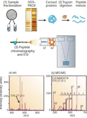
There are two main approaches for the identification of proteins using MS. Top-down proteomics which analyses intact proteins without the need of proteolytic digestion of the sample, while its counterpart, bottom-up proteomics, requires protein cleavage into short peptides prior to MS analysis. One of the most applied methods for the identification and quantification of proteomes is Liquid chromatography coupled to tandem mass spectrometry (LC-MS/MS) [136] (Figure 1.4).
Figure 1.4 Generic workflow schematization of a mass spectrometry (MS) proteomics experiment: (1) Sample fractioning, starting with isolation of proteins to be analysed from cell lysates and a final step of sodium dodecyl sulfate-polyacrylamide gel electrophoresis (SDS-PAGE) for further separation of proteins of interest; (2) proteolytic digestion (e.g. trypsin digestion); (3) Peptide separation (e.g. high pressure liquid chromatography-HPLC) and ionization using ion sources such as electrospray ionization (ESI) or matrix-assisted laser desorption/ionization (MALDI); (4) Acquisition of mass spectra; (5) Additional round of MS (i.e. tandem MS) [123].
Following bottom-up proteomics, LC-MS/MS starts with the proteolytic cleavage of the sample into short peptides which are then separated by liquid chromatography (Figure 1.4). LC-MS/MS can be operated using two different strategies: discovery/shotgun proteomics and targeted proteomics. In discovery proteomics, the MS is performed in information-dependent acquisition mode (IDA), where a subset of precursor ions (commonly the more abundant) formed at a first stage of MS (MS1) are selected and fragmented by collision resulting in fragment ions (MS2). The resulting MS/MS spectra are then analysed by sequence database search. Targeted proteomics is operated in selected reaction monitoring (SRM) where a limited number of predetermined ions of a specific mass are selected in the first stage of the MS1 and are fragmented in a second MS for detection. To overcome the limitations of IDA and SRM, such as low sensitivity to less abundant proteins and the limited number of target proteins per run, respectively, a data-independent acquisition (DIA) strategy has been emerging in this field (reviewed in [137]). In the DIA strategy, every ion (and not just a subset) in the chosen m/z range
INTRODUCTION
is fragmented and analysed in the second MS stage. An example of DIA method is Sequential Windowed Acquisition of All Theoretical Fragment Ion Mass Spectra (SWATH-MS) which is a recent developed MS-based proteomic tool to identify and quantify proteins [138].
1.5.1. SWATH-MS
There are several MS-based methods used in the quantification of proteomes, including chemical tagging methods such as isobaric tags for relative and absolute quantification (iTRAQ), isobaric labelling with tandem mass tags (TMT) and isotope-coded affinity tags (ICAT) and metabolic labelling, namely stable isotope labelling with amino acids (SILAC) (reviewed in [139]). In contrast, SWATH-MS is a novel approach of MS-based quantification of peptide ions in a sample that doesn’t require labelling [140]. First introduced by Gillet et. al., SWATH-MS appeared to combine the high throughput advantages of shotgun proteomics with the consistency and reproducibility of SRM. Its high-resolution MS data is obtained using a mass analyzer combination of a quadrupole and TOF (time-of-flight) instruments (Q-TOF) through repeated analysis of sequential windows (called swaths) throughout the chromatographic elution range [138]. The procedures of the tagging methods usually contribute to loss of protein which is avoided in SWATH-MS [141]. Being label-free, also removes the errors in quantification caused by incomplete labelling [142].
Proteomic tools have already been applied in studies regarding stem cells, including CSCs. For example, in the unveiling of hCSCs’ receptome [143] (i.e. plasma membrane receptors) or in the phenotypic characterization upon scale-up production and expansion of hCSCs [144]. More recently, using label-free quantitative proteomic tools (in this case, MaxLFQ), the human heart proteome has been mapped for the first time, making it easier to identify differences between diseased and healthy hearts in the future [145].
AIM OF THE THESIS
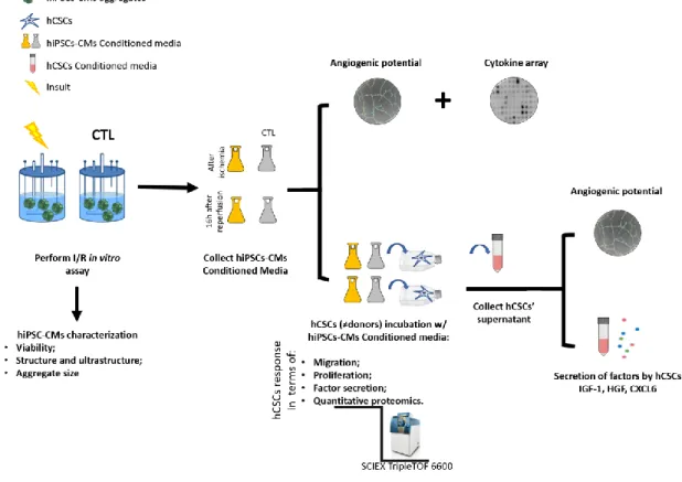
2. Aim of the thesis
This thesis was divided in two major parts. The first goal was to develop and characterize a novel human cell-based myocardial in vitro I/R injury model by combining both 3D culture of hiPSC-CMs and stirred tank bioreactor technology. This approach aimed at filling the gap between in vitro and in vivo research tools by better mimicking human CM AMI pathophysiology. The second aim of the thesis was to investigate the mechanism of action of hCSCs upon contact with paracrine factors from hiPSC-CMs subjected to I/R injury. Since it is known that paracrine cross-talk plays a key role on the activation of these cells, it is important to understand how hCSCs respond to the secretome of CMs in AMI context, aiding in the development of novel therapies based on the activation of the endogenous heart regeneration capacity.
The rational of the thesis is demonstrated in the figure below (Figure 2.1):
Figure 2.1 Thesis rational. Outlined strategy and major readouts. hiPSCs-CMs: human induced pluripotent stem cell derived cardiomyocytes; hCSCs: human cardiac stem cells.
![Figure 1.3 Examples of different bioreactor systems used in cell culture: a) rotating wall vessel; b) stirred-tank bioreactor (STBR); c) airflow bioreactor (adapted from [125]); d) hollow fiber bioreactor (adapted from [122]) and e) wave b](https://thumb-eu.123doks.com/thumbv2/123dok_br/15880626.1089220/36.892.177.752.290.676/figure-examples-different-bioreactor-rotating-bioreactor-bioreactor-bioreactor.webp)