Introduction
Epidemiologic studies indicate that cardiovascular diseases ac-count for approximately 30% of all deaths in global population, turning it the leading cause of mortality worldwide1. Among the cardiovascular diseases, coronary artery disease (CAD) deserves special attention for its exponential increase in working-age adults1.
CAD is characterized for the reduction or interruption in
coronary blood low to a speciic area of the cardiac muscle,
which generates a myocardial ischemia2. Given that the revers-ibility and extension of the tissue damage are directly related to the duration of ischemia, the main therapeutic goal is to
restore blood low, allowing reperfusion as quickly as possible2. Although thrombolytic therapies and percutaneous coronary intervention are considered the treatment of choice for reduc-ing the infarct area3, they may cause several damages, includ-ing functional and structural impairments and cellular death. Together these alterations are commonly known as ischemia reperfusion (IR) injury2,3.
On the other hand, previous data reinforce the hypothesis that regular physical exercise induces cardioprotection4-8 and may reduce by up to 30% the mortality risk for cardiovascular
diseases6. Besides reducing cardiovascular disease risk factors, such as hypertension, diabetes mellitus, obesity and dyslipidemia7, it is well described that exercise also promotes cardioprotection against IR injury through a direct effect on the myocardium4,7,9,10.
In 1978, McElroy et al11 demonstrated that regular physical activity could provide cardioprotection. In that study, mice were subjected to physical training for 5 weeks and after irreversible occlusion of the left coronary artery, a 30% reduction of the infarcted area was observed in trained mice when compared with the sedentary control.
Interestingly, it has been already demonstrated that perform-ing a sperform-ingle aerobic exercise session prior to an IR injury is
suf-icient to promote increment in cardiac output and ameliorate the
cardiac function during and after a cardiac insult12. Nonetheless, considering that aerobic training implies several adaptations that are not observed after a single exercise session, it would be reasonable to assume that cardioprotection following exercise
training is superior than that achieved after an unique exercise
session. However, despite the lack of interventional settings
focused on this speciic matter, previous reviews claim that
exercise-induced cardioprotection following few exercise ses-sions is similar than long-term physical training13,14. Clarifying Original Article (short paper)
Aerobic exercise training induces superior
cardioprotection following myocardial ischemia
reperfusion injury than a single aerobic exercise
session in rats
Juliana Pereira Borges Guilherme de Oliveira França
Universidade Estadual do Rio de Janeiro, Rio de Janeiro, RJ, Brasil
Mariana Delgado Cruz Rômulo Lanza
Alessandro Rodrigues do Nascimento Marcos Adriano Lessa
Instituto Oswaldo Cruz, Rio de Janeiro, RJ, Brasil
Abstract — Aim: To compare the amount of cardioprotection induced by a single exercise session with those achieved
after an 8-week aerobic exercise training following ischemia reperfusion injury in rats. Methods: Twenty-ive male
Wistar rats (250-300g) were assigned into a group submitted to physical training (TR; n=12) or a single maximal exercise session (EXE; n=13). Following sedentarism or physical training (8 weeks, 5 sessions/wk, 1h/session at 70% of maximal speed) both groups performed a maximal exercise test. Then, groups were submitted to ischemia reperfusion injury (30 min/1h) through an isolated heart protocol, in which left ventricle developed pressure was measured. Results: The TR group presented greater maximal oxygen consumption compared to the EXE group (77.25±20.41 vs 41.32±25.86 ml/Kg/min; P=0.003). Regarding left ventricle developed pressure, no differences were detected between groups at baseline (TR: 89.78±24.40 vs EXE: 81.37±31.84 mmHg; P=0.48). However, after reperfusion, the TR group presented superior intraventricular pressure than EXE group (37.94±18.34 vs 21.59±13.67 mmHg; P=0.03). Conclusion: Eight-week aerobic training induced greater cardioprotection against ischemia reperfusion injury in rats compared to a single exercise session, due to an increased cardiac function. This suggests that exercise-induced cardioprotection is a multifactorial process that may involve different mediators according to the exercise duration.
this issue would be important as the potential mechanisms involved in exercise-induced cardioprotection are still largely debated; therefore, data in this sense could provide insights about these mechanisms that could help understanding them.
Therefore, the purpose of the present study was to compare the amount of cardioprotection induced by a single exercise ses-sion with that achieved after an 8-week aerobic exercise training following ischemia reperfusion injury in rats.
Methods
Study Design
All procedures described in the present study were approved by the Oswaldo Cruz Foundation Animal Welfare Committee (protocol # LW-6/12) and are consistent with the USA National Institutes of Health Guide for the Care and Use of Laboratory Animals
(NIH Publication No. 85-23, revised 1996). Twenty-ive male
Wistar rats (250-300g) were housed with controlled light (12:12 h light-dark cycle) and temperature (22 ± 1°C) with free access to water and standard rat chow. Rats were randomly divided into a group submitted to an 8-week aerobic exercise training (TR; n=12) or a single maximal aerobic exercise session (EXE; n=13).
Exercise Protocol
Initially, the TR group was familiarized to treadmill running us-ing a low-speed, motor-driven rodent treadmill (HT 2.0, Hectron
Fitness Equipment, RJ, Brazil), on which the animals walked
at 12 m/min (0% grade) for 15 min/day on three consecutive days. Following this brief period of familiarization, a maximal exercise testing was performed in TR group to allow the exercise training prescription, which corresponded to 5 sessions/week of 60 min/session on treadmill at 70% of maximal velocity for 8 weeks. Whilst the TR group was submitted to exercise training, the EXE group was maintained sedentary.
Twenty-four hours after the end of exercise training or seden-tarism, maximal oxygen consumption (VO2max) was measured in both groups during a maximal exercise testing. The exercise test protocol began with rats running at 10 m/min (0% grade) with the treadmill speed increasing by 3 m/min every 3 min until the animals could no longer maintain the desired running speed. VO2max
was measured by assessing the total airlow through the treadmill
chamber and assessing the oxygen content of the expired gas using an electronic oxygen analyzer (AVS Projetos, SP, Brazil).
After 72h of the exercise test, all animals underwent the surgical procedures to induce IR injury in isolated heart proto-col. Knowing that exercise-induced cardioprotection persists practically unchanged for at least 9 days following an acute exercise15, the 72h between the exercise test and isolated heart
preparation was chosen to preclude the inluence of different
acute effects other than cardioprotection per se in the results, such as post-exercise hypotension.
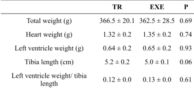
Table 1: Biometric parameters
TR EXE P
Total weight (g) 366.5 ± 20.1 362.5 ± 28.5 0.69 Heart weight (g) 1.32 ± 0.2 1.35 ± 0.2 0.74 Left ventricle weight (g) 0.64 ± 0.2 0.65 ± 0.2 0.93 Tibia length (cm) 5.2 ± 0.2 5.0 ± 0.1 0.06 Left ventricle weight/ tibia
length 0.12 ± 0.0 0.13 ± 0.0 0.61
Results are expressed as means ± SD. TR. group submitted to exercise training; EXE. group submitted to a single exercise session.
Isolated Heart Preparation
Following 72 hours of the maximal test, all animals, previously
heparinized (500 i.u. kg−1, I.P.), were killed by CO2 and cervical dislocation, and the excised hearts were immediately cannulated throughout the aorta according to the method of Langendorff
and perfused via the coronary circulation at a constant low rate of 10 ml.min−1 with modiied Krebs–Henseleit solution (mM:
118 NaCl, 4.7 KCl, 1.2 MgSO4, 1.2 KH2PO4, 25 NaHCO3,
10 glucose and 1.8 CaCl2, pH 7.2; gassed with 95% O2 – 5% CO2, 36±0.5 ◦C). A latex balloon was inserted in the left ventricle
through the left atrium and adjusted to an end-diastolic pressure
of 5–10 mmHg at baseline. After 30 min of baseline perfusion, all
hearts underwent a period of 30 min of sustained global ischemia followed by 1 h of reperfusion. Left ventricle pressure (monitored via the latex balloon) was recorded at baseline and at the end of the reperfusion period. For analysis, we used the ANCAD data recording software (AVS Projetos, São Paulo, Brazil).
Statistical Analysis
All results are expressed as mean ± SD. Comparisons between groups were performed with the Student t test. Bonferroni post
test was used to localize the signiicant differences. Correlation
between left ventricle developed pressure and VO2max was performed by Pearson correlation. P-values of <0.05 were
considered statistically signiicant. All calculations were made
by computer-assisted analysis using a commercially available statistical package (Graphpad Prism, Graphpad Software, San Diego, CA).
Results
Biometric Parameters
Maximal Exercise Capacity
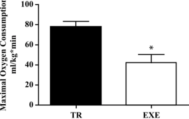
Table 2 presents the maximal exercise testing results. The TR group showed higher distance (546.14 ± 141.84 vs. 263.22 ± 65.91 m; P < 0.001), maximal velocity (35.0 ± 3.5 vs. 25.0 ± 3.0 m/seg; P < 0.001) and duration (28 ± 4 vs. 18 ± 3 min; P < 0.001) than the EXE group. As expected, the TR group showed greater VO2max than the EXE group (77.25 ± 20.41 vs. 41.32 ± 25.86 ml/ Kgmin; P = 0.003; Figure 1).
Table 2: Parameters obtained in maximal exercise testing in experi-mental groups.
TR EXE P
Distance (m) 546.14 ± 141.84 263.22 ± 65.91 < 0.001 Maximal velocity
(m/sec) 35.0 ± 3.5 25.0 ± 3.0 < 0.001 Duration (min) 28 ± 4 18 ± 3 < 0.001
Results are expressed as means ± SD. TR. group submitted to exercise
training; EXE. group submitted to a single exercise session.
Cardiac Function
Left ventricle developed pressure in baseline condition did not differ between groups (TR: 89.78 ± 24.40 vs. EXE: 81.37 ± 31.84 mmHg; P = 0.48; Figure 2). However, after 60 min of reperfusion, the TR group presented superior left ventricle developed pressure in comparison to the EXE group (37.94 ± 18.34 vs 21.59 ± 13.67 mmHg; P=0.03; Figure 2). There was no correlation between left ventricle developed pressure obtained at the end of reperfusion and VO2max in the TR and EXE groups (P = 0.30 and P = 0.11; respectively; Figure 3).
Discussion
The aim of the present study was to compare the amount of cardioprotection induced by a single exercise session with that
Regarding the duration of exercise training, several previous studies have demonstrated that 8 to 12 weeks of exercise provide myocardial protection against IR injury in rats16-18. Meanwhile, it is also well documented that few exercise sessions of 60 min alter the cardioprotection phenotype9,18. Hamilton et al19 showed
that ive exercise sessions reduce the incidence of ventricular
arrhythmias after IR injury in vivo. Similarly, Demirel et at12 assessed the cardiac function following IR injury of rats exer-cised for 60 min/day during 3 or 5 days at 60 or 70% of VO2max (respectively) and observed that exercised rats maintained superior intraventricular pressure than sedentary rats. Although it is already clear that short-term exercise provides cardiopro-tection, so far no original study compared the protective effect
*
TR EXE
0 20 40 60 80 100
Maximal Oxygen Consumption
ml/kg*min
Figure 1: Maximal oxygen consumption in animals exercised for eight weeks (TR) and a single aerobic exercise session (EXE). * P = 0.003.
*
TR EXE
Baseline After reperfusion 120
100
80
60
40
20
0
Left ventricle developed pr
essur
e
mmHg
Figure 2: Left ventricle developed pressure in animals exercised for eight weeks (TR) and for a single session (EXE) at baseline and fol-lowing ischemia reperfusion injury. * P = 0.03.
0 20 40 60 80 100
0 10 20 30 40 50 60 70
VO2max (ml/kg*min)
Left ventricle developed pr
essur
e
(mmHg)
TR EXE
Figure 3: Correlation between left ventricle developed pressure ob-tained at the end of reperfusion and maximal oxygen consumption (VO2max) in animals exercised for eight weeks (TR; P = 0.30) and for a single session (EXE; P = 0.11).
achieved after an 8-week aerobic exercise training following ischemia reperfusion injury in rats. In this sense, our major
inding was that the long-term training provided greater car -diac function measured through the left ventricle developed pressure, in comparison to a single exercise session following IR injury. In addition, we also observed a greater maximal ex-ercise capacity in trained vs. single-exex-ercised rats that was not correlated to the cardiac function. Therefore, our data support
the idea that besides the beneits in cardiorespiratory function,
induced by long and short-term exercise. Considering that the potential mechanisms involved in this response are still largely debated, data in this sense could provide insights regarding these mechanisms that could help clarifying this issue. Given that
our experiments reveal for the irst time that 8-week exercise
training provides greater cardioprotection against IR injury than a single exercise session, some assumptions in regards to the mechanisms involved in this response could be raised.
Considering that exercise-induced cardioprotection occurs even after few exercise sessions, several adaptations exclusively
observed after long-term training (8 to 12 weeks) are frequently
neglected as mechanisms of cardioprotection, such as increased collateral circulation20. Indeed, these training-induced
adapta-tions probably are not a prerequisite to achieve cardioprotection,
but given our results of increased cardiac function of long-term training following IR injury, they could minimally play a role in this response. This reinforces the hypothesis that exercise-induced cardioprotection is a multifactorial process or even that it involves different mediators according to the exercise duration.
To illustrate this, pertinent literature documents well that aerobic training causes an enhancement in the antioxidant enzyme activity in various tissues21. This is an physical training adapta-tion process that only happens because of the transient release of reactive oxygen species during exercise sessions, acting as signaling molecules21. This stimulates the gene expression and, hence, increases production of key antioxidant enzymes that help minimizing the oxidative stress process involved in IR in-juries22-25. Therefore, it is feasible to think that long-term training leads to greater amount of antioxidant enzymes production than acute exercise, which could justify the superior cardioprotection observed after exercise training. Another potential mechanism largely discussed in exercise-induced cardioprotection is the change in the coronary arteries, which includes increased conduit artery diameters, arteriolar densities, and diameters of arteriolar26. Considering that this adaptation requires several weeks of exercise training27, it could also account for our results.
As in relation to the exercise training intensity, previous data have demonstrated that this is an important issue when it comes to exercise-induced cardioprotection effect8,28. For instance, Starnes et al28 have found that 16 weeks of exercise training bellow 55 to 60% of VO2max did not attenuate the dam-age caused by IR injury. On the other hand, Lennon et al.8 have concluded that exercise training at low (50% of VO2max) and moderate (70% of VO2max) intensity are equally protective. In this sense, in our study, we chose to apply the aerobic exercise training at 70% of maximal velocity (which corresponds ap-proximately to 70% of VO2max 29 ) to ensure the cardioprotection afforded by exercise training.
The results of the present study should be interpreted con-sidering certain limitations. First, the major limitation is the lack of a control group not submitted to exercise. Data in this
sense would establish reference values and subsequently allow
a more precise conclusion upon the acute exercise-induced cardioprotection. However, although we cannot assume that acute exercise induced cardioprotection in comparison to sedentarism, the lack of a control group did not jeopardize the comparison between the cardioprotective effect between short
and long-term exercises, which is the major aim of the present study. Second, the assessment of tissue damage following IR injury, such as myocardial infarct size, would contribute to our results. Nonetheless, maintaining cardiac function is a key component involved in cardioprotection.
In addition, marked physiological differences are notably observed between different species; thus, direct extrapolation
of these indings from rats to humans should be approached
with caution. Nonetheless, experimental settings investigating
issues related to IR injury are well accepted due to the dificulty
in developing such studies with humans.
Conclusion
Our results indicate that eight weeks of exercise training lead to greater cardioprotection against IR injury in rats due to an increased cardiac function than a single exercise session. This reinforces the hypothesis that exercise-induced cardioprotection is a multifactorial process that may involve different mediators according to the exercise duration. However, further research is necessary to obtain more consistent conclusions, especially in regards to the mechanisms involved in short and long-term exercise-induced cardioprotection.
References
1. Go AS, Mozaffarian D, Roger VL, Benjamin EJ, Berry JD, Blaha MJ, et al. Heart disease and stroke statistics--2014 update: a report from the American Heart Association. Circulation. 2014;129(3):e28-e292.
2. Evora PR, Pearson PJ, Seccombe JF, Schaff HV. Ischemia-reperfusion lesion. Physiopathologic aspects and the importance of the endothelial function. Arq Bras Cardiol. 1996;66(4):239-45. 3. Hausenloy DJ, Yellon DM. Myocardial ischemia-reperfu-sion injury: a neglected therapeutic target. J Clin Invest. 2013;123(1):92-100.
4. Powers SK, Quindry JC, Kavazis AN. Exercise-induced cardio-protection against myocardial ischemia-reperfusion injury. Free Radic Biol Med. 2008;44(2):193-201.
5. Ferdinandy P, Schulz R, Baxter GF. Interaction of cardio-vascular risk factors with myocardial ischemia/reperfusion injury, preconditioning, and postconditioning. Pharmacol Rev. 2007;59(4):418-58.
6. Berlin JA, Colditz GA. A meta-analysis of physical activity in the prevention of coronary heart disease. Am J Epidemiol. 1990;132(4):612-28.
7. Powers SK, Lennon SL, Quindry J, Mehta JL. Exercise and car-dioprotection. Curr Opin Cardiol. 2002;17(5):495-502.
8. Lennon SL, Quindry JC, French JP, Kim S, Mehta JL, Powers SK. Exercise and myocardial tolerance to ischaemia-reperfusion. Acta Physiol Scand. 2004;182(2):161-9.
10. Golbidi S, Laher I. Molecular mechanisms in exercise-induced cardioprotection. Cardiol Res Pract. 2011;2011:972807. 11. McElroy CL, Gissen SA, Fishbein MC. Exercise-induced
reduc-tion in myocardial infarct size after coronary artery occlusion in the rat. Circulation. 1978;57(5):958-62.
12. Demirel HA, Powers SK, Zergeroglu MA, Shanely RA, Hamilton K, Coombes J, et al. Short-term exercise improves myocardial tolerance to in vivo ischemia-reperfusion in the rat. J Appl Physiol (1985). 2001;91(5):2205-12.
13. Powers SK, Sollanek KJ, Wiggs MP, Demirel HA, Smuder AJ. Exercise-induced improvements in myocardial antioxidant capac-ity: the antioxidant players and cardioprotection. Free Radic Res. 2014;48(1):43-51.
14. Kavazis AN. Exercise preconditioning of the myocardium. Sport Med. 2009;39(11):923-35.
15. Lennon SL, Quindry J, Hamilton KL, French J, Staib J, Mehta JL, et al. Loss of exercise-induced cardioprotection after cessation of exercise. J Appl Physiol (1985). 2004;96(4):1299-305.
16. Libonati JR, Gaughan JP, Hefner CA, Gow A, Paolone AM, Houser SR. Reduced ischemia and reperfusion injury following exercise training. Med Sci Sport Exercise. 1997;29(4):509-16. 17. Libonati JR, Kendrick ZV, Houser SR. Sprint training improves
postischemic, left ventricular diastolic performance. J Appl Physiol (1985). 2005;99(6):2121-7.
18. Soui FG, Saber MM, Ghiassie R, Alipour M. Role of 12-week resistance training in preserving the heart against ischemia-reperfusion-induced injury. Cardiology journal. 2011;18(2):140-5. 19. Hamilton KL, Quindry JC, French JP, Staib J, Hughes J, Mehta JL,
et al. MnSOD antisense treatment and exercise-induced protection against arrhythmias. Free Radic Biol Med.
20. Wiggs MP, Duarte AF, Powers SK. Exercise can protect against a broken heart. Curr Sports Med Rep. 2015;14(1):6-8.
21. Gomes EC, Silva AN, de Oliveira MR. Oxidants, antioxidants, and the beneicial roles of exercise-induced production of reactive species. Oxid Med Cell Longev. 2012;2012:756132.
22. Borges JP, Lessa MA. Mechanisms Involved in Exercise-Induced Cardioprotection: A Systematic Review. Arq Bras Cardiol. 2015;105(1):71-81.
23. Powers SK, Demirel HA, Vincent HK, Coombes JS, Naito H, Hamilton KL, et al. Exercise training improves myocardial tol-erance to in vivo ischemia-reperfusion in the rat. Am J Physiol. 1998;275(5 Pt 2):R1468-77.
24. Hamilton KL, Powers SK, Sugiura T, Kim S, Lennon S, Tumer N, et al. Short-term exercise training can improve myocardial tolerance to I/R without elevation in heat shock proteins. Am J Physiol Heart Circ Physiol. 2001;281(3):H1346-52.
25. Hamilton KL, Staib JL, Phillips T, Hess A, Lennon SL, Powers SK. Exercise, antioxidants, and HSP72: protection against myocardial ischemia/reperfusion. Free Radic Biol Med. 2003;34(7):800-9. 26. Powers SK, Smuder AJ, Kavazis AN, Quindry JC. Mechanisms
of exercise-induced cardioprotection. Physiology (Bethesda). 2014;29(1):27-38.
27. White FC, Bloor CM, McKirnan MD, Carroll SM. Exercise training in swine promotes growth of arteriolar bed and capillary angiogenesis in heart. J Appl Physiol (1985). 1998;85(3):1160-8. 28. Starnes JW, Taylor RP, Ciccolo JT. Habitual low-intensity exercise
does not protect against myocardial dysfunction after ischemia in rats. Eur J Cardiovasc Prev Rehabil. 2005;12(2):169-74. 29. Lawler JM, Powers SK, Hammeren J, Martin AD. Oxygen cost
of treadmill running in 24-month-old Fischer-344 rats. Med Sci Sports Exerc. 1993;25(11):1259-64.
Corresponding author
Juliana Pereira Borges
Laboratório de Atividade Física e Promoção à Saúde (LABSAU) Rua São Francisco Xavier, 524, sala 8133F, Maracanã, Rio de Janeiro, RJ. Email: [email protected]
Manuscript received on September 11, 2016 Manuscript accepted on November 07, 2016