Design and methods of the Ludwig-McGill
longitudinal study of the natural history of
human papillomavirus infection and cervical
neoplasia in Brazil
Eduardo Franco,
1Luisa Villa,
2Thomas Rohan,
3Alex Ferenczy,
4Maria Petzl-Erler,
5and Greg Matlashewski,
6for the Ludwig-McGill Study Group
7This article reports on a large longitudinal study, begun in 1993, of the natural history of human papillomavirus (HPV) infection and cervical neoplasia in a population of low-income women in São Paulo, Brazil, a city with one of the highest risks worldwide for cervical cancer. Known as the Ludwig-McGill cohort study, the epidemiological investigation focuses on persistent infec-tion with oncogenic HPV types as the precursor event leading to cervical neoplasia.
The objectives of this study are to: 1) study the epidemiology of persistent cervical HPV in-fection in asymptomatic women, 2) investigate whether persistent HPV inin-fection increases risk of low-grade and high-grade cervical lesions, 3) search for determinants of persistent HPV infection, 4) search for molecular variants of HPV that may be associated with an increased risk of lesions, 5) investigate whether viral burden is correlated with persistent infections and with lesion risk, 6) study the antibody response to HPV as a predictor of persistence and le-sion progresle-sion, and 7) examine the role of HLA typing and codon 72 p53gene polymorphism in mediating HPV persistence and lesion severity.
The study accrued 2 528 female subjects through March 1997. Subjects were followed up every 4 months in the first year, with twice-yearly return visits to take place in subsequent years. Participants undergo a questionnaire-based interview, have a cervical specimen taken for Pap cytology and HPV testing, and have a blood sample drawn for HPV antibody testing. A cervicography is performed once in the first year and every two years thereafter. In this arti-cle we describe the design and methods of the study, provide baseline cohort characteristics, and present a preliminary assessment of the prognostic value of baseline HPV status.
ABSTRACT
Although few would dispute now-adays that human papillomavirus
(HPV) infection is the central cause of cervical cancer, most of the epidemio-logic data have come from retrospec-tive, case-control studies (1), which do not provide information on the dy-namics of cumulative exposure to cer-vical HPV infection. Considering the public health and economic impor-tance of cervical cancer and the current widespread interest in the develop-ment of HPV vaccines and in using HPV testing to augment existing
cytol-1 McGill University, Department of Oncology, and
Department of Epidemiology, Montreal, Quebec, Canada. Address correspondence and reprint re-quests to: Prof. E. Franco, Department of Oncol-ogy, McGill University, 546 Pine Avenue West, Montreal, QC, Canada H2W 1S6. Phone: (514) 398-6032; fax: (514) 398-5002; e-mail: eduardof@oncol-ogy.lan.mcgill.ca
2 Ludwig Institute for Cancer Research, São Paulo,
São Paulo, Brazil.
3 University of Toronto, Department of Public
Health Sciences, Toronto, Ontario, Canada.
4 McGill University, Department of Pathology,
Montreal, Canada, and Sir Mortimer B. Davis Jew-ish General Hospital, Montreal, Canada.
5 Universidade Federal do Paraná, Curitiba, Brazil.
6 McGill University, Department of Parasitology,
Montreal, Canada.
7 Additional collaborators: Maria Baggio, Otávia
ogy screening programs, there is a clear need for prospective, long-term multidisciplinary studies of the nat-ural history of HPV infection as it pro-gresses to preinvasive cervical lesions. In 1993, we began a large longitudi-nal study of the natural history of HPV infection and cervical neoplasia in a population of low-income women in São Paulo, Brazil, a city with one of the highest risks worldwide for cervical cancer. Our study was designed to an-swer questions that have not yet been addressed in epidemiologic investiga-tions of this neoplastic disease. The Brazilian investigation focuses on per-sistent infection with oncogenic HPV types as the precursor event leading to cervical neoplasia. The investigation will attempt to understand attributes of the natural history of viral infection that may be instrumental to the design of primary and secondary strategies for preventing cervical cancer. As a unique feature of our cohort study, persistence of HPV infection is moni-tored by molecular variant analysis (2) and measurement of viral burden (3), which together provide a much finer level of detail than simple HPV testing and may improve the prediction of the likelihood of lesion progression.
The Ludwig-McGill cohort study is designed to further our understanding of the etiopathogenesis of cervical neo-plasia by focusing on the following specific objectives:
• measuring the prevalence and inci-dence of persistent cervical HPV in-fection in asymptomatic women • testing the hypothesis that
persis-tent HPV infection increases risk of low-grade and high-grade cervical lesions
• identifying the epidemiologic deter-minants of persistent cervical HPV infection
• identifying specific molecular vari-ants of oncogenic types of HPV that are associated with increased risk of cervical lesions
• testing the hypothesis that viral bur-den in the cervix may be correlated with persistent infections and with low- and high-grade lesions
• studying the humoral immune re-sponse to HPV as a predictor of per-sistent cervical HPV infection and of risk of progression of lesion severity • searching for specific human leuko-cyte antigen (HLA) alleles or haplo-types associated with HPV persis-tence and lesion severity
• testing the hypothesis that a specific
p53gene polymorphism may confer increased resistance against viral per-sistence and consequently against development of cervical lesions
The purpose of this article is to pre-sent a detailed description of the de-sign and methods of the Ludwig-McGill longitudinal study, to describe the characteristics of the cohort at baseline, and to present a preliminary assessment of the prognostic value of baseline HPV status.
MATERIALS AND METHODS
The study described here is a longi-tudinal study. It involves repeated measurements on the individual par-ticipants over time of such risk factors as lifestyle, nutritional and behavioral attributes, and reproductive health and hygiene variables; intermediate end-points (cervical HPV infection mea-sured via direct DNA detection as well as indirectly, via serologic testing for HPV antibodies); and outcome (prein-vasive cervical neoplasia).
Study population and setting
The study population is derived from women attending a comprehen-sive maternal and child health pro-gram catering to low-income families in the city of São Paulo, Brazil. The city has a population of some 12 million and is the capital of the state of São Paulo, the most populous and most in-dustrialized state of Brazil. The clinic setting where subjects were accrued and are being followed up is the Mater-nidade Escola Dr. Mario de Moraes Al-tenfelder Silva Municipal Hospital, which is part of a network of primary,
secondary, and tertiary health care in-stitutions maintained by the municipal health department. This clinic is also known by its shorter, unofficial name, “Maternidade Escola Vila Nova Ca-choeirinha” (MEVNC).
Eligibility
Women were eligible to participate if they: 1) were between 18 and 60 years old, 2) were permanent residents of the city of São Paulo, 3) ) were not currently pregnant and had no intention of be-coming pregnant during the next 12 months, 4) had an intact uterus and no current referral for hysterectomy, 5) re-ported no use of vaginal medication in the previous 2 days, and 6) had not had treatment for cervical disease by electro-coagulation, cryotherapy, or conization (the prevailing methods at MEVNC) in the previous 6 months. In addition to these criteria, women were considered ineligible if they were not interested in complying with all scheduled returns, at least for the subsequent 2 years.
Subject recruitment
Two nurses were employed and trained specifically for the study. They recruited subjects by selecting women at random from the daily lists of outpa-tients in the family medicine, gynecol-ogy, and family planning clinics at MEVNC. The nurse-interviewers ap-proached each selected patient to deter-mine eligibility and to explain the gen-eral purpose and nature of the study. Women who were potentially eligible were then given a more detailed overview of the study, including the need for cervical and blood specimens, cervicographies, and interviews deal-ing with sensitive questions, all durdeal-ing multiple scheduled visits to the clinic. The nurses then explained the meal ticket system for compensating partici-pants for the time that they would in-vest in the study (see the section below on encouraging compliance).
study procedures and the informed consent were approved by the institu-tional review boards and ethical com-mittees of the participating institu-tions: McGill University, Montreal, Quebec, Canada; the University of Toronto, Toronto, Ontario, Canada; and the Ludwig Institute for Cancer Research and the MEVNC clinic, both in São Paulo, Brazil.
Scheduled returns and procedures
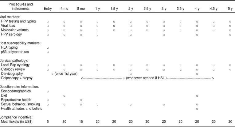
Subjects enrolled into the study are being followed up over a 5-year period in prescheduled return visits, which will extend into the year 2002. All par-ticipants were seen every 4 months in the first year, and are to be seen twice yearly thereafter. In the first four visits and on the annual returns, subjects complete an interviewer-administered structured questionnaire specific for the current visit and have cervical spec-imens taken for Pap cytology and HPV testing. A 10-cm3blood sample is also drawn by venipuncture into a vacu-tainer tube without anticoagulant. Dur-ing the semester returns between an-nual follow-up visits, only the cervical specimen is collected for cytology and HPV testing. A cervicography is per-formed once in the first year during any one of the quadrimester visits. Ad-ditional cervicographies are performed at 24 and 48 months.
Encouraging compliance with scheduled follow-up returns
Because of the importance of retain-ing subjects for the entire duration of the study, participants were told at en-rollment that they would receive cash-equivalent incentives. We opted to use meal tickets, which are widely used in Brazil as employee benefits for salaried workers. Meal tickets have an infla-tion-adjusted cash value and come in various denominations, being honored in nearly all commercial establish-ments in exchange for meals, groceries, and other goods. The cash-equivalent incentives begin at US$ 5.00 at the
en-rollment visit and increase $5 per sub-sequent visit to a maximum of $20, which is then given for all subsequent appointments that are kept by the par-ticipant. This strategy has ensured ex-cellent rates of follow-up compliance despite the complexity of the proce-dures used in the study and the re-quirement for blood specimens.
Questionnaires
In-person interviews are adminis-tered by the study nurses according to the schedule described above. Prior to commencement of the study, the nurses underwent extensive training in interview strategies. During these sessions we used role playing, mock interviews, and case studies to train the nurses in how to establish rapport with the respondent, to earn her re-spect, to avoid being judgmental, and to facilitate recollection of life events, some of which are sensitive. The infor-mation that is collected during the in-terviews covers all classes of risk fac-tors for HPV infection and cervical neoplasia, that is, sociodemographics, reproductive health, sexual practices, smoking, and diet.
Cervical cell specimens
An Accelon biosampler (Medscand, Inc., Hollywood, FL, United States of America) is used to collect a sample of ectocervical and endocervical cells at each of the visits. After the smear is prepared on a glass slide and fixed in 95% ethanol, the sampler containing the exfoliated cells is immersed in a tube containing Tris-EDTA buffer pH 7.4. The tube is agitated to release the cells from the sampler, and the sam-pler is discarded. Samples are kept at 4 °C at the clinic for 5 days at most and then brought to the laboratory at the Ludwig Institute, where they are kept frozen at –20 °C until testing. The Pap smears are fixed in absolute ethanol, stained, and read at the Ludwig Insti-tute’s cytopathology laboratory for an initial diagnosis. They are then
shipped to Montreal, where they are coded and then sent to the laboratory of Dr. Alex Ferenczy, at the Jewish General Hospital, one of McGill Uni-versity’s teaching hospitals. Cyto-pathology reports produced locally in São Paulo are based on the old Papa-nicolaou class system, which is the re-port format preferred by local physi-cians. The Montreal cytopathology reports are based on the Bethesda sys-tem for cytological diagnoses (4).
Cervicography
Because of possible concerns with false negative cytology results, all women also undergo a cervicography during one of the visits in their first year of participation in the study—at a time that is mutually convenient for the participant and for the nurses— and then at 24 and 48 months. Cer-vicography is being used to detect clinically relevant lesions that are visu-ally identifiable, thus providing a “safety net” to supplement the infor-mation obtained from the two cytolog-ical readings in the study (see the sec-tion below about management of lesions). Cervicography was proposed as a cervical cancer screening tool by Stafl (5) and plays a useful role in large-scale studies in high-risk popula-tions, particularly in remote areas, where well-trained colposcopists can-not be recruited easily. Log sheets and rolls of film are prepared according to the instructions from the provider, Na-tional Testing Laboratories (NTL), and then shipped monthly to Fenton, Mis-souri, United States of America, for de-velopment and evaluation by NTL’s expert colposcopists. The list of results are then mailed to the project manager in Montreal for computer data entry.
Management of lesions
also triggered if the cytopathology re-view performed in Montreal reveals a diagnosis of a high-grade squamous intraepithelial lesion (HSIL) or worse. Similarly, cervigrams indicating high-grade lesions or worse are grounds for recall. Since the slide review and the cervicography result are first available in Montreal, we use electronic mail to notify the nurses in São Paulo when-ever an HSIL is found. Cumulative lists of HSIL-positive cases are subse-quently transferred via a file transfer protocol (FTP) computer connection from Montreal to São Paulo for further verification. This ensures a rapid and safe means of recalling patients with clinically relevant lesions. In the inter-est of expediency, our own nurses take responsibility for scheduling appoint-ments for the colposcopy, which is per-formed by the MEVNC gynecologist on duty. At colposcopy, if any lesional tissue is present, a biopsy is taken for histopathological assessment and, if indicated, these women are treated ac-cording to the local prevailing proto-col. Women with lesions continue to be followed in the cohort if they so wish but do not contribute further person-time to the cohort. All histopathologi-cal slides are read lohistopathologi-cally and then sent to Dr. Ferenczy’s laboratory in Mon-treal for review.
HPV DNA detection
DNA samples are purified by spin column chromatography. Early in the study we extracted DNA from all cer-vical specimens using digestion with 100 µg/mL proteinase K for 3 h at 55 °C, followed by organic extraction and ethanol precipitation. Cervical specimens are tested for the presence of HPV DNA by a previously described polymerase chain reaction (PCR) pro-tocol amplifying a highly conserved 450 bp segment in the L1 viral gene (flanked by primers MY09/11) (6, 7). Typing of the amplified products is performed by hybridization with indi-vidual oligonucleotide probes specific for all 27 HPV genital types whose nu-cleotide sequences for probes within
the MY09/11 fragment have been pub-lished in the literature. Twenty-four of these have received a taxonomic entry, as HPV types 6/11, 16, 18, 26, 31, 33, 35, 39, 40, 42, 45, 51, 52, 53, 54, 55, 56, 57, 58, 59, 66, 68, and 73. Three additional gen-ital types (MM4, MM7, and MM8) are still awaiting taxonomic classification (7). Amplified products that hybridize with the generic probe but with none of the type-specific probes are considered positive for HPV of unknown type(s). These products are tested further, to distinguish among unknown HPVs, by restriction fragment length polymor-phism (RFLP) analysis of the L1 frag-ment (8). Use of the RFLP analysis ex-tends the range of identifiable HPV types in our study to over 40 genital types, by allowing additional detection of HPVs 32, 34, 44, 62, 64, 67, 69, 70, 72, CP6108, CP8061, CP8304, IS39, and other unknown types. To verify the specificity of the hybridizations, we in-clude more than 30 type-specific posi-tive controls in all membranes. In order to check the integrity of the host DNA material extracted from the specimens, assays also include an additional set of primers (GH20 and PC04) to amplify a 268 bp region of the b-globin gene (6). All HPV assays are done blindly on coded specimens, with no identifica-tion linking specimens from the same woman. Appropriate precautions are taken to reduce the possibility of speci-men contamination.
Molecular variant analysis
For HPVs 16 and 18, we use a PCR sequencing method with primers flank-ing a 364 bp segment (nucleotide posi-tions 7478 to 7841) within the LCR re-gion, a hypervariable genomic segment of HPV (9, 10). The amplified products are cloned using the SureClone™ plas-mid cloning kit (Pharmacia Biotech, Uppsala, Sweden), according to the manufacturer’s instructions and are in-troduced into E. coliXL1-Blue. Recom-binant plasmid DNAs are isolated from positive clones and submitted to DNA sequencing by the dideoxy method. Due to possible misincorporation of
bases by Taq polymerase and the possi-bility of multiple coexisting variants, a minimum of five clones are sequenced for each isolate. We also test for molec-ular variants of HPV 16 in the E6 gene by targeting a 456 bp segment with nearly the same intratype variability as that in the LCR region that we sequence in our standard protocol. We use the method described by Yamada et al. (11) for sequencing this E6 fragment. By fo-cusing on two genomic sequences of HPV 16, the most common oncogenic type in our population, we will be able to obtain an extra level of confidence in assigning the putative phylogenetic re-latedness for a given variant.
Measuring viral burden
di-rectly proportional to the logarithm of the amount of HPV DNA in the indi-vidual samples. Proper quantification is obtained by linear interpolation in a standard curve constructed with the results from the control mixtures.
Serologic testing for HPV antibodies
Serum samples are separated from the clotted blood specimens and stored at –20 °C until testing. An ELISA technique described previously (13) is used for the semiquantitative assessment of immunoglobulin G (IgG) antibodies to L1 and L2 HPV capsid antigens. Antigen preparations consist of self-assembled empty virus-like particles (VLP) produced from a baculovirus system expressing L1 and L2 (14). A sufficiently large batch of HPV 16 VLP antigen was donated by Dr. John Schiller, Laboratory of Cellu-lar Oncology, U.S. National Institutes of Health. This initial batch is being used to define seropositivity at base-line and initial follow-up visits. Subse-quent batches are being prepared at the Ludwig Institute using strict ad-herence to Dr. Schiller’s expression system protocol. Seropositivity is arbi-trarily based on the cutpoint for the 90th percentile of the distribution of reactivity for control specimens from women free of lesions and testing con-sistently negative for HPV DNA dur-ing all four first-year visits. Advances in VLP technology permitting, we will incorporate in our assay system VLP antigens from any other HPV types that become available.
Testing for host susceptibility markers
One of the most important objectives of research on the prevention of cervi-cal cancer is to identify host cofactors that could influence HPV carcinogene-sis. We are testing for two such cofac-tors, the woman’s HLA makeup and
p53 polymorphism in codon 72. HLA class II alleles or haplotypes may play
a role in the HPV infection and genetic susceptibility to cervical cancer, proba-bly regulating the immune response against HPV infection and ultimately interfering in the establishment of pro-ductive persistent infections.
One pivotal step in the etiology of HPV-induced cervical neoplasia seems to be the binding of the viral E6
gene product to the p53 tumor sup-pressor protein. This promotes degra-dation of the latter, exempting the cell to undergo p53-mediated control. In the general population, there are two common and distinct wild-type alleles of the p53 tumor suppressor gene that are not functionally equivalent with respect to HPV. These alleles represent a polymorphism. One encodes argi-nine at amino acid residue 72 (Arg al-lele), and the other encodes a proline at amino acid residue 72 (Pro allele). At the molecular level, HPVs can me-diate the degradation of the p53Arg protein more efficiently than the p53Pro protein (15), raising the possi-bility that individuals homozygous for the p53Argalleles would be more sus-ceptible to HPV-associated pathology than individuals with p53Pro alleles. We hypothesize that women with the homozygous p53Arggenotype will be more susceptible to the acquisition of persistent HPV infection and, ulti-mately, to the development of cervical intraepithelial neoplasia than those homozygous or heterozygous for the
p53Pro allele. It is possible that in women with the homozygous p53Arg
genotype, HPV infection will be estab-lished more productively with colo-nization of the cervical epithelium be-cause the virus will be more successful in inhibiting p53-mediated apoptosis in infected cells. Hence, there is the ex-pectation that these women will be more susceptible to persistent HPV detection.
The hypothesis regarding the role of a p53 polymorphism is at present highly controversial. The original as-sociation (15) was not confirmed by subsequent studies, such as by Hilde-sheim et al. (16). However, we have obtained preliminary data from a case-control study conducted in Brazil that
indicates a greater cervical cancer risk among women with the homozygous
p53Arg form. Nevertheless, we were able to identify the association only after restricting the analysis to subjects with concordant genotype information obtained in three different laborato-ries. This suggests that misclassifica-tion of genotypes could be statistically diluting the association, thus prevent-ing it from beprevent-ing measured in epi-demiologic studies (Makni et al., man-uscript submitted).
HLA typing. Molecular typing of HLA alleles will be performed in the DNA extracted from the enrollment cervical specimen, or, when that speci-men is depleted or insufficient, from any of the cervical specimens collected in subsequent visits. The HLA class II DRB1 and DQB1 high-resolution typ-ing will be performed by PCR and se-quence-specific oligonucleotide probe hybridization following the standard international nomenclature (17). All HLA typing will be performed blindly on coded samples.
P53 polymorphism analysis. The
p53 genotype for codon 72 will be de-termined on the enrollment DNA sam-ples (or using subsequent ones if the first specimen is depleted) using a re-cently described PCR technique (15). In brief, two PCR reactions will be car-ried out on each sample, one with a primer set specific to the p53Pro allele and the other with a primer set specific to the p53Arg allele. The p53Pro allele yields a 277 bp fragment, and the
X-ray film as described previously (15). All p53 genotyping will be per-formed blindly on coded samples.
Cohort management
At each visit, the nurse first collects the cervical specimen and prepares the Pap smear, followed by the cervical cell suspension for HPV testing. She then performs the cervicography, if prearranged for that visit, followed by the venipuncture to collect the blood sample. The final step is the interview, using the questionnaire specific for that visit. Table 1 summarizes the var-ious study procedures and instru-ments used at the initial visit and dur-ing each of the prescheduled returns for all subjects.
After each visit the nurses schedule the patient’s subsequent return at the expected date according to the study plan. Delays in returning for a given appointment cause the following one to be postponed correspondingly, so
that there is always a full between-visit period of 4 or 6 months. Returns are scheduled for 4 months later for a first-year cohort member, and for 6 months later for those beyond the first four visits. The nurses call the patients a few days before the scheduled re-turns to remind them of the pending visits. Those who miss appointments are contacted by phone and/or letter. These attempts at contacting subjects are repeated once a month until an ap-pointment can be scheduled or the woman explicitly states that she wishes to drop out of the study.
RESULTS
Subject recruitment and follow-up compliance
Between November 1993 and March 1997 our nurses made 6 188 contacts with 4 990 patients to ascertain eligi-bility and invite their participation. Of these, 3 589 women were deemed
eli-gible and 2 528 were enrolled into the study during that period, representing a response rate of 70.4%. Table 2 shows the status with respect to par-ticipation and reasons for lack of eligi-bility in each category. Pregnancy or a previous hysterectomy together ac-counted for more than 50% of the in-stances of noneligibility.
As of mid-November 1998, the 2 528 participants had logged 14 094 visits since enrollment, for a total of 71 017 woman-months of follow-up (average per subject of 5.6 visits and 28.1 months of follow-up). Allowing for delays in follow-up, actuarial rates of compli-ance with all prescheduled clinic visits are 73%, 69%, 70%, and 77% at 12, 24, 36, and 48 months, respectively.
Selected characteristics of the cohort
Table 3 shows selected sociodemo-graphic, lifestyle, and reproductive health characteristics as reported at en-rollment. The mean age was 32.9 years
TABLE 1. Study procedures and instruments applied at enrollment and at different prescheduled follow-up returns in the Ludwig-McGill cohort study, São Paulo, Brazil, accrual period 1993–1997
Procedures and
instruments Entry 4 mo 8 mo 1 y 1.5 y 2 y 2.5 y 3 y 3.5 y 4 y 4.5 y 5 y
Viral markers:
HPV testing and typing u u u u u u u u u u u u
Viral load u u u u u u u u u u u u
Molecular variants u u u u u u u u u u u u
HPV serology u u u u u u u u
Host susceptibility markers:
HLA typing u
p53 polymorphism u
Cervical pathology:
Local Pap cytology u u u u u u u u u u u u
Cytology review u u u u u u u u u u u u
Cervicography u(once 1st year) u u
Colposcopy + biopsy < u(whenever needed if HSIL) >
Questionnaire information: Sociodemographics u
Diet u u
Reproductive health u u
Sexual behavior, smoking u u u u u u u
Health attitudes and beliefs u
Compliance incentive:
(median: 32; mode: 30 years). Most subjects were white and of European ancestry (Portuguese, Italian, German, Arab, Spanish). More than 80% of the women had not attended high school. However, reflecting the clinic-based sampling used for recruitment, the vast majority of the subjects reported hav-ing had Pap tests previously. A little over one-third of all women reported being current smokers (Table 3), which suggests that this study will attain a relatively high statistical power to in-vestigate the role of smoking in the epi-demiology of cervical neoplasia, a cur-rently unresolved issue.
Baseline cervical cytology and cervicography results
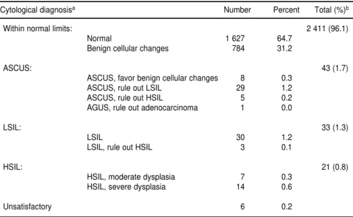
Enrollment cervical specimens were evaluable for 2 514 women. For the re-maining 14 women, the specimen was insufficient to enable appropriate cyto-logical review to be performed, or the slides were lost or broken. Table 4 shows the distribution of subjects with respect to their baseline cytology re-sults, as determined from the review
of the enrollment Pap smears at McGill University. The prevalence of SILs of both low and high grade was 2.2%.
A blind assessment of the diagnostic reproducibility was done in Montreal with random samples of 109 normal and 103 SIL (74 LSIL, 29 HSIL) smears. The kappa statistic was 0.847, with only 19 of 212 smears (9.0%) classified outside of their original diagnostic cat-egories (normal, LSIL, HSIL), and only 10 of 212 (4.7%) if diagnostic mis-matches were based on a broader, any-grade SIL category.
The local cytology reading in São Paulo was based on the old Papanico-laou classification system. The per-centages by class were as follows: I (normal): 26.7%; II (inflammatory): 71.2%; III (not otherwise specified): 1.3%; IIIa (mild dysplasia): 0.7%; IIIc (severe dysplasia = 1 case): 0.0%; IV (carcinoma in situ): 0.1%; and V (inva-sive cancer = 1 case): 0.0%. Of the 86 cases of SIL detected either by the local smear reading or by the cytology re-view in Montreal, only 23 were de-tected at both opportunities.
Cervicography was introduced in the study only in December 1994,
which prevented 628 women from being examined during the first year. The remaining 1 886 women were clas-sified as follows: 1 616 (85.7%) within normal limits, 214 (11.3%) low-grade lesions A or B, 9 (0.5%) high-grade le-sions, and 47 (2.5%) unsatisfactory. Of the 223 low-grade or high-grade le-sions detected by cervicography, only 4 (1.8%) were also diagnosed as Pap classes III or higher by the local cytol-ogy, and 9 (4.0%) were labeled as LSIL or HSIL by the cytology review.
Baseline HPV status
As of August 1998, we had com-pleted HPV DNA testing and typing for 4 935 specimens from 1 430 women, representing the first-year specimens from those accruing long-term follow-up. The prevalence of HPV infection by any type at entry was 13.8%. The most prevalent types were HPV 16 (2.8%), HPV 53 (1.5%), HPV 58 (1.2%), HPV 6/11 (1.0%), HPV 31 (1.0%), HPV 70 (0.9%), and HPV 18 (0.8%). Among HPV-positive specimens, the mean viral copy number was 3.0 per cell, with most specimens having 1 or fewer viral copies per cell. Only 11% of the HPV-positive specimens at enrollment had more than 100 copies per cell.
Determination of molecular variants of HPV by sequencing is the most time-consuming procedure in the study. We have thus far found seven different variants of HPV 16 in 97 iso-lates from 65 subjects. These variants are grouped phylogenetically as fol-lows: E branch: prototype, B-12; AA branch: B-2, B-2A, one new (related to B2); Af2 branch: S21-A; and As branch: AC-2. Four variants of HPV 18 were identified in 27 isolates from 11 sub-jects: B18-2, B18-6, one new (related to B18-2), and T18-17.
Preliminary assessment of the prognostic value of baseline HPV status
We analyzed the risk of any-grade, incident SIL in the cohort as a function of HPV positivity at enrollment. Data
TABLE 2. Characteristics of subject participation and reasons for ineligibility at time of initial enrollment, São Paulo, Brazil, accrual period 1993–1997
% (in relation to)
Status Category Number Status Overall
Ineligible Presently pregnant or intends to become 411 33.0 8.2
Total or partial hysterectomy 285 22.9 5.7
Residence outside São Paulo or intention to
move soon 223 17.9 4.5
Age < 18 or > 60 158 12.7 3.2
Reasons invalidating Pap smear 91 7.3 1.8
Recent surgery preventing participation 40 3.2 0.8
Virgin 20 1.6 0.4
Mental retardation or deafness 17 1.4 0.3
Total 1 245 100 24.9
Unknown Refused to be approached 138 88.5 2.8
Did not provide information when approached 18 11.5 0.4
Total 156 100 3.1
Eligible Enrolled in the study 2 528 70.4 50.7
Refused participation 1 061 29.6 21.3
Total 3 589 100 71.9
Total (all categories) 4 990 NAa 100
on cytological outcomes were based on smears read through August 1997. Only the 887 women who were free of SIL at enrollment and had been tested for HPV were included in the analysis. Two separate incidence curves were computed using actuarial techniques, one for cytological outcome
deter-mined by the smear reading by the local hospital provider in São Paulo (Figure 1, top) and the other by the cy-tology review in Montreal (Figure 1, bottom). The actuarial curves of SIL in-cidence based on the local cytology were largely overlapping, but not the ones based on the Montreal slide
re-view. Using the research-quality cytol-ogy (smear readings provided by Dr. Ferenczy’s Montreal laboratory), the relative risk (RR) of SIL associated with HPV positivity was 5.8 (95% con-fidence interval [CI]: 3.0–11.1) (using the Cox proportional hazards model). Based on the local reading of the same smears, the equivalent RR of SIL was a considerably lower 1.4, with a 95% CI of 0.6–3.3.
DISCUSSION
Although there is currently great en-thusiasm concerning the possible ap-plication of HPV testing as an adjunct to Pap cytology screening for cervical cancer, there are several problems that need to be solved before any current secondary prevention programs can be augmented. Positive predictive val-ues of HPV testing are low in most asymptomatic women because of the relatively high prevalence of subclini-cal HPV infection in the general popu-lation. Most of these infections are transient and are probably of little sig-nificance. The concern resides, how-ever, with the small proportion of women who harbor persistent HPV infections. These women have a much greater risk of subsequent cervical neoplasia (18, 19), indicating that per-sistent HPV infections, rather than transient ones, are the actual biologic precursor in cervical carcinogenesis. Research on the epidemiology of viral persistence and on its determinants will help formulate algorithms and policies for inclusion of some form of HPV testing as an adjunct to screening for cervical cancer prevention. More importantly, however, we believe that the present study will provide new in-sights into the pathogenic mechanisms of HPV infection leading to cervical cancer, which will eventually aid in ef-forts at vaccine development and pri-mary prevention.
As far as we know, the Ludwig-McGill longitudinal study is one of the largest ongoing epidemiologic investi-gations of the natural history of cervi-cal neoplasia to employ a powerful array of laboratory techniques to
doc-TABLE 3. Distribution of selected characteristics at enrollment for participants in the Ludwig-McGill cohort, São Paulo, Brazil, accrual period 1993–1997
Variable Categories Numbera %
Age 18–24 485 20.3
25–34 944 39.6
35–44 686 28.8
≥45 270 11.3
Ethnicity White 1 535 64.4
Mestiza 511 21.4
Black 301 12.6
Other 36 1.5
Education Less than elementary 543 22.8
Elementary 1 393 58.5
High school 381 16.0
College/university 66 2.8
Smoking Never 1 137 47.7
Current 834 35.0
Former 414 17.4
Age at first intercourse ≤15 652 27.3
16–17 610 25.6
18–19 501 21.0
≥20 622 26.1
Lifetime number of sexual partners 1 1 058 44.4
2 502 21.1
3 331 13.9
4 192 8.1
5 115 4.8
≥6 186 7.8
Duration of oral contraceptive use Never 387 16.2
≤5 years 1 308 54.8
> 5 years 690 28.9
Number of pregnancies 0 45 1.9
1 353 14.9
2 505 21.3
3 500 21.1
4–5 568 24.0
≥6 397 16.8
Previous number of Pap tests 0 124 5.2
1–2 534 22.4
3–4 509 21.3
5–6 315 13.2
≥7 903 37.9
ument the onset of precursor lesions. It is also one of the few ongoing cohort studies with multiple measurements of HPV markers over time, thus allow-ing determination of the true incidence rate of viral infection. Since it is being conducted in a population at high risk for cervical cancer, the levels of statis-tical power for cytological endpoints might be higher than if the same study were conducted in a typical North American population.
Subject recruitment was completed in March 1997, after the cohort reached the sample size that we had estimated was needed to attain sufficient statisti-cal power to assess associations of in-terest. Actuarial rates of compliance with all follow-up visits have stabi-lized at around 75%. Such a rate of long-term adherence can be considered as very high in a protocol with proce-dure-intensive follow-up that requires multiple hospital visits frequently last-ing longer than one hour. This compli-ance rate has been attained despite the fact that participation is dependent upon agreeing to donate a blood sam-ple and a cervical cell specimen and to
undergo lengthy interviews dealing with sensitive life events in most clinic visits.
An ongoing cohort study in Costa Rica also includes repeated testing of subjects for HPV and cervical neopla-sia over time, but the focus of that study is on the role of HPV in the eti-ology of HSIL and on the evaluation of new cervical cancer screening strate-gies (20). Unlike other cohort studies that use viral type as a taxonomic unit, we have chosen the innovative ap-proach of testing for molecular vari-ants and measuring viral load in the cervix to study viral persistence. We will further document the natural his-tory of cervical HPV infection as the precursor event for SIL by correlating the viral findings with the subject’s immune response to specific capsid antigens of HPV and by correlating the latter with risk of subsequent SIL.
The focus of our investigation is on the dynamics of the natural history of HPV infection and cervical neoplasia. Although it is not one of our main ob-jectives, our cohort study will provide a limited assessment of the relative
diag-nostic performance of Pap cytology, cervicography, and HPV testing, as well as their combinations, using the biopsy results as the gold standard. We will calculate the sensitivity and speci-ficity for each combination of test and lesion severity. Women who were re-called as a result of the algorithm process described earlier in the “Man-agement of lesions” subsection but who did not have lesional tissue to be biop-sied will be considered disease-free for the purposes of the latter calculation.
Our preliminary assessment of cyto-logic outcomes in the study indicates that there was substantial underesti-mation of the prognostic value of HPV status at entry with respect to lesion outcome using the nonresearch-quality smear reading, presumably as a reflec-tion of outcome misclassificareflec-tion. This empirical illustration of the effects of cytological misclassification prompted us to stop using the cytology diagnoses from the local hospital provider for
TABLE 4. Distribution of cervical cytological results (based on slide review) at enrollment among cohort participants, São Paulo, Brazil, accrual period 1993–1997
Cytological diagnosisa Number Percent Total (%)b
Within normal limits: 2 411 (96.1)
Normal 1 627 64.7
Benign cellular changes 784 31.2
ASCUS: 43 (1.7)
ASCUS, favor benign cellular changes 8 0.3
ASCUS, rule out LSIL 29 1.2
ASCUS, rule out HSIL 5 0.2
AGUS, rule out adenocarcinoma 1 0.0
LSIL: 33 (1.3)
LSIL 30 1.2
LSIL, rule out HSIL 3 0.1
HSIL: 21 (0.8)
HSIL, moderate dysplasia 7 0.3
HSIL, severe dysplasia 14 0.6
Unsatisfactory 6 0.2
aAbbreviations: ASCUS = atypical squamous cells of undetermined significance; AGUS = atypical glandular cells of
undeter-mined significance; LSIL = low grade squamous intraepithelial lesions; HSIL = high grade squamous intraepithelial lesions. bPercent totals by diagnostic grouping exclude unsatisfactory smears from the denominator. Overall and diagnostic group
per-cent totals do not necessarily add up because of rounding errors.
triage purposes. An accredited cy-totechnologist has joined our team to provide a timely and reliable first screen of all smears generated in the study, to prevent delays in referring for colposcopy any cases of HSIL.
The traditional epidemiologic study designs of single-opportunity assess-ment of exposure and outcome do not permit assessment of questions of viral persistence, fluctuation in viral load, regression of cervical lesions, and the dynamics of risk factor changes over time (e.g., acquisition of
new sexual partners) (21). To under-stand the role and mechanism of such dynamic changes in the natural his-tory of the disease one must conduct studies that repeatedly collect data on risk factors, HPV, and cervical lesions on multiple occasions during follow-up. A longitudinal, repeated-measure-ment cohort investigation, such as the one used in the Ludwig-McGill study, is the only design that permits acrate and unbiased assessment of cu-mulative HPV exposure and lesion outcome history.
Acknowledgments. This project is supported by an intramural grant from the Ludwig Institute for Cancer Research and by grants from the U.S. National Cancer Institute, the Medical Research Council of Canada, and the National Cancer Institute of Canada. The authors are indebted to Drs. Maria Nozaki and Lúcia Aoki for the local cytology readings during the initial phase of the investigation. The authors are grateful for the kind contributions of materials by Dr. E. M. de Villiers and by Dr. John Schiller.
1. IARC Working Group. Human papillo-maviruses. Lyon, France: International Agency for Research on Cancer; 1995. (IARC Monographs on the Evaluation of Carcino-genic Risks to Humans. Vol. 64).
2. Franco EL, Villa LL, Rahal P, Ruiz A. Molecu-lar variant analysis as an epidemiological tool to study persistence of cervical human papil-lomavirus infection. J Natl Cancer Inst 1994; 86(20):1558–1559.
3. Caballero OL, Villa LL, Simpson AJG. Low stringency-PCR (LS-PCR) allows entirely in-ternally standardized DNA quantitation. Nu-cleic Acid Res 1995;23:192–193.
4. Solomon D. The 1988 Bethesda system for re-porting cervical/vaginal cytologic diagnoses. Developed and approved at the National Cancer Institute Workshop, Bethesda, Mary-land, USA, December 12–13, 1988. J Clin Cytol Cytopathol 1989;33:567–574.
5. Stafl A. Cervicography: a new method for cer-vical cancer detection. Am J Obstet Gynecol 1981;139:815–825.
6. Bauer HM, Ting Y, Greer CE, Chambers JC, Tashiro CJ, Chimera J, et al. Genital human papillomavirus infection in female university students as determined by a PCR-based method. JAMA 1991;265:472–477.
7. Hildesheim A, Schiffman MH, Gravitt PE, Glass AG, Greer CE, Zhang T, et al. Persis-tence of type-specific human papillomavirus infection among cytologically normal women. J Infect Dis 1994;169:235–240.
8. Bernard HU, Chan SY, Manos MM, Ong CK, Villa LL, Delius H, et al. Identification and assessment of known and novel human papil-lomaviruses by polymerase chain reaction amplification, restriction fragment length poly-morphisms, nucleotide sequence, and phylo-genetic algorithms. J Infec Dis 1994;170: 1077–1085.
9. Ho L, Chan SY, Chow V, Chong T, Tay SK, Villa LL, et al. Sequence variants of human pa-pillomavirus type 16 in clinical samples per-mit verification and extension of epidemiolog-ical studies and construction of a phylogenetic tree. J Clin Microbiol 1991;29:1765–1772. 10. Ong CK, Chan SY, Campo MS, Fujinaga K,
Mavromaranazos P, Labropoulou V, et al. Evo-lution of human papillomavirus type-18: an an-cient phylogenetic root in Africa and intratype diversity reflect coevolution with human eth-nic groups. J Virol 1993;67: 6424–6431. 11. Yamada T, Wheeler CM, Halpern AL, Stewart
ACM, Hildesheim A, Jenison SA. Human pa-pillomavirus type 16 variant lineages in United States populations characterized by nucleotide sequence analysis of the E6, L2, and L1 coding segments. J Virol 1995;69:7743–7753. 12. VanDenBrule AJC, Snijders PJF, Gordijn RLJ, Bleker OP, Meijer CJLM, Walboomers JMM. General primer-mediated polymerase chain reaction permits the detection of sequenced and still unsequenced human papillomavirus genotypes in cervical scrapes and carcinomas. Int J Cancer 1990;45: 644–649.
13. Kirnbauer R, Hubbert NL, Wheeler CM, Becker TM, Lowy DR, Schiller JT. A virus-like particle enzyme-linked immunosorbent assay detects serum antibodies in a majority of women infected with human papillomavirus type 16. J Natl Cancer Inst 1994;86:494–499. 14. Kirnbauer R, Taub J, Greenstone H, Roden R,
Durst M, Gissmann L, et al. Efficient self-assembly of human papillomavirus type 16 L1 and L1-L2 into virus-like particles. J Virol 1993;67:6929–6936.
15. Storey A, Thomas M, Kalita A, Harwood C, Gardiol D, Mantovani F, et al. Role of a p53 polymorphism in the development of human papillomavirus-associated cancer. Nature 1998;393(6682):229–234.
16. Hildesheim A, Schiffman M, Brinton L, Frau-meni Jr J, Herrero R, Bratti MC, et al. p53 poly-morphism and risk of cervical cancer. Nature 1998;396(6711):531–532.
17. Bodmer JG, Marsh SG, Albert ED, Bodmer WF, Bontrop RE, Charron D, et al. Nomencla-ture for factors of the HLA system, 1996. Vox Sanguinis 1997;73(2):105–130.
18. Koutsky LA, Holmes KK, Critchlow CW, Stevens CE, Paavonen J, Beckmann AM, et al. A cohort study of the risk of cervical intraep-ithelial neoplasia grade 2 or 3 in relation to pa-pillomavirus infection. N Engl J Med 1992; 327:1272–1278.
19. Ho GYF, Burk RD, Klein S, Kadish AS, Chang CJ, Palan P, et al. Persistent genital human pa-pillomavirus infection as a risk factor for per-sistent cervical dysplasia. J Natl Cancer Inst 1995;87:1365–1371.
20. Herrero R, Schiffman MH, Bratti C, Hilde-sheim A, Balmaceda I, Sherman ME, et al. De-sign and methods of a population-based nat-ural history study of cervical neoplasia in a rural province of Costa Rica: the Guanacaste Project. Rev Panam Salud Publica 1997;1(5): 362–375.
21. Franco EL, Rohan T, Villa L. Epidemiologic evidence and human papillomavirus infec-tion as a necessary cause of cervical cancer. J Natl Cancer Inst 1999;91:506–511.
Manuscript received on 11 January 1999. Revised version accepted for publication on 11 June 1999.
Este artículo describe un amplio estudio longitudinal, iniciado en 1993, acerca de la historia natural de la infección por papilomavirus humanos (PVH) y las neoplasias cervicales en una población de bajos recursos económicos de São Paulo, Brasil, una de las ciudades con mayor riesgo de cáncer cervical en todo el mundo. Esta investigación epidemiológica, conocida como el estudio de cohorte Ludwig-McGill, se centra en la infección persistente por tipos oncogénicos de PVH como el acontecimiento precursor que conduce a la neoplasia cervical. Sus objetivos consistieron en: 1) estudiar la epi-demiología de la infección cervical persistente por PVH en mujeres asintomáticas; 2) investigar si la infección persistente por PVH incrementa el riesgo de lesiones in-traepiteliales escamosas cervicales de bajo y alto grado; 3) identificar los determi-nantes de la infección persistente por PVH; 4) buscar variantes moleculares de los PVH que puedan estar asociadas a un aumento del riesgo de lesiones; 5) determinar si la carga vírica está correlacionada con la infección persistente y el riesgo de lesiones; 6) averiguar si la respuesta de anticuerpos frente a los PVH permite predecir la per-sistencia de la infección y la progresión de las lesiones, y 7) investigar el papel de los tipos HLA y del polimorfismo del codón 72 del gen p53como mediadores de la per-sistencia de los PVH y de la gravedad de las lesiones. Hasta marzo de 1997 se reclu-taron 2 528 mujeres que fueron examinadas cada cuatro meses en el primer año y cada seis meses en los años subsecuentes. Las participantes se someten a una entrevista basada en un cuestionario; a la obtención de una muestra cervical para citología de Papanicolaou y detección de PVH, y a la extracción de una muestra de sangre para identificar anticuerpos anti-PVH. Además, en el primer año se les realiza una cer-vicografía, que después se repite cada dos años. En este artículo se describen el diseño y los métodos del estudio, se presentan las características basales de la cohorte y se re-aliza una evaluación preliminar del valor pronóstico del estado basal de los PVH.
RESUMEN