Design and methods of a population-based natural
history study of cervical neoplasia in a rural province
of Costa Rica: the Guanacaste Project
1
Rolando Herrero,
2,3,4Mark H. Schiffman,
4Concepción Bratti,
3Allan Hildesheim,
4Ileana Balmaceda,
5Mark E. Sherman,
6Mitchell Greenberg,
7Fernando Cárdenas,
5Víctor Gómez,
5Kay Helgesen,
8Jorge Morales,
5Martha Hutchinson,
9Laurie Mango,
10Mario Alfaro,
5Nancy W. Potischman,
4Sholom Wacholder,
11Christine Swanson,
4and Louise A. Brinton
4This paper reports on the enrollment phase of a population-based natural history study of cer-vical neoplasia in Guanacaste, a rural province of Costa Rica with consistently high rates of invasive cervical cancer. The main goals of the study are to investigate the role of human papil-lomavirus (HPV) infection and its co-factors in the etiology of high-grade cervical neoplasia, and to evaluate new cervical cancer screening technologies. To begin, a random sample of cen-sal segments was selected and enumeration of all resident women 18 years of age and over was conducted with the aid of outreach workers of the Costa Rican Ministry of Health. Of the 10 738 women who were eligible to participate, 10 049 (93.6%) were interviewed after giving written informed consent. After the interview on cervical cancer risk factors was administered, a pelvic examination was performed on those women who reported previous sexual activity. The pelvic examination included a vaginal pH determination and collection of cervical cells for cytologic diagnosis using three different techniques. Additional cervical cells were collected for determination of the presence and amount of DNA from 16 different types of HPV, and two photographic images of the cervix were taken and interpreted offsite by an expert colposcopist. Finally, blood samples were collected for immunologic and micronutrient assays. Women with any abnormal cytologic diagnosis or a positive Cervigram, as well as a sample of the whole group, were referred for colposcopy, and biopsies were taken when lesions were observed. The enrollment screening will serve as the basis for a prevalent case-control study, and the mem-bers of the cohort free from serious disease will be followed actively, at intervals of no more than a year, to study the natural history of HPV infection and the origins of high-grade squamous intraepithelial lesions (HSIL). Details of the field operation are outlined, with particular refer-ence to the realization of this kind of study in developing countries. Descriptive data on the prevalence of disease and exposure to various risk factors are also presented.
ABSTRACT
21A Spanish version of this article will also be
pub-lished in this journal. The project was supported by contract N01-CP-21081 and N01-CP-31061 of the National Cancer Institute, National Institutes of Health, Bethesda, Maryland, U.S.A.
22Address reprint requests to Dr. Rolando Herrero,
International Agency for Research on Cancer, 150 Cours Albert Thomas, 69372 Lyon, Cedex 08, France.
23Ministerio de Salud, San José, Costa Rica.
24Environmental Epidemiology Branch, National
Cancer Institute, Bethesda, Maryland, U.S.A.
25Caja Costarricense de Seguro Social, San José,
Costa Rica.
26Department of Pathology, Johns Hopkins
Univer-sity, Baltimore, Maryland, U.S.A.
27Department of Gynecology, Graduate Hospital,
Philadelphia, Pennsylvania, U.S.A.
28Information Management Services Inc., Rockville,
Maryland, U.S.A.
29Department of Pathology, Tufts University,
Boston, Massachusetts, U.S.A.
10 Neuromedical Systems, New York, New York,
U.S.A.
11 Biostatistics Branch, National Cancer Institute,
Cervical cancer is the second most common cancer among women world-wide, with an estimated 471 000 new cases every year (1). The highest reported incidence rates are from de-veloping countries. Extensive knowl-edge has accumulated about risk fac-tors for the disease and its precursors, called cervical intraepithelial neoplasia (CIN) or squamous intraepithelial lesions (SIL). Consistent associations with sexual activity suggested early on that cervical cancer might be a late sequela of a sexually transmitted dis-ease (2–4). Recently, infection with cer-tain genital types of human papillo-mavirus (HPV) has been established as the central cause that can initiate the cascade of events leading to cervical neoplasia, including cancer (5). Thus, there is the possibility of eventually developing vaccines against this glob-ally devastating disease (6).
Although HPV infection appears to be a necessary condition, it is unlikely to be sufficient in itself to cause the development of high-grade SIL (HSIL) and cancer. Etiologic research has therefore focused on determining why only a small fraction of women ex-posed to this apparently common infection develop high-grade precur-sors and cancer. In addition, many other issues related to transmission, natural history, and host response to these viruses remain unknown. Until successful vaccines are developed, it is imperative that screening efforts be optimized in both resource-poor and wealthier nations.
A large, multicenter case-control study of invasive cervical cancer was conducted from 1985 to 1987 by the National Cancer Institute (NCI) of the United States of America and by local research centers in four Latin Ameri-can countries, producing extensive in-formation on the risk factors operating in the areas studied (7–10). As a con-tinuation of that effort, a population-based natural history study of cervical neoplasia in a rural province of Costa Rica is now under way. This project is part of a substantial NCI initiative to study the epidemiology of cervical cancer and purposely parallels in many respects two other NCI-funded
investigations being conducted in Oregon, U.S.A. (11), and Copenhagen, Denmark (12). In addition, the study addresses some of the same issues dealt with in other important investi-gations that have been conducted in the Americas (13, 14). This article de-scribes the main design elements, methodological aspects, and univariate results of the enrollment phase of the field work of the Guanacaste Project, including descriptive data on the prevalence of HPV and other risk fac-tors for cervical cancer, as well as the prevalence of cervical disease.
MATERIALS AND METHODS
Overview of design
The main objective of the study is to investigate the role of HPV and other host and environmental factors in the etiology of SIL and cervical cancer. Another objective is to determine the comparative efficacy of different screening techniques for cervical can-cer and its precursors. A sample of one-fifth of the adult female population of Guanacaste Province, Costa Rica, was selected randomly and invited to par-ticipate in the enrollment phase, which consisted of an interview and a pelvic exam including vaginal pH determina-tion, visual inspection of the cervix, preparation of a conventional Pap smear, preparation of a liquid buffer-based ThinPrep slide, collection of additional cells for DNA studies of HPV, and performance of a Cervigram picture (see below). In addition, a blood sample was collected for im-munologic, nutritional, and genetic studies. Women with any abnormal results and a small sample of the entire cohort were invited to a colposcopic evaluation, where punch biopsies were obtained as appropriate. When needed, cone biopsies were performed to clarify worrisome cytologic-histologic dis-crepancies. Expert collaborators sup-ported each technical aspect of the study. To allow for study of the epi-demiology of invasive cancer in this context, all Guanacaste residents diag-nosed with invasive cervical cancer
during the period of enrollment were also included in the sample.
In order to meet the first objective of the study, the enrollment phase will generate information on prevalence of infection with type-specific HPVs and on prevalence of different stages of cervical neoplasia, including cancer. In addition, it will allow for a prevalent case-control study of the determinants of HPV infection and risk factors for each stage of the disease. Potential factors to be studied include age, edu-cation, sexual behavior, number of pregnancies, use of hormonal contra-ceptives, smoking, dietary consump-tion of micronutrients, exposure to pesticides, medical history, family his-tory of cancer, vaginal pH, serum lev-els of micronutrients, and specific human leukocyte antigen (HLA) hap-lotypes. With respect to the second objective, the performance of each screening test will be compared to the final diagnosis to evaluate the efficacy of different screening methods for control of cervical cancer in high-incidence settings.
The follow-up phase will consist of periodically rescreening a carefully selected subcohort of 3 000 women for at least four years to determine the ori-gins of HSIL, which is the main out-come of the prospective component of the study. Incidence of disease will be determined and compared between groups exposed to different levels of the above-mentioned factors.
Choice of study site
Guanacaste is a rural province bor-dering the Pacific Ocean in northwest Costa Rica (Figure 1). The estimated total population of the province in 1993 was 240 000 inhabitants (8% of the population of Costa Rica), 58% of whom were at least 18 years old. The area is 12 241 square kilometers (24% of the area of Costa Rica), with a pop-ulation density of 20.5 inhabitants per km2. The main economic activities in
14.6 per 1 000 children born alive; and life expectancy was 73.6 years.
Guanacaste was selected as the study site because it reports consis-tently high incidence rates of invasive cervical cancer. Table 1 shows inci-dence data from the population-based National Cancer Registry of Costa Rica, which has received mandatory reports of all cancer cases occur-ring in the country since 1981 (15). Age-adjusted incidence rates in the province of Guanacaste have averaged about 33 per 100 000 in the last 12 years, higher than the average for Costa Rica and about 4–5 times higher than comparable rates in the United States (16). Previous research estab-lished that the difference between high- and low-incidence areas in Costa Rica is related more to geographic variation in the prevalence of risk fac-tors than to different intensities of screening (17).
Guanacaste, like most of Costa Rica, has a well-established health infra-structure, including two regional
hos-pitals, 11 clinics operated by the Costa Rican Social Security Fund (Caja Costarricense de Seguridad Social, CCSS), and 95 health posts operated by the Ministry of Health. There are 0.56 physicians per 1 000 inhabitants. The smallest health posts have electricity, examination facilities, and waiting rooms and are staffed with an out-reach worker from the Ministry of Health, who makes periodic visits to every house in the area to promote vaccination, prenatal care, and other preventive programs. As these health facilities are used only sporadically by visiting teams from the Ministry of Health, they were available for use in the study.
Cervical cancer screening services are provided in the area under the uni-versal health care system. However, the coverage of the program has tradi-tionally been low and the target popu-lation generally restricted to women attending family planning and prena-tal care clinics. In addition, the labora-tories have limited quality control
sys-tems and there are difficulties in the follow-up and referral of cases de-tected. Some of these problems, in addition to the high prevalence of risk factors, probably explain the persis-tently high rates of cervical cancer in Guanacaste.
Organization of the study
The study is being conducted under a contract between the NCI and the Costa Rican Foundation for Education in Medical Sciences (FUCODOCSA). The latter is a part of the CCSS, which is the main government health pro-vider, with universal coverage in the country. In addition, collaborative agreements were established with the Ministry of Health for use of their facil-ities and with the Pan American Health Organization for tax exemption of imported equipment and materials. Assistance was also received from other government agencies including the Postal Service, which provided free delivery of letters under the Ministry of Health’s postal franchise. A plan-ning period of more than a year allowed time to complete these agree-ments, select and train the staff, and prepare the equipment and materials necessary for the study.
Central headquarters were estab-lished in San José, the capital city of
Atlantic Ocean
Panama San José
Pacific Ocean Nicaragua
Nicoya GUANACASTE
Liberia
FIGURE 1. Map of Costa Rica TABLE 1. Incidence of invasive cervical
cancer in Guanacaste Province, Costa Rica, by year, 1982–1992
Crude Adjusted
Year Cases rate ratea
1982 20 21.1 30.0
1983 24 25.0 37.0
1984 25 25.7 37.1
1985 23 23.3 35.4
1986 21 21.1 31.2
1987 19 18.9 26.3
1988 24 23.7 32.5
1989 34 33.3 45.1
1990 22 21.4 28.4
1991 25 24.2 33.7
1992 18 17.3 23.5
Source:National Tumor Registry, Ministry of Health, Costa Rica.
aIncidence per 100 000 women, age-adjusted to the world
Costa Rica, and two field offices were set up, one at each of the regional hos-pitals in Guanacaste in the towns of Liberia and Nicoya, which are about 250 and 300 km from San José, respec-tively (Figure 1). Key staff included a principal and a co-principal investiga-tor and an experienced field supervi-sor. Each study site was staffed by a registered nurse; a nurse’s aide; a supervisor, chosen among national census supervisors with extensive experience in field studies and person-nel management; three or more inter-viewers; a keypuncher; a driver; and an office manager. During the most intensive part of the study, a third team operated from the Liberia field office. To encourage subject participa-tion, all the nurses, nurse’s aides, and interviewers were women. Each office was equipped with a four-wheel drive vehicle, a –30 °C freezer for specimen storage, a computer to support the information system, and a –20 °C freezer for storage of ice-packs. In addition, a mobile colposcopic clinic was staffed by a colposcopist, a nurse’s aide, and two interviewers, and equipped with a Zeiss 2000 colposcope and a computerized image system (see below). Study vehicles and offices were identified with Social Security logos and equipped with radio com-munications within the Social Security network. The materials and equipment necessary for the daily field trips were transported every day to the local clin-ics where study procedures were per-formed (see below). Manuals for all the procedures were developed, and detailed training sessions were con-ducted with all members of the staff. A component of this training was site visits by expert collaborators. A com-puterized information system was developed by IMS (Information Man-agement Services Inc., Silver Spring, Maryland, U.S.A.) to keep track of data on every participant (appointments, diagnoses, samples, etc.).
Sample selection and census
The primary sample unit used in this study was the censal segment, of
which there are 1 038 in Guanacaste. For the last national census, which was conducted in 1984, each censal seg-ment was designed by the Bureau of the Census to comprise between 40 and 60 households. However, since 1984, significant changes have oc-curred in the size and distribution of the population, and some segments with new developments may now have substantially more dwellings than the census count, while others may have lost some of their popula-tion. Maps with details of each seg-ment, including the location of houses (Figure 2), were available although somewhat outdated, and a prelimi-nary effort was made to identify cen-sus tracts with new housing develop-ments. In order to improve sampling efficiency as well as the distribution of daily workloads, segment bound-aries were redrawn before the selec-tion to create segments of more homo-geneous size in terms of number of houses. Then, a sequential random sample of the redrawn censal seg-ments was selected, resulting in 208 segments. This selection was expected to produce a targeted sample size of 10 000 women.
Enumeration was conducted during February and March 1993, by visiting every house in the segments selected. For this purpose, 95 outreach workers from the Ministry of Health were trained by study supervisors and the study demographer in one-day ses-sions that included practice and evalu-ation. In three groups they conducted the census of their areas, taking approximately two weeks for each. All women 17 years of age or older who reported living in households within the selected segments were listed; for each the name, date of birth, complete mailing address, and cédula (national identification number) were recorded, although the name and address were considered sufficient to send letters of invitation. Most women were at home during the day, but houses were often visited several times or in the evenings to assure completeness of the census. Only women who were 18 years of age or older by 1 July 1993 were eligible to participate in the study.
Enrollment visit procedures
All women in the sample were ini-tially sent a personal letter inviting them to participate in the study and giving them an appointment at the closest local clinic. Those who did not come to the initial appointment were visited at home, given a more detailed explanation of the study, and offered transportation in study vehicles. Travel expenses were paid for some women in an effort to increase partici-pation, but no clinical examinations were done at home.
logic checks were conducted to ensure accuracy of the data. When necessary, hard copies of the questionnaires were reviewed to resolve apparent discrep-ancies. No data retrieval was sought by visiting the subjects again.
Pelvic examination and biological samples
Specially trained study nurses per-formed pelvic exams on women reporting previous sexual intercourse. First, the vulva, vagina, and cervix were inspected for the presence of gross abnormalities and, when such abnormalities were present, patients were referred to the study
colpo-scopist for evaluation. For patients with clinical signs of severe trichomo-niasis, candidiasis, or severe atrophy, the remainder of the exam was post-poned until after specific treatment of these conditions.
After insertion of a nonlubricated sterile speculum, a pHydrion strip (Micro Essential Laboratories, New York) with a pH range of 3.0 to 5.5 was placed against the lateral vaginal wall until wet. Vaginal pH was determined by comparing the color change of the strip with a colorimetric scale, and the measurement was recorded.
Following pH determination, exfoli-ated cervical cells were collected by firmly rotating a Cervex brush (Uni-mar, Connecticut) five times (1 800°) in
the ecto- and endocervical area (18). A conventional smear was prepared and the sample immediately spray-fixed with ethanol and carbowax Pap Per-fect fixative (Medscand, Florida). After the smear was made, the Cervex brush was swirled in 20 mL of methanol-based PreservCyt solution and then discarded. The solution was kept refrigerated until sent to the United States at ambient temperature for the preparation of monolayer cervical smears (ThinPreps).
Additional cervical cells were col-lected for virologic assays with a Dacron swab, which was swabbed over the ectocervix and rotated in the endocervical canal. The swabs were stored in Virapap DNA transport
medium tubes, which were kept in coolers in the field until frozen at –30 °C at the regional study offices. After collection of the materials men-tioned above, the cervix was rinsed with 5% acetic acid and two photo-graphic images of the cervix (Cervi-grams) were taken. The undeveloped film was sent to National Testing Lab-oratories in Fenton, Missouri, along with the patient’s log sheet, for devel-oping, processing, and evaluation. Finally, each participant was asked to donate 15 mL of blood. These samples were collected with standard proce-dures in heparinized tubes and kept at 1–4 °C in coolers in the field. The sam-ples were transported daily to the field stations, centrifuged for 20 minutes at 900 G, and then aliquotted into 2 mL prelabeled Nalge vials of plasma (usu-ally 5 aliquots), buffy coat (one), and red blood cells (one). Aliquots were frozen at –30 °C at the field stations and, along with the Virapap tubes, transported weekly on ice packs to San José, stored at –70 °C, and sent period-ically on dry ice to the NCI repository.
Interpretation of cytologic screening tests and Cervigrams
The conventional smears were stained and interpreted locally by a team of cytotechnologists and cyto-pathologists. Because of early variabil-ity, the staining process was stan-dardized in mid-enrollment. After interpretation in Costa Rica, which usually took one week, slides were sent to Neuromedical Systems in New York for computerized screening and prepa-ration of PapNet videotapes, which were interpreted at the Johns Hopkins Hospital, Baltimore, Maryland.
The PreservCyt vials were sent weekly to Tufts University (Boston, Massachusetts), where ThinPreps were prepared, and a third cytology diagnosis was sent back to the field for keying. Results were reported by all three cytology laboratories on stan-dard study forms and classified with the Bethesda System (19). The quality of smears was reported as adequate, adequate but limited, or inadequate.
The cytologic diagnosis was reported as one of the following: normal; nor-mal with reactive changes; atypical squamous cells of unknown signifi-cance (ASCUS); low-grade squamous intraepithelial lesion (LSIL); high-grade squamous intraepithelial lesion (HSIL), further classified as CIN 2 or CIN 3; squamous carcinoma; and ade-nocarcinoma.
Cervigrams were interpreted at Graduate Hospital in Philadelphia, Pennsylvania. The categories of evalu-ation were “negative” (no lesion seen), “atypical” (a lesion was seen, but the site and/or morphology of the lesion was such that colposcopy was not rec-ommended), “positive” (colposcopy recommended), and “technically de-fective” (not positive and unable to be properly evaluated). The “positive” category had the following classifica-tions: P0, probably normal variant, but colposcopy preferable to rule out seri-ous neoplasia; P1-P1A, compatible with trivial disease, but colposcopy recommended because part of the lesion extends into the canal; P1B, com-patible with CIN 1; P2, comcom-patible with high-grade disease (CIN 2 or CIN 3); P3, compatible with cancer. Evalua-tion reports were sent back to the field (usually within three weeks) for key-ing, along with a color photograph that included a mark in the most significant area to designate the recommended biopsy site for the colposcopist. The system aimed at the timely receipt of all the reports so that they would be useful for clinical management.
Colposcopy clinic visit
All patients with abnormal results in any of the three Pap smears (ASCUS or worse) or positive Cervigrams (P0, P1, P2, P3) were referred for colposcopy and biopsy of any visible lesions. In addition, any patients for whom eval-uation was considered urgent on the basis of visual inspection and a con-trol group consisting of 1 randomly selected woman in every 50 were referred for colposcopy. A second interview was administered at the time of colposcopy, covering the same
time period in the history of the patient as the enrollment interview and including more extensive ques-tions on medical history, family his-tory of gynecological cancer, douch-ing, details about sexual history, sexual behavior of the partner, alcohol consumption, occupational history, and exposure to pesticides. A food fre-quency questionnaire concerning the 42 most common sources of selected vitamins in the Guanacastecan diet was also included.
Following the interview, a colpo-scopic examination was performed by the study colposcopist to determine the presence and possible nature of cervical lesions and, when such lesions were detected, to take biopsies of the most relevant areas for histologic con-firmation. To assist the colposcopist in this process, Cervigram photographs with significant areas marked were available. The colposcopist registered with a precoded system the character-istics and topography of the transfor-mation zone and used a modified Reid index (20) to describe the severity of the lesions based on distal margins, color of acetowhitening, vascular pat-terns, and iodine staining. He also drew and described the location of the lesion with a grid system and recorded five images of the examination using a computer-assisted digital-imaging sys-tem (Den-Vu). The images recorded were the following: low magnification before and after application of acetic acid, high magnification after acetic acid, high magnification after iodine staining, and biopsy site if one was taken.
determina-tions, which were performed from blood drawn from the vacutainer tubes.
Biopsies were fixed in 10% buffered formalin, embedded in paraffin, sec-tioned, stained, and read at a local lab-oratory in Costa Rica. Slides and paraf-fin blocks were sent to Johns Hopkins Hospital for review by a pathologist. All other materials obtained during subsequent procedures (e.g., cone biopsies, hysterectomies) were also retrieved for review. Cone biopsies were mapped to follow the evolution of lesions over time and to correlate histopathology with cervicography.
Final diagnosis and treatment of cases
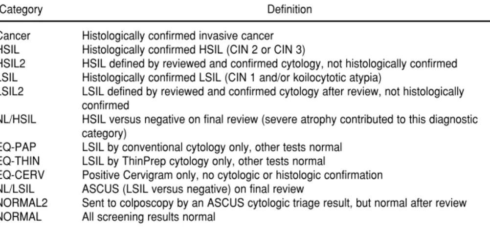
Histology alone cannot be consid-ered the gold standard, because a biopsy can be taken from an area that is not the most representative or severe, and a lesion may have disap-peared by the time a biopsy is taken. Therefore, a variety of diagnostic cate-gories were created (Table 2) based on a diagnostic protocol that included review of all cytology and pathology data available from all cases (i.e., cytol-ogy slides, biopsy slides, cone biop-sies, and hysterectomies).
All women with histologically con-firmed HSIL and those with cytologic evidence of HSIL in at least two
reports were managed by the Social Security clinicians according to local treatment protocols, most of them with large loop excision of the transforma-tion zone (LLETZ or LEEP) or cold knife cone (LEEP equipment was donated by Utah Medical). For women with only one cytologic report of HSIL who had normal colposcopy or whose biopsies were LSIL or less, the diag-nostic materials were sent for pathol-ogy review; those judged to be possi-ble HSIL were also referred for treatment. As a conservative measure, those initially called HSIL who were classified as LSIL, ASCUS, or normal upon further review were also referred to the local system and excluded from subsequent study follow-up.
Supplemental sample of invasive cancer cases
Incidence data from the National Cancer Registry indicated that only a small number of invasive cancer cases would be expected in a popula-tion of 10 000 women. Therefore, in an effort to increase the sample size, a rapid detection system was estab-lished to enroll all the cases of invasive cervical cancer diagnosed between June 1993 and November 1994 among women residing in Guanacaste. Per-sonnel from the Departments of Sta-tistics of three regional and three
tertiary care hospitals in San José (San Juan de Dios, Calderón Guardia, and México) performed daily reviews of admission and discharge diagnoses and visited oncology and gynecology wards in search of potential cases. In addition, the National Tumor Registry and the cytology laboratory providing services to the region notified study personnel immediately of any new cases. Upon notification, study staff visited the hospitals, determined eligi-bility of the cases, reviewed and abstracted charts, administered both the enrollment and colposcopy visit interviews, and collected the bio-specimens (Virapap and 15 mL blood, processed with a protocol similar to colposcopy blood samples). They also obtained relevant biopsy specimens from the hospitals for histologic con-firmation.
HPV testing
The primary technique used in the study to determine the presence of HPV in the Dacron swab samples collected at the enrollment visit was the hybrid capture method (Digene) (21). Briefly, with this technique the specimens are first hybridized with a full-length genomic HPV RNA probe. The resultant RNA:DNA hybrid is captured onto the surface of a tube coated with an anti-RNA:DNA hybrid antibody. Immobilized hybrids are then reacted with an hybrid anti-body conjugated to alkaline phos-phatase, and the light which is emitted is measured as relative light units (RLUs) on a luminometer. The inten-sity of the light emitted is proportional to the amount of target DNA in the specimen. Viral types were grouped as HPV 16, other high-risk (HPV 18, 45, and 56), intermediate-risk (HPV 31, 33, 35, 39, 51, 52, and 58), and low-risk (HPV 6, 11, 42, 43, and 44) types, based on an analysis of their respective asso-ciation with cancer (22). Additional testing with an L1 consensus primer basis polymerase chain reaction (PCR) technique, which is a more sensitive method (23), is under way for 3 000 selected specimens.
TABLE 2. Definition of diagnostic categories used for classifying cervical samples in the Guanacaste Project
Category Definition
Cancer Histologically confirmed invasive cancer HSIL Histologically confirmed HSIL (CIN 2 or CIN 3)
HSIL2 HSIL defined by reviewed and confirmed cytology, not histologically confirmed LSIL Histologically confirmed LSIL (CIN 1 and/or koilocytotic atypia)
LSIL2 LSIL defined by reviewed and confirmed cytology after review, not histologically confirmed
NL/HSIL HSIL versus negative on final review (severe atrophy contributed to this diagnostic category)
EQ-PAP LSIL by conventional cytology only, other tests normal EQ-THIN LSIL by ThinPrep cytology only, other tests normal
EQ-CERV Positive Cervigram only, no cytologic or histologic confirmation NL/LSIL ASCUS (LSIL versus negative) on final review
Selection of subjects for active follow-up
All women with HSIL in any of the screening tests, regardless of final diagnosis, were excluded from follow-up and referred to the Social Security health services for management ac-cording to local protocols.
During the follow-up phase of the study, women with histologic or cyto-logic evidence of LSIL will be re-screened every six months, and women with ASCUS, those who test positive for HPV by hybrid capture, those reporting five or more sexual partners, and a sample of women with normal results will be screened every year. The same clinical procedures and biological specimen collections per-formed at enrollment will be included in the follow-up examination, in addi-tion to a new interview focusing on behavioral changes during the obser-vation period, in particular regarding sexual practices, smoking, and use of hormonal contraceptives.
RESULTS
Sample selection and census
Census data for the original seg-ments selected (before redrawing, as no census data were available for the
redrawn segments) were compared (in combination) to the 1984 census data for the whole province of Guanacaste regarding all the census variables (e.g., age group, province of birth, national-ity, social security affiliation, province of residence five years earlier, educa-tion level, marital status, labor force participation, and children currently alive). Based on the 1984 data, the combined segments did not appear different from Guanacaste for any of the variables examined.
Enumeration of all adult women in the 208 selected segments was con-ducted between February and April 1993, producing a total sample of 14 750 women. However, this number was unexpectedly high and such a sample would have required more funding than was available; thus, it was decided to exclude enough women to obtain a final sample of about 12 000. This was accomplished by excluding randomly one out of every seven segments in the sample, leaving a total of 178 segments and 11 742 women.
Participation rates in the interview component of the study
Enrollment started in June 1993 and was completed in December 1994. Of the 11 742 women in the final sample
(Table 3), 2.6% were pregnant when contacted and would not have com-pleted three months postpartum when the enrollment phase ended, 1.6% had mental/language problems or were physically incapacitated or had died, and 4.4% had moved out of the area, leaving a total of 10 738 eligible women. Of the eligibles, 1.1% refused participation, and 5.2% repeatedly agreed to participate when contacted by study personnel but never showed up at the clinics. A total of 10 049 women were interviewed, correspond-ing to 93.6% of the eligibles.
Initially, all women were given ap-pointments by mail; those who did not show up for their appointments were visited at home by study personnel for rescheduling, usually around the day of the appointment. Several attempts were made to encourage participation, and sometimes outreach workers of the Ministry of Health and even “vol-unteer ladies” of the Liberia Hospital were involved in this effort.
Participation rates were slightly lower in the extreme age groups (Table 4), with women under 25 hav-ing rates of about 90% of the eligibles and women over 75 around 85%.
TABLE 3. Participation rates in the enrollment phase of the Guanacaste Project
No. %
Women in the census 11 742 100.0
Ineligible
Pregnanta 308 2.6
Mental/language problems 84 0.7
Physically incapacitated 56 0.5
Dead 42 0.4
Moved out of Guanacaste 514 4.4
Total eligible for interview 10 738 100.0
Refusal 120 1.1
No show 555 5.2
Other 14 0.1
Interviewed 10 049 93.6 (of eligibles)
Total blood sample taken 9 967 99.2 (of those interviewed)
aDeferred to a date out of enrollment period.
TABLE 4. Participation rates during the enrollment phase, Guanacaste Project, by age
Age
group Eligible Intervieweda %
18–19 461 417 90.5
20–24 1 263 1 147 90.8
25–29 1 474 1 396 94.7
30–34 1 463 1 407 96.2
35–39 1 267 1 210 95.5
40–44 1 051 1 004 95.5
45–49 847 802 94.7
50–54 651 614 94.3
55–59 584 544 93.2
60–64 497 470 94.6
65–69 430 385 89.5
70–74 321 309 96.3
75–79 191 169 88.5
80+ 214 175 81.8
Unknown 24 0
Total 10 738 10 049 93.6
aThis age distribution is based on the age of the women at
Younger women were more difficult to enroll mainly because many were not yet sexually active. Older women were much more difficult to mobilize from their homes and many were sim-ply not interested.
Participation was slightly higher among rural residents than among those of urban areas, where women are more likely to work outside their homes and to have their own doctors. Participation rates in the largest city (Liberia) remained lower than in the rest of the cantones, although study teams made additional recruitment efforts in that area to increase partici-pation.
Appraisal by the interviewers of the quality of the interviews indicated that overall quality was considered unreli-able for only 0.2% of participants, gen-erally reliable for 14.9%, and highly reliable for 84.9% of the subjects. Fig-ures were similar for specific sections of the interview.
Participation in the pelvic exam and blood component of the study
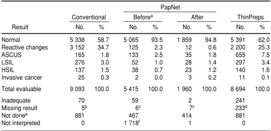
Of the 10 049 women interviewed, a total of 9 466 women reported previ-ous sexual experience and were there-fore eligible for a pelvic examination (Table 5). Such an exam was per-formed on 9 175 women (96.9%). The 291 women on which the pelvic exam was not performed either refused that specific component (n = 213) or had medical problems which precluded the examination, most of them associ-ated with old age. Results of conven-tional cytology and vaginal pH mea-surements were obtained for virtually all subjects examined. Computer-assisted (PapNet) results were avail-able for only 7 375 women, because an unacceptable backlog in reporting was produced in mid-enrollment. Thus, it was decided to exclude a number of smears that would have been reported undesirably late. In addition, this tech-nique was observed to add very little to the sensitivity of the screening, at least in part because the overall quality of the conventional smears was subop-timal for automated cytology. In
par-ticular, nuclear staining tended to be light, and a large proportion of smears showed some degree of inflammation and blood, as is common in this popu-lation. The staining process of the smears was optimized after additional training was provided in March 1994.
Table 6 shows results for each of the three Pap readings. Important differ-ences are evident in the percentages of specific abnormalities and in the appraisal of the quality of the smears done with the different techniques. The conventional and ThinPrep tech-niques appeared to diagnose very similar percentages of abnormalities (LSIL or worse), while PapNet
re-vealed lower percentages. However, this comparison does not provide information on the agreement of indi-vidual results, and differences may be related to the interpreter or to the tech-nique. This question will be the subject of detailed analysis, with final diagno-sis used as the gold standard.
Almost 99% of patients with a pelvic exam had a Cervigram, and a similar percentage of the Cervigrams per-formed were considered technically adequate. About 5% of Cervigrams were reported as “positive” or abnor-mal (Table 7). A blood sample was donated by 99.2% of the women who were interviewed, and a total of 89 877
TABLE 6. Results of cytologic screening tests during enrollment phase, Guanacaste Project
PapNet
Conventional Beforea After ThinPreps
Result No. % No. % No. % No. %
Normal 5 338 58.7 5 065 93.5 1 859 94.8 5 391 62.0
Reactive changes 3 152 34.7 125 2.3 12 0.6 2 200 25.3
ASCUS 165 1.8 133 2.5 35 1.8 655 7.5
LSIL 276 3.0 52 1.0 28 1.4 297 3.4
HSIL 137 1.5 38 0.7 23 1.2 140 1.6
Invasive cancer 25 0.3 2 0.0 3 0.2 11 0.1
Total evaluable 9 093 100.0 5 415 100.0 1 960 100.0 8 694 100.0
Inadequate 70 59 2 241
Missing result 5b 6c 7c 233d
Not donee 881 467 414 881
Not interpreted 0 1 718f 1 0
aBefore and on 7 March versus after 7 March 1994, when Pap smear staining was optimized. bReports with mistakes that had already been sent to the United States when the mistakes were noticed. cThe majority represent a group of slides broken during transportation.
dThe majority represent a group of slides lost during shipment.
eIncludes 583 virgins and 298 women who refused the pelvic exam (291) or for whom the collection of the Pap smear was
technically impossible (7) (see text).
fIntentionally not interpreted because of a backlog (see text).
TABLE 5. Participation rates in pelvic exam component during enroll-ment phase, Guanacaste Project
No. %
Total eligible for pelvica 9 466
Total pelvic doneb 9 175 96.9 (of those eligible for pelvic) Total Cervigrams 9 062 98.8 (of exams done)
Total Virapap 9 159 99.8 (of exams done)
Total final
diagnosis 9 175 100.0 (of exams done)
aExcludes 583 virginal women.
aliquots were sent to the NCI reposi-tory. All the shipments on dry ice were received frozen in the United States, reflecting very careful preparation and tracking of the international shipments.
Colposcopy
A total of 2 199 patients were referred to colposcopy; 1 736 were referred because of an abnormal Pap smear (ASCUS or worse) or Cervi-gram (P0 or worse), 287 were referred by the nurses because of abnormal clinical findings, and 176 were sent to colposcopy as controls. The controls were selected randomly from the whole cohort, regardless of screening diagnosis, to verify that the extensive screening was detecting all colposcop-ically evident cervical neoplasia. In fact, no disease was diagnosed among the 144 controls with a negative screening diagnosis.
A colposcopy interview was ob-tained from 2 134 patients (97% of referred women) and a colposcopy was performed on 2 129 (96.8%). The median period between enrollment visit and colposcopy visit was 13 weeks with a range of 4 to 65 weeks. Among women with a final diagnosis of cancer or HSIL, the median period was 9.7 weeks, with a range of 1 to 50 weeks. There were sometimes sub-stantial delays in the reporting of the cytologic diagnoses from the United States, and delayed referrals to
colpo-scopic evaluation were produced when prompted by those diagnoses alone.
Supplemental sample of invasive cervical cancer
The informants from the different hospitals reported 121 potential cases, 31 of which were eligible after deter-mination of their residence, histologic diagnosis, and date of diagnosis. Of these, 27 (87%) were interviewed, a Virapap sample was collected from 24 (77%), a blood specimen was collected from 25 (81%), and biopsies were available for review from 29 (94%).
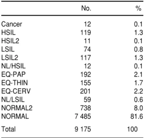
Final diagnosis and prevalence of HPV
Table 8 shows the frequencies and percentages of the final diagnoses. According to the case definition used in this study, all cancers were histolog-ically confirmed, compared to 92% of the HSILs and 39% of the LSILs. The lesions that were not confirmed histo-logically were included in the specific diagnostic group after extensive re-view of the materials available, and separate analyses are warranted for the different subgroups. The equivocal cat-egories indicate lesions suggested by different techniques that were not con-firmed after review and that will also be the subject of individual analyses.
The overall prevalence of HPV in the population, as determined by the hybrid capture technique, was 8.6%. The prevalences of HPV 16, other high-risk types, intermediate-risk types, and low-risk types were 1.7%, 2.0%, 5.4%, and 1.7%, respectively. These figures were similar when women with a history of hysterectomy were excluded (n = 621) and when women with a history of cryotherapy, cervical cauterization, or cervical surgery (n= 322) were also excluded. Detected prevalence declined sharply with age to a low around age 40 (Table 9). However, a partial rebound in prevalence was observed after age 60, which may be related to a cohort
effect or to re-emergence of latent infections. These prevalence figures are not directly comparable with other studies in Latin America because of differences in study design and HPV detection techniques. A sample of these specimens is being reanalyzed with the PCR technique, but more comparative studies are necessary.
Demographic and social characteristics of the cohort
Tables 10 and 11 show some of the main demographic characteristics of the cohort enrolled. The median age was 37 years, with a range of 18–97.
TABLE 7. Results of Cervigram evaluation during the enrollment phase, Guanacaste Project
Resulta No. %
Negative 7 718 85.2
Atypical 857 9.5
P0 (probably normal) 146 1.6
P1 (LSIL) 291 3.2
P2 (HSIL) 32 0.3
P3 (cancer) 18 0.2
Total evaluable 9 062 100.0
Technically defective 159b
aSee text for definition of the categories. bCorresponds to 1.7% of Cervigrams taken.
TABLE 8. Frequency (number and percent-age) of diagnoses of cervical abnormalities
No. %
Cancer 12 0.1
HSIL 119 1.3
HSIL2 11 0.1
LSIL 74 0.8
LSIL2 117 1.3
NL/HSIL 12 0.1
EQ-PAP 192 2.1
EQ-THIN 155 1.7
EQ-CERV 201 2.2
NL/LSIL 59 0.6
NORMAL2 738 8.0
NORMAL 7 485 81.6
Total 9 175 100
TABLE 9. Prevalence of hybrid capture HPV detection, by age, among sexually active women,aGuanacaste Province, Costa Rica
Age Percentage positive
18–19 24.0
20–24 14.7
25–29 9.7
30–34 8.4
35–39 6.2
40–44 5.3
45–49 5.8
50–54 5.3
55–59 5.2
60–64 7.4
65–69 9.1
70–75 10.5
75+ 8.6
Almost 60% had attended only ele-mentary school, 8.8% had never attended school, and only 12.7% had gone beyond high school. Eighty per-cent of the women were born in Gua-nacaste, and almost 90% of those born in other places moved to Guanacaste before age 30. The majority of the par-ticipants reported very low annual
incomes. Over 60% reported income below the poverty line for 1993 by Costa Rican standards (366 684 colones
or US$ 2 820). However, many of these patients were not aware of actual household income and oftentimes received supplemental income in the form of goods. Regarding household facilities, 88.5% reported electricity in the home, 62.4% had a refrigerator, 67.4% had a toilet inside the house, 80.8% had a television set, and 86.1% had running water (data not shown in table). The majority of these women reported being married or living as married. The median age at first sexual intercourse was 17 years, and initia-tion of sexual activity after age 26 was rare. Fifty-four percent reported hav-ing had only one sexual partner, and almost 90% reported having three or fewer. Ninety-five percent of the women had been pregnant, with a median age at first pregnancy of 19 years. This is a highly parous popula-tion, with around 42% of the women reporting five or more pregnancies. A history of cesarean section was given by 16.9% of the participants and 6.8% reported hysterectomies. Only 11.0% reported having smoked; in contrast, 36.3% of their husbands were smokers. Notably, a large percentage reported having had a Pap smear (87.8%).
DISCUSSION
The Guanacaste Project is a large natural history study of cervical neo-plasia in a rural community in Central America where cervical cancer re-mains a major public health problem, as it does in most developing regions. The enrollment phase was completed in December 1994, and initial analyses will provide information on the preva-lence of HPV and other risk factors for cervical neoplasia. In addition, inclu-sion of multiple screening techniques will permit an accurate assessment of the sensitivity and specificity of each method and their combinations. The second phase of the study will consist of the follow-up of 3 000 women with normal results or mild abnormalities to investigate the origin of HSIL and
TABLE 10. Demographic characteristics of the women in the cohort,a Guanacaste
Province, Costa Rica, 1993–1994
Characteristic No. %
Age (years)
18–19 229 2.5
20–24 917 10.0
25–29 1 301 14.2
30–34 1 358 14.8
35–39 1 167 12.7
40–44 972 10.6
45–49 780 8.5
50–54 591 6.5
55–59 517 5.6
60–64 443 4.8
65–69 355 3.9
70–74 277 3.0
75+ 268 2.9
Years of education
0 804 8.8
1–3 1 786 19.5
4–6 3 591 39.0
7–9 1 053 11.5
10–11 777 8.5
12–16 639 7.0
17+ 524 5.7
Born in Guanacaste
Yes 7 300 79.6
No 1 875 20.4
Age when first moved to Guanacaste (if not born there)
0–9 778 41.7
10–19 483 25.8
20–29 391 21.0
30–39 136 7.3
40–49 53 2.8
50–59 14 0.7
60+ 12 0.6
Yearly income in US dollars
<1 128 2 592 28.3
1 129–2 628 3 489 38.0
2 629–4 116 1 365 14.9
4 117–5 628 494 5.4
5 628–7 129 285 3.1
>7 129 368 4.0
Unknown/no response 582 6.3
aRestricted to 9 175 women who received a pelvic exam and
a final diagnosis. Unknowns for specific variables excluded, unless specified.
TABLE 11. Selected characteristics of women in the cohort,a Guanacaste
Province, Costa Rica, 1993–1994
Characteristic No. %
Marital status
Single 1 022 11.1
Married or living as married 7 167 78.1 Separated or divorced 518 5.7
Widowed 468 5.1
Age at first sexual intercourse
<13 113 1.2
13–14 704 7.7
15–16 2 183 23.8
17–18 2 595 28.3
19–20 1 601 17.5
21–25 1 473 16.1
26–30 377 4.1
30+ 122 1.3
Number of sexual partners
1 4 948 53.9
2 1 960 21.4
3 1 182 12.9
4 473 5.2
5 250 2.7
6+ 361 3.9
Ever been pregnant
Yes 8 737 95.2
No 438 4.8
Age at first pregnancy
<15 302 3.5
15–19 4 661 53.4
20–24 2 686 30.8
25–29 792 9.1
30–34 213 2.4
35–39 65 0.7
40+ 11 0.1
Total number of pregnancies
1–2 2 599 29.8
3–4 2 490 28.5
5–6 1 343 15.4
7–8 789 9.0
9–10 597 6.8
11–12 432 5.0
13–14 257 2.9
15+ 230 2.6
History of cesarean section
(in women with live births or stillbirths)
Yes 1 463 16.9
No 7 215 83.1
Had a hysterectomy
Yes 621 6.8
No 8 554 93.2
Ever had a Pap smear
Yes 8 038 87.8
No 1 122 12.2
Ever smoked
Yes 1 013 11.0
No 8 157 89.0
Smoking by husband
Yes 2 956 36.3
No 5 191 63.7
aRestricted to 9 175 women who received a pelvic exam and
cancer among women harboring spe-cific types of HPV.
Epidemiologic research in Gua-nacaste has offered advantages diffi-cult to find in combination elsewhere, including excellent participation of study subjects, low costs, and direct applicability of the findings to a region where cervical cancer is common. Guanacaste is a relatively poor area of Costa Rica. However, the infrastruc-ture is adequate for the logistics of a large study like this one. The universal health system offers the availability of many small clinics with basic require-ments for the study procedures. Also, local health authorities recognize the importance of these activities and offer full collaboration, it is possible to recruit educated and enthusiastic per-sonnel, most of the roads are in good condition, and there are excellent tele-phone and radio communications.
One of the disadvantages was the dearth of experienced investigators and administrators, particularly with regard to contract negotiation and management. This situation hampered the process of establishing the contract and initiating disbursement of funds. Another major difficulty was the preparation of equipment and materi-als, many of which were imported from the United States. The process of their importation and timely release from customs was a source of delays and additional cost. It was sometimes difficult to standardize procedures, particularly laboratory methods that were unfamiliar to the staff. The track-ing and shipment of thousands of bio-logical samples was a nearly over-whelming task, given the multiple permits and requisites established by the governments and airlines. For example, frozen samples are sent on dry ice, which is considered a danger-ous substance, and only airlines with specially trained staff are allowed to transport it.
Despite these difficulties, the partici-pation rate was above 93%, a figure rarely obtained in studies conducted in developed countries. A reason for the success of this phase of the study was that women in Guanacaste were will-ing to participate, not only in the
inter-views but also in the exams and sam-ple collection. The “project,” as it came to be known in the community, gained a reputation as a unique opportunity to obtain excellent cervical cancer screen-ing; often, people not selected in the sample approached the staff request-ing to be included, but such requests were denied with regret. The study staff did not hesitate to make special efforts to enhance participation of the women in the sample, because some of the field procedures may serve as a model for mass screening programs. In fact, the Costa Rican health sector is launching a nationwide cervical cancer screening initiative which will imple-ment some of the essential field meth-ods used in this study. This initiative will be facilitated in Guanacaste by the increased awareness generated by the study both in the community and among health personnel.
One of the concerns was to obtain a true population-based sample in order to determine prevalence of HPV infec-tion and cervical disease, which can also be useful for the planning of pre-ventive interventions, including the design of vaccines. The existence of a census office and detailed political divisions made it possible to select a random sample of censal segments. The large sample fraction, the similar-ity of the segments selected within the province, and the high participation rates lend certainty to the claim that this is a true population-based sample. The fact that both the interview com-ponents and the biological samples had similar participation rates nearly eliminates the possibility of important bias associated with differences be-tween subjects who donated speci-mens and those who did not.
Another key aspect was to screen as thoroughly as possible to guarantee that the disease-free cohort that will be followed was truly disease-free ini-tially. Screening with three different cytologic readings and the Cervigram was believed to constitute a very sen-sitive overall screening. Women in whom an abnormality was not de-tected by any technique are very unlikely to have serious disease. In addition, participation in the
colpo-scopic evaluation was virtually com-plete, and a final diagnosis was ob-tained on all participants.
In terms of etiologic research the main goal is to determine the origin of HSIL, given HPV infection. In the prevalent case-control study, compari-son of cases of HSIL or cancer with dif-ferent control groups will allow the study of risk factors for progression from normal to LSIL, from LSIL to HSIL, or directly from normal to HSIL, in particular among HPV-positive women. This issue will also be addressed more properly in a prospec-tive way during the follow-up phase of the study.
screening tool is HPV testing, which could select certain groups of patients at highest risk either for referral to col-poscopy or for more intensive screen-ing. We are starting formal evaluation of all these techniques, including cost-benefit analyses, to determine their potential role alone or in combination in screening programs in different socioeconomic contexts.
High participation in investigations of this kind is common in developing areas (8), and the follow-up phase is continuing with similarly high compli-ance. It is expected that, in return, the results of the study will yield answers that will benefit the health of women in Guanacaste and elsewhere.
Acknowledgments. We wish to
acknowledge the collaboration of Drs. Fernando Berdugo, Pierre Gaby Bien-Aimé, Federico Di Paola, Francisco Fuster, Seidy Herrera, Jaime Jenkins, Enrique Jiménez, Manuel Jiménez, Danilo Medina, Saeed Mekbel, Gabriel Odio, Jessie Orlich, Mario Pacheco, Vinicio Pérez, Alvaro Salas, Alfredo Santiesteban, Rafaela Sierra, Rodrigo Urcuyo, Gonzalo Vargas, Herman Weinstok, Lics. Aylin Carmona and Gonzalo Elizondo, and all the person-nel at clinics and hospitals in Costa Rica who made this project possible. Study staff: Manuel Barrantes, Fer-nando Cárdenas, and Elmer Pérez (supervisors); Lidia Ana Morera, Iris
Ugarte, and Pacífica Valdés (nurses); Jenny Díaz, Lidia Pastrana, Dalila Per-alta, and Elizabeth Sánchez (nurse’s aides); Sonia Avila, Pricila Bolandi, Marta Chaves, Lucía González, María Gutiérrez, Franco Mainieri, Dorian Miranda, Minor Miranda, Roberto Monge, Ana Lieth Moreno, Maribel Obando, María A. Pizarro, Esperanza Ramírez, Rebeca Sibaja, Hugo Viales, Nidia Viales, Jorge Umaña, and Kattia Umaña. In the United States, assis-tance in the interpretation of cytologic screening tests and Cervigrams was provided by Deidra Kelly, Karen Plowden, and Dr. M. Campion.
1. Pisani P, Parkin DM, Muñoz N, Ferlay J. Can-cer and infection: estimates of the attributable fraction for 1990. Cancer Epidemiol Biomarkers Prev[in press].
2. Beral V. Cancer of the cervix: a sexually trans-mitted infection? Lancet1974;110:37–40. 3. Brinton LA, Hamman RF, Huggins GR.
Sex-ual and reproductive risk factors for invasive squamous cell cervical cancer. J Natl Cancer Inst 1987;79:23–30.
4. Herrero R, Brinton LA, Reeves WC, Brenes MM, Tenorio F, de Britton RC, et al. Sexual behavior, venereal diseases, hygiene prac-tices, and invasive cervical cancer in a high-risk population. Cancer1990;65:380–386. 5. International Agency for Research on Cancer
(IARC). Monograph on the evaluation of carcino-genic risks to humans: human papillomaviruses. Lyon, France: IARC; 1995. (IARC Scientific publications, Vol 64).
6. Dillner J. Immunobiology of papillomavirus; prospects for vaccination. Cancer J 1992;5(4): 181–187.
7. Brinton LA, Reeves WC, Brenes MM, Herrero R, Gaitan E, Garcia M, et al. Parity as a risk factor for cervical cancer. Am J Epidemiol 1989;130(3):486–496.
8. Brinton LA, Herrero R, Brenes MM, Montal-van P, de la Guardia ME, Avila A, et al. Con-siderations for conducting epidemiologic case-control studies of cancer in developing countries. Bull Pan Am Health Organ 1991; 25(1):1–15.
9. Reeves WC, Brinton LA, Garcia M, Brenes MM, Herrero R, Gaitan E, et al. Human papil-lomavirus (HPV) infection and cervical cancer
in Latin America. N Eng J Med 1989;320: 1347–1441.
10. Herrero R, Brinton LA, Reeves WC, Brenes MM, Tenorio F, de Britton RC, et al. Risk fac-tors for invasive carcinoma of the uterine cervix in Latin America. Bull Pan Am Health Organ 1990;24(3):263–283.
11. Schiffman MH, Bauer HM, Hoover RN, Glass AG, Cadell DM, Rush BB, et al. Epidemiologic evidence showing that human papillomavirus infection causes most cervical intraepithelial neoplasia. J Natl Cancer Inst1993;85:958–964. 12. Krüger Kjaer S, van der Brule AJC, Bock JE, et
al. Human papillomavirus—The most signifi-cant risk determinant of cervical intraepithe-lial neoplasia. Int J Cancer 1996;65(5):601–606. 13. Eluf-Neto J, Booth M, Muñoz N, Bosch FX, Meijer CJLM, Walboomers JMM. Human papillomavirus and invasive cervical cancer in Brazil. Br J Cancer1994;69:114–120. 14. Muñoz N, Bosch FX, de San Jose S, Tafur L,
Izarzugaza I, Gili M, et al. The causal link between human papillomavirus and invasive cervical cancer: a population-based case-control study in Colombia and Spain. Int J Cancer1992;52:743–749.
15. Parkin DM, Muir CS, Whelam SL, et al. (eds.) Cancer incidence in five continents, Volume VI. Lyon, France: IARC; 1992. (IARC Scientific publications, number 120).
16. Ries LAG, Miller BA, Hankey BF. (eds.) SEER Cancer Statistics Review: 1973–1991. Bethesda, MD: National Cancer Institute; 1994. (NIH Pub. 94-2789).
17. Herrero R, Hartge P, Brinton LA, Reeves WC, Brenes MM, Urcuyo R, et al. Determinants of
the geographic variation of invasive cervical cancer in Costa Rica. Bull Pan Am Health Organ 1993;27(1):15–25.
18. Ferris DG, Berrey MM, Ellis KE, Petry LJ, Voxnaes J, Beatie RT. The optimal technique for obtaining a Papanicolaou smear with the Cervex brush. J Fam Pract 1992;34:276–280. 19. National Cancer Institute Workshop. The
1988 Bethesda System for reporting cervi-cal/vaginal cytologic diagnoses. JAMA1989; 262:931–934.
20. Reid R, Herschman BR. A colposcopic index for differentiating subclinical papilloma viral infection from cervical intraepithelial neopla-sia. Am J Obstet Gynecol 1984;149:815. 21. Schiffman MH, Kiviat N, Burk RD, Shah K,
Daniel R, Lewis R, et al. Accuracy and inter-laboratory reliability of HPV DNA testing by hybrid capture. J Clin Microbiol 1995;33: 545–550.
22. Lorincz AT, Reid R, Jenson AB, Greenberg MD, Lancaster W, Kurman RJ. Human papil-lomavirus infection of the cervix: relative risk associations of 15 common anogenital types. Obstet Gynecol1992;79:328–337.
23. Hildesheim A, Gravitt P, Schiffman MH, Kur-man RJ, Barnes W, Jones S, et al. Determinants of genital human papillomavirus infection in low-income women in Washington, DC. Sex Transm Dis1993;20:279–285.
Manuscript received on 30 November 1995. Revised ver-sion accepted for publication on 15 May 1996.
En el presente trabajo se describe la fase de reclutamiento de un estudio poblacional sobre la historia natural de las neoplasias de cuello uterino en Guanacaste, provincia rural costarricense donde las tasas de cáncer cervicouterino invasor son invariable-mente altas. Las metas principales del estudio son investigar el papel que desem-peñan la infección por el virus del papiloma humano (VPH) y sus cofactores en la eti-ología de las neoplasias cervicouterinas de alto grado, y evaluar las nuevas tecnologías empleadas en el tamizaje del cáncer cervical. Para empezar se seleccionó una muestra aleatoria de segmentos censuales y, con la ayuda de trabajadores de acción comunitaria del Ministerio de Salud de Costa Rica, se hizo un recuento de todas las habitantes de 18 años de edad o mayores. De las 10 738 mujeres que cumplían con los requisitos para participar, 10 049 (93,6%) fueron entrevistadas después de haber dado su consentimiento informado por escrito. Después de la entre-vista sobre los factores de riesgo del cáncer cervicouterino, se hizo un examen pélvico a las mujeres que dijeron haber tenido actividad sexual. El examen pélvico incluyó la determinación del pH vaginal y la obtención de células para análisis citológico medi-ante tres técnicas distintas. También se obtuvieron células cervicales para determinar la presencia y cantidad de ADN de 16 tipos de VPH diferentes y se tomaron dos fotografías del cérvix que fueron interpretadas en un local distinto por un experto en colposcopia. Por último, se sacaron muestras de sangre para hacer ensayos inmunológicos y determinaciones de micronutrientes. Las mujeres con un diagnóstico citológico anormal o un cervigrama positivo, más una muestra del grupo en general, fueron remitidas para hacerles colposcopia y se tomaron biopsias cuando se obser-varon lesiones. El tamizaje con fines de reclutamiento servirá de base para un estudio de prevalencia de casos y controles, y las integrantes de la cohorte sin enfermedad avanzada tendrán un seguimiento activo a intervalos mínimos de un año, con el propósito de estudiar la historia natural de la infección por VPH y los orígenes de las lesiones escamosas intraepiteliales de alto grado. Se describe en detalle la operación de campo y se hace especial alusión a la realización de estudios de este tipo en países en desarrollo. También se presentan datos descriptivos sobre la prevalencia de la enfermedad y la exposición a diversos factores de riesgo.
RESUMEN