Ar
ti
cl
e
0103 - 5053 $6.00+0.00
*e-mail: [email protected]
Diphenyl Ditelluride Induces Neurotoxicity and Impairment of Developmental
Behavioral in Rat Pups
Simone Pinton,a Cristiane Luchese,a Eluza C. Stangherlin,a Silvane S. Romana,b and
Cristina W. Nogueira*,a
a Departamento de Química, Centro de Ciências Naturais e Exatas, Universidade Federal de Santa Maria,
97105-900 Santa Maria-RS, Brazil
b Departamento de Ciências da Saúde, Universidade Regional Integrada do Alto Uruguai e das Missões,
Campus de Erechim, 99700-000 Erechim-RS, Brazil
O objetivo deste estudo foi investigar se a exposição aguda ao ditelureto de difenila [(PhTe)2]
causaria prejuízo no desenvolvimento comportamental de ilhotes de ratos. Os ilhotes receberam uma injeção subcutânea de (PhTe)2 (0,1 mg kg-1, 3 mL kg-1) ou veículo (3 mL kg-1) no 14º dia
após o nascimento (DPN). Do 15º ao 21º DPN, foram realizados testes comportamentais nos ilhotes e, imediatamente após estes testes, os ilhotes foram submetidos à eutanásia. As análises histológicas, a determinação do conteúdo de mielina e a atividade da acetilcolinesterase (AChE) foram realizadas. O período crítico de intoxicação ocorreu no 4º e 5º dias após a injeção de (PhTe)2, quando os sinais de toxicidade foram mais evidentes, caracterizados pelo aparecimento de sinais sistêmicos de toxicidade, disfunções comportamentais, neurotoxicidade e uma alteração no sistema colinérgico. Desta forma, conclui-se que o (PhTe)2 induziu neurotoxicidade e prejuízos no desenvolvimento comportamental de ilhotes de ratos.
The purpose of the present study was to investigate if acute exposure to diphenyl ditelluride (PhTe)2 causes impairment of developmental behavioral performance in rat pups. Rat pups received a single subcutaneous injection of (PhTe)2 (0.1 mg kg-1, 3 mL kg-1) or vehicle (3 mL kg-1) at 14th
postnatal day. After exposure to (PhTe)2, the general parameters of neurotoxicity, behavioral
tasks, cerebral myelin content, histological analysis and acetilcholinesterase (AChE) activity were performed during seven days. The appearance of classic signs of toxicity, behavioral alterations and the reduction in myelin content were dependent on the time after (PhTe)2 exposure to pups. Neuronal damage, reduction of myelin content, and the increase in AChE activity occurred mainly at 4th and 5th day after (PhTe)
2 exposure, indicating that the critical period of neurotoxicity
is coincident with the major behavioral alterations. In conclusion, exposure to (PhTe)2 induced
neurotoxicity and impairment of developmental behavioralin rat pups.
Keywords: tellurium, neurotoxicity, developmental behavioral, myelin, acetylcholinesterase
Introduction
Tellurium (Te) is a metalloid with increasing utilization in rubber, metallurgic and electronics industry because of its unique chemical and physical properties. Similarly, the use of organic Te compounds will increase due to its importance
in organic synthesis.1,2 Thus, the incidence of occupational
exposure to Te either in elemental, organic or inorganic form has growing rapidly, but the biochemistry and clinical signiicance of such exposure are poorly understood.
Diphenyl ditelluride (PhTe)2 is an organotellurium
compound widely used as intermediate in organic
synthesis.2 Besides its use as an organic reactant, (PhTe)
2
has been reported as a neurotoxic compound in adult
rodents.3-5 (PhTe)
2, administered to dams, is teratogenic
to rat fetuses, changes behavioral parameters related to neural function and causes oxidative stress in cerebral areas
in young rats.6-8 Moretto and co-workers9 demonstrated
that (PhTe)2 impaired function of calcium channels in the
can interact directly with low molecular thiols, oxidizing
them to disulides.10
Additionally, there has been reported that rats exposed to tellurium develop a demyelination process following
a remyelination process.11,12 The effects of tellurium
intoxication on the nervous system have been established as suggestive of peripheral neuropathy during a period of active myelinogenesis affecting transcription of the myelin proteins
at the gene level and inhibiting the cholesterol synthesis.13-16
Acetylcholinesterase (AChE) is one of the most efficient biological catalysts known and plays a key role in cholinergic neurotransmission by hydrolyzing the transmitter acetylcholine (ACh), thus terminating
its action.17,18 However, the biological role of AChE is
not limited to cholinergic transmission. AChE has been implicated in several non-cholinergic actions including cell proliferation, neurite outgrowth and other responses to
various insults including stress and amyloid formation.19-21
Although, the neurotoxic effect of (PhTe)2 has been
reported, the interaction of this organotellurium compound with cholinergic system has not been documented.
Based on these considerations, the purpose of the present
study was to investigate if acute exposure to (PhTe)2 causes
impairment of developmental behavioral performance in
rat pups. To better understand the effect of (PhTe)2 in the
developmental period of life (14 to 21 days), parameters of neurotoxicity, such as histology, myelin content and activity of AChE, were studied in brain of rat pups.
Experimental
(PhTe)2 was synthesized in our laboratory according
to the method described in the literature by Petragnani.22
Analysis of the 1H NMR and 13C NMR spectrum showed
that (PhTe)2 obtained presented analytical and spectroscopic
data in full agreement with its assigned structure. The chemical purity of compound (99.9%) was determined by GC or HPLC. This drug was dissolved in canola oil, which was obtained from a standard commercial supplier. All other chemicals were of analytical grade and obtained from standard commercial suppliers.
Animals
Virgin female Wistar rats (180-240 g) from our own breeding colony were used. The animals were kept on a 12 h light/dark cycle, at a room temperature of 22 ± 2 °C, with free access to food and water. The animals were used according to the guidelines of the Committee on Care and Use of Experimental Animal Resources, the Federal University of Santa Maria, Brazil. Sexually virgin female rats were mated
with male (three females and one male in each cage). The onset of pregnancy was conirmed by the presence of sperm in vaginal smears (day 0 of pregnancy) and pregnant dams were immediately housed in individual cages.
The day of delivery was counted as postnatal day (PND) 0. At birth, all litters were culled to eight pups (four males and four females if possible) to ensure good nutrition. The rat pups were kept with their mothers during the period of the experiment.
Procedure
Previously, a toxicological pilot study with (PhTe)2 was
performed in rat pups. In this pilot study, three different doses of compound were used to determine the dose that is not lethal but causes signs of toxicity in rat pups. Based
on this pilot study, the dose of 0.1 mg kg-1 of (PhTe)
2 was
chosen.
Thirty-two litters were used in this study, as shown in Figure 1, each litter was divided into two groups: control
(canola oil, 3 mL kg-1) and tellurium groups (0.1 mg kg-1
(PhTe)2, 3 mL kg-1). The rat pups received a single
subcutaneous injection of (PhTe)2 or vehicle (canola oil) at
14th PND. In the following days, from day 1 to 7 (Figure 1,
PND 15 to 21) after exposure, behavioral tests were performed in rat pups. Rat pups were euthanized daily (from day 1 to 7) immediately after the behavioral tests. Brains were quickly removed for enzymatic assay and myelin isolation. The brain and cerebellum were used to histological analysis.
Signs of tellurium toxicity
Signs of tellurium toxicity were observed in rat pups
from the 1st day (PND 15) after exposure to 0.1 mg kg-1
(PhTe)2 to the end of the experimental period. The signs
of toxicity, such as garlic-like odor, partial or complete paralysis of hind legs, diarrhea, tremors, body hair loss and reduction of the body weight were recorded.
Behavioral tests
Negative geotaxis
Rat pups were placed in the middle of sandpaper covered 30° inclined surface plane, in a head down position and the latency to turn 180° to a head-up position was recorded. Negative geotaxis relects vestibular function,
motor development and activity.23
Forelimb support
Rat pup forepaws were placed on a horizontally suspended wire (1 mm in diameter), 47 cm above a soft foam rubber landing area. Each pup was timed from the moment it was placed on the wire until it was unable to
remain on the wire.24 This test relects muscular strength
in neonate rats.
Open ield
Open-ield was made of plywood and surrounded by walls 30 cm in height. The loor of the open-ield, 45 cm in length and 45 cm in width, was divided by masking tape markers into 9 squares (3 rows of 3). Each animal was placed individually at the centre of the apparatus and observed for 4 min to record the locomotor (number of segments crossed with the four paws) and exploratory activities (expressed by
the number of time rearing on the hind limbs).25 The open
ield test was only carried out in rat pups belonging to the
7th day after exposure to (PhTe)
2 group.
Ex vivo assay
Acetylcholinesterase (AChE) activity
For AChE activity assay, samples of cerebral cortex
were homogenized in 0.25 mol L-1 sucrose buffer
(1:10, m/v) and centrifuged at 2,400 × g at 4 °C for 15 min.
Activity of AChE was carried out according to the method
of Ellman and co-workers,26 using acetylthiocholine as
substrate. The activity of AChE was spectrofotometrically measured at 412 nm. The activity of AChE was expressed
as nmol h-1 mg protein-1.
Protein determination
Protein concentration was measured according to the
method of Lowry et al.27
Myelin content
The myelin isolation was conducted using the method
of Norton and Poduslo.28 Brains were homogenized in
approximately 20 vol. (m/v) of 0.32 mol L-1sucrose. The
myelin was isolated using discontinuous sucrose gradient
of 0.32 to 0.85 mol L-1. After isolation, myelin was dry until
the weight of the sample was stabilized. Myelin density was expressed as mg myelin/g brain.
Histological analysis
Samples of brain and cerebellum of each group from four different litters were randomly selected for histological study. The brain, cerebellum and spinal cords were placed in a ixative solution of 10% buffered formalin, processed by routine histological techniques, and embedded in parafin. The histological evaluation was made on 4 μm tissue sections of brain and cerebellum. The tissue sections were stained by haematoxylin-eosin (HE) to further analysis in a light microscope. The analyzed areas of the cerebellum were nigra and white matter.
Statistical analysis
The results are presented as means ± S.E.M. Comparisons
between experimental and control groups were performed by one-way analysis of variance (ANOVA) followed by the
Duncan’s test when appropriate. P values less than 0.05
(P < 0.05) were considered as indicative of signiicance.
Results
Signs of tellurium toxicity
After exposure to (PhTe)2 rat pups presented the
following signs of toxicity: garlic-like odor and diarrhea
(started at 1st day and persistent through the end of the
experiment), partial or complete paralysis of hind legs and
tremors (started at 3rd day and persistent through 5th day),
body hair loss (5th day) and gradual recovery of paralysis
(6th and 7th day). The animals exposed to (PhTe)
2
demonstrated a signiicant reduction in the body weight
gain from 3rd day to the end of the experiment (Figure 2).
Figure 2. Effect of (PhTe)2 on the body weight of rat pups. Data are
Behavioral tests
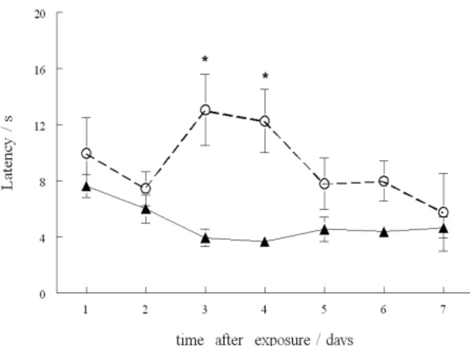
Negative geotaxis
At 3rd and 4th day after exposure to (PhTe)
2, rat pups
presented a signiicant increase in the latency to correct body position when compared to the respective control
groups. From 5th day to the end of the experiment, the
latency for animals to correct body position returned to control levels. Negative geotaxis was not altered in rat pups from all other time points (Figure 3).
Forelimb support
Forelimb support latency of rat pups was similar in
(PhTe)2 and the respective control groups up to 5th day after
exposure. From 6th day to the end of the experiment, the
latency of forelimb support was higher in the respective
control groups than in (PhTe)2 groups (Figure 4).
Figure 3. Effect of (PhTe)2 on the negative geotaxis test. Data are reported
as means ± S.E.M. of 5 litters per group. (*) Denotes p < 0.05 as compared to the respective control groups (one-way ANOVA/Duncan); () control and () (PhTe)2 groups.
Figure 4. Effect of (PhTe)2 on the forelimb support test. Data are reported
as means ± S.E.M. of 4 to 5 litters per group. (*) Denotes p < 0.05 as compared to the respective control groups (one-way ANOVA/Duncan); () control and () (PhTe)2 groups.
Figure 5. Effect of (PhTe)2 on AChE activity in cerebral cortex of rat
pups. Data are reported as means ± S.D. of 5 litters per group. (*) Denotes p < 0.05 as compared to the respective control groups (one-way ANOVA/Duncan).
Open ield
At 7th day after (PhTe)
2 exposure, rat pups presented a
signiicant decrease in locomotor (crossing: control = 31.2 ±
4.1 and (PhTe)2 = 15.1 ± 2.8) and exploratory (rearing:
control = 17.9 ± 2.1 and (PhTe)2 = 6.7 ± 3.1) activities when
compared to the respective control groups.
Ex vivo assay
AChE activity
AChE activity was increased in cerebral cortex of rat
pups at 4th and 5th day after exposure to (PhTe)
2 when
compared to the respective control groups. There was no alteration in AChE activity in cerebral cortex of rat pups from all other groups (Figure 5).
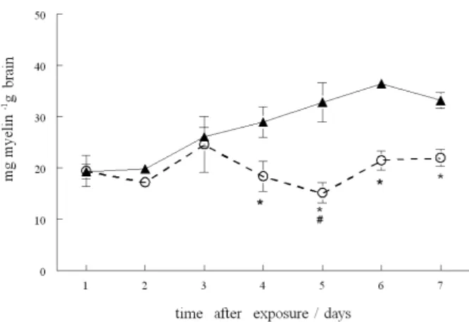
Myelin content
Exposure to (PhTe)2 caused a signiicant reduction
in cerebral myelin weight of rat pups from 4th day after
exposure to the end of the experiment. At 5th day after
exposure to (PhTe)2, rat pups presented the lowest cerebral
myelin content (Figure 6).
Histological analysis
In the cerebellar cortex of animals at 3th day after
exposure to (PhTe)2, the Purkinje cell monolayer, located
between the molecular and granular layers, demonstrated moderated chromatolysis, a sign of histological alteration (compare Figures 7 A and B).
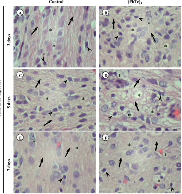
Cerebellar white matter in the animals exposed to
(PhTe)2 at 4th but mainly at 5th day after exposure presented
the widened endoneural space between individual axons and inflammatory cells in some areas of parenchyma (compare Figures 8 C and D). In the brain areas,
of neurons, such as eosinophilic cells, chromatolysis, loss of multipolar appearance and Nissl substance migration towards the cell periphery (compare Figures 7 C and D). The neurons clearly showed nucleus and nucleoli with retraction, hyperchromasia and pycnosis. Some of these morphological changes, although with less intensity, were
observed at 3rd day after exposure to (PhTe)
2.
At 6th and 7th day after exposure to (PhTe)
2, the disorder
in the cerebellar white matter induced by this compound
was smaller than that of presented at 4th and 5th days,
demonstrated by the appearance of illed spaces between axons, with linear axons of uniform thickness and cellular organization with a central nucleus (Figure 8).
Discussion
The present study showed that exposure to (PhTe)2
caused systemic toxicity in rat pups, demonstrated by a
garlic-like odor, theclassic signoftelluriumintoxication in
rats.29 Hind leg paralysis, which could be related to neuronal
damage, accompanied by behavioral alterations were also
observed in rat pups exposed to (PhTe)2.
The appearance of signs of toxicity, neurotoxicity and behavioral alterations were dependent on the time after
(PhTe)2 exposure to rat pups. The critical toxic period was at
4th and 5th day after (PhTe)
2 exposure, in which histological
analysis revealed alterations in the morphology of cerebral and cerebellar tissues and a reduction in the myelin content. Besides, the increase in AChE activity also occurred at
4th and 5th day after (PhTe)
2 exposure, indicating that the
critical period of neurotoxicity is related to the action of this compound in the cholinergic system.
Abnormal myelin deposition could potentially be the result of an insult to one or more of several different stages
Figure 6. Effect of (PhTe)2 on myelin content. Data are reported as means
± S.E.M. of 4 litters per group. (*) Denotes p < 0.05 as compared to the respective control groups and (#) denotes p < 0.05 as compared to all other groups (one-way ANOVA/Duncan); () control and () (PhTe)2 group.
Figure 7. Photomicrography of brain and cerebellum showed different degrees of injury with detail on the right. A) Cerebellar cortex of control pups shows
of development. In the rat brain, most oligodendrocytes complete a proliferation phase between approximately 1-2 weeks after birth, which is followed by a period of a very rapid rate of myelination (16-30 days after birth). The rate of myelination increases to a maximum at about 20 days of age and then declines to a low level that appears to be maintained throughout the life of the rats. In the myelination period, in the developing rat model, the biochemical mechanism of hypomyelination features a speciic reduction in myelin membrane synthesis rather
than an increased turnover.30
In this experimental protocol, (PhTe)2 was given during
physiological very rapid rate of myelination period when the CNS (central nervous system) is not yet myelinated in
the rat pups. Thus, one interpretation for the data found in
this study is that (PhTe)2 caused hypomyelination rather
than demyelination. The data presented in Figure 6 provide further support for this idea. It is clearly demonstrated in Figure 6 that there is no major decrease in myelin content but rather stagnation with any new myelin being formed. The fact that tellurium blocks cholesterol synthesis and causes
down-regulation of myelin gene expression31-34 could help to
explain the stagnation of myelin synthesis seen in rat pups
exposed to (PhTe)2. The unusual sensitivity of the enzyme to
tellurium is due to reaction with sulfhydryls and the binding
to the vicinal cysteines on squalene monooxygenase.31
In the current study, histological examination revealed
neuronal damage in rat pups exposed to (PhTe)2.
Figure 8. Photomicrography of the cerebellar white matter of: A,C,E) control group shows normal axons (arrow), myelin sheath (*) and oligodendrocytes
Neuronal damage was demonstrated by the appearance of chromatolysis; widened endoneural space between individual axons; inflammatory cells in some areas of parenchyma; eosinophilic cells; loss of multipolar appearance and Nissl substance migration towards the cell periphery; nucleus and nucleoli with retraction;
hyperchromasia and pycnosis. Schwab and Bartholdi35
have demonstrated morphological changes related to chromatolysis, a reaction to axon injury, present in most neurons of the pons nuclei. Several authors have reported that these changes are caused by the alteration of the cytoplasm and of the nuclear pH and to the presence of a
metabolic stage.36-39 Moreover, the appearance of widened
endoneural space between individual axons, demonstrated by histological examination of cerebellar white matter and brain, could be interpreted as a neural damage since edema could make this effect on tissue.
Regarding behavioral performance, rat pups exposed to
(PhTe)2 presented partial or complete paralysis of hind legs
from 3rd to 5th day after exposure, a period coincident with
the reduction in myelin content. The reduction of myelin content was accompanied by the most severe histological changes. Therefore, it is plausible to assume that paralysis of hind legs is associated with both events. Similarly, the results obtained on negative geotaxis, demonstrated a
commitment of the motor and sensorial functions at 3rd
and 4th day after exposure to (PhTe)
2. It is important to note
that the group exposed to (PhTe)2 presented similar results
in negative geotaxis when compared to control animals, at
latter stages after exposure (6th and 7th day), suggesting that
(PhTe)2-exposed pups reversed toxicity. These results are
supported, at least in part, by those obtained by Lampert
and co-workers.11,16 Lampert reported that weanling rats
less than three weeks of age became paralyzed when fed with a diet containing elemental tellurium, rat pups showed paralysis resulted from segmental demyelination of sciatic nerves and spinal roots and remyelination took place despite continued ingestion of tellurium, suggesting that whatever induced the neuropathy is effective only during a very limited but critical age period, the period of most active myelinogenesis.
The neurobehavioral response depends on developmental stage of the offspring and their CNS. Besides, the muscular maturation is also very important. Accordingly, the absence of increasing in forelimb support latency in rat pups at
6th and 7th day after (PhTe)
2 exposure supports the idea
that development of the pups muscular maturation was
delayed when they were subjected to exposure to (PhTe)2.
It is important to point out that no difference was found in the latency in the forelimb support between control and
exposed groups until 5th day after exposure to (PhTe)
2,
indicating that rat pups from PND 15 to 19 are still in
developmental stage of muscular strength. At 7th day after
exposure to (PhTe)2, the content of myelin in brain of rat
pups is still lower than that of control group at the same period, which would be consistent with the behavioral performance of rat pups in the open ield test. In fact,
rat pups at 7th day after exposure to (PhTe)
2 presented a
signiicant decrease in locomotor and exploratory activities in the open ield apparatus when compared to the respective control groups. The cholinergic neurotransmission is
vital to normal behavior and muscular function.39 In this
way, AChE plays a major role in the regulation of several
physiological events.40 Alterations in the activity of AChE
have been evidenced in several neurological disorders and
intoxication with organophosphorus.41 However, this is
the irst study aiming to investigate the in vivo effect of an
organotellurium, (PhTe)2, on AChE activity. The results
demonstrated an increase in AChE activity in cortex of rat
pups at 4th and 5th day after (PhTe)
2 exposure, which suggest
that some functions mediated by cholinergic system were
affected by (PhTe)2 exposure. In this context, we believe
that the increase in AChE activity could be explained as: i) a
direct effect of (PhTe)2 on this enzyme or ii) a consequence
of neuronal damage caused by (PhTe)2.
From a mechanistic point of view, the increase in AChE activity leads to a reduction of cholinergic neurotransmission efficiency due to a decrease in acetylcholine levels in the synaptic cleft, thus contributing to neurological dysfunctions. It is well recognized that altered membrane functions in several tissues including brain occur due to an enhancement of free radical formation which promotes increased lipid peroxidation, having as major consequence oxidative deterioration of the cellular
membranes.42 AChE is a signiicant biological component
of the membrane that contributes to its integrity and changes in permeability during synaptic transmission and
conduction.42 Because (PhTe)
2 causes oxidative stress in
brain,8 alterations in the lipid membrane could lead to
the increase in AChE activity. Thus, AChE activation in
(PhTe)2-exposed rat pups is probably precipitated by free
radical generation and consequent oxidative stress in the brain.
Conclusions
In summary, rat pups exposed to (PhTe)2 showed signs
of tellurium toxicity and impairment of developmental
behavioral performance. (PhTe)2 caused neuronal damage
in rat pups, which was revealed by histological examination and reduction of myelin content. Additionally, AChE
Acknowledgments
This study was supported in part by the FAPERGS/ CNPq (PRONEX) research grant # 10/0005-1 and by grants from the Brazilian National Research Council CNPq and CAPES. The inancial support from UFSM is also gratefully acknowledged.
References
1. Zeni, G.; Ludtke, D. S.; Panatieri, R. B.; Braga, A. L.; Chem. Rev.2006, 106, 1032.
2. Ogra, Y.; Anal. Sci. 2009, 25, 1189.
3. Nogueira, C. W.; Rotta L. N.; Perry, M. L.; Souza, D. O.; Rocha, J. B. T.; Brain Res. 2001, 906, 157.
4. Nogueira, C. W.; Rotta, L. N.; Zeni, G.; Souza, D. O.; Rocha, J. B. T.; Neurochem. Res. 2002, 27, 283.
5. Moretto, M. B.; Rossato, J. I.; Nogueira, C. W.; Zeni, G.; Rocha, J. B. T.; J. Biochem. Mol. Toxicol.2003, 17, 154.
6. Stangherlin, E. C.; Favero, A. M.; Zeni, G.; Rocha, J. B. T.; Nogueira, C.W.; Toxicology2005, 207, 231.
7. Stangherlin, E. C.; Favero, A. M.; Zeni, G.; Rocha, J. B. T.; Nogueira, C.W.; Brain Res. Bull. 2006, 69, 311.
8. Stangherlin, E. C.; Ardais, A. P.; Rocha, J. B. T.; Nogueira, C. W.; Arch. Toxicol.2009, 83, 485.
9. Moretto, M. B.; Thomazi, A. P.; Godinho, G.;Roessler, T. M.; Nogueira, C. W.; Wofchuk, S.; Rocha, J. B. T.; Toxicol. in Vitro
2007, 21, 639.
10. Nogueira, C. W.; Zeni, G.; Rocha, J. B. T.; Chem. Rev. 2004, 104, 6255.
11. Lampert, P.; Garro, F.; Pentschew, A.; Acta Neuropathol. 1970,
15, 308.
12. Harry, G. J.; Goodrum, J. F.; Bouldin, T. W.; Wagner-Recio, M.; Toews, A. D.; Morell, P.; J. Neurochem. 1989, 52, 938. 13. Duckett, S.; Said, G.; Streletz, L. G.; White, R. G.; Galle, P;.
Neuropathol. Appl. Neurobiol.1979, 5, 265.
14. Morell, P.; Toews, A. D.; Wagner, M.; Goodrum, J. F.;
Neurotoxicology1994, 15, 171.
15. Toews, A. D.; Hostettler, J.; Barrett, C.; Morelli, P.; Neurochem. Res.1997, 22, 1271.
16. Lampert, P.; Garrett, R. S.; Lab. Invest.1971, 25, 380. 17. Soreq, H.; Seidman, S.; Nat. Rev. Neurosci. 2001, 19, 294. 18. Mesulan, M. M.; Guillozet, A.; Shaw, P.; Levey, A. Duysen, E.
G.; Lockridge, O.; Neuroscience2002, 110, 627. 19. Appleyard, M. E.; Biochem. Soc. Trans.1994, 22, 749. 20. Chacón, M. A.; Reyes, A. E.; Inestrosa, N. C.; J. Neurochem.
2003, 87, 195.
21. Grisaru, D.; Sternfeld, M.; Eldor, A.; Glick, D.; Soreq, H.; Eur. J. Biochem.1999, 264, 672.
22. Petragnani, N.In Tellurium in Organic Synthesis; Katritzky, A. R.; Meth-Cohn, O.; Rees, C. W., eds.; Academic Press: London, 1994, ch. 3.
23. Crozier, W. J.; Pincus, G.; J. Gen. Physiol.1926, 10, 257. 24. Fox, W. M.; Anim. Behav.1965, 13, 234.
25. Walsh, R. N.; Cummins, R. A.; Psychological Bull.1976, 83, 482.
26. Ellman, G. L.; Courtney, D. K.; Andres, V. Jr.; Feather-Stone, R. M.; Biochem. Pharmacol.1961, 7, 88.
27. Lowry, O. H.; Rosebrough, N. J.; Farr, A. L.; Randall, R. J.;
J. Biol. Chem.1951, 193, 265.
28. Norton, W. T.; Poduslo, S.; J. Neurochem.1973, 21, 749. 29. Jackson, K. F.; Hammang, J. P.; Worth, S. F.; Duncan, I. D.;
Acta Neuropathol.1989, 78, 301.
30. Royland, J. E.; Wiggins, R. C.; Konat, G. W.; Brain Res. 1993,
207, 113.
31. Laden, B.; Porter, T.; J. Lipid. Res.2001, 42, 235.
32. Toews, A. D.; Roe, E. B.; Goodrum, J. F.; Bouldin, T. W.; Weaver, J.; Goines, N. D.; Morell, P.; Mol. Brain Res.1997,
49, 113.
33. Morell, P.; Toews, A. D.; Neurotoxicology1996, 17, 685. 34. Calle, E.; Berciano, M. T.; Fernández E.; Lafarga, M.; Acta
Neuropathol. 1999, 97, 143.
35. Schwab, M. E.; Bartholdi, D.; Physiol. Rev.1996, 76, 319. 36. Yamaoka, Y.; Shimohama, S.; Kimura, J.; Fukunaga, R.;
Taniguchi, T.; Exp. Toxicol. Pathol.1993, 45, 205. 37. Rosenblum, W. I.; J. Neurotrauma1997, 14, 313.
38. McD Anderson, R.; Opeskin, K.; Am. J. Forensic.Med.Pathol.
1998, 19, 1.
39. Payne, J. F.; Mathieu, A.; Melvin, W.; Fancey, L. L.; Mar. Pollut. Bull.1996, 32, 225.
40. Milatovic, D.; Dettbarn, W. D.; Toxicol. Appl. Pharmacol. 1996,
136, 20; Schetinger, M. R. C.; Porto, N. M.; Moretto, M. B.; Morsch, V. M.; Rocha, J. B. T.; Vieira, V.; Moro, F.; Neis, R. T.; Bittencourt, S.; Bonacorso, H. G.; Zanatta, N.; Neurochem. Res.2000, 25, 949.
41. Parry, A. M. M.; Scott, R. B.; Palace, J.; Smith, S.; Matthews, P. M.; Brain2003, 126, 2750; Christodoulou, C.; Melville, P.; Scherl, W. F.; Mc Allister, W. S.; Elkins, L. E.; Krupp, L. B.; J. Neurol. Sci.2006, 245, 127; Delino, R. T.; Ribeiro, T. S.; Figueroa-Villar, J. D.; J. Braz. Chem. Soc. 2009, 20, 407; Zanata, N.; Borchhardt, D. M.; Carpes, A. D.; Marchi, T. M.; Andricopulo, A. D.; Salum, L. B.; Schetinger M. R. C.; Bonacorso, H. G.; Martins M. A. P.; Flores, A. F. C.; J. Braz. Chem. Soc. 2009, 19, 1118.
42. Halliwell, B.; Gutteridge, J. M.; Free Radic. Biol. Med.1992,
12, 93; Das, A.; Dikshit, M.; Nath, C.; Life Sci. 2001, 68, 1545.
Submitted: March 29, 2010