Joint positioning sense, perceived force level and
two‑point discrimination tests of young
and active elderly adults
Priscila G. Franco1, Karini B. Santos1, André L. F. Rodacki1
ABSTRACT | Background: Changes in the proprioceptive system are associated with aging. Proprioception is important to maintaining and/or recovering balance and to reducing the risk of falls. Objective: To compare the performance of young and active elderly adults in three proprioceptive tests. Method: Twenty-one active elderly participants (66.9±5.5 years) and 21 healthy young participants (24.6±3.9 years) were evaluated in the following tests: perception of position of the ankle and hip joints, perceived force level of the ankle joint, and two-point discrimination of the sole of the foot. Results: No differences (p>0.05) were found between groups for the joint position and perceived force level. On the other hand, the elderly participants showed lower sensitivity in the two-point discrimination (higher threshold) when compared to the young participants (p < 0.01). Conclusion: Except for the cutaneous plantar sensitivity, the active elderly participants had maintained proprioception. Their physical activity status may explain similarities between groups for the joint
position sense and perceived force level, however it may not be suficient to prevent sensory degeneration with aging.
Keywords: physical activity; aging; proprioception; movement.
HOW TO CITE THIS ARTICLE
Franco PG, Santos KB, Rodacki ALF. Joint positioning sense, perceived force level and two-point discrimination tests of young and active elderly adults. Braz J Phys Ther. 2015 July-Aug; 19(4):304-310. http://dx.doi.org/10.1590/bjpt-rbf.2014.0099
1 Departamento de Educação Física, Universidade Federal do Paraná (UFPR), Curitiba, PR, Brazil
Received: Sep. 04, 2014 Revised: Dec. 27, 2014 Accepted: Feb. 27, 2015
Introduction
Physiological changes associated with aging lead to decreased functionality and reduced independence1.
Physiological changes include progressive reduction of the visual, vestibular, and proprioceptive systems that are essential to maintaining and/or recovering balance and reducing the risk of falls2. Falls are a
serious health problem3, as its complications are the
leading cause of hospitalization and death among individuals over 65 years4. Moreover, falls directly
affect the quality of life of elderly adults because of
their inluence on lifestyle and health5.
Changes in the central and peripheral nervous systems decrease the ability of the elderly to identify several stimuli in the environment and to select appropriate responses6. Losses in the proprioceptive
system reduce the ability to continuously monitor motor sequences and interfere in coordination and balance. Proper functioning of the proprioceptive system is crucial to adequate responses and correct actions of the segments involved in the movement7. Thus, these changes can inluence the level of physical activity
and functional capacity needed to perform daily tasks
independently2. Furthermore, proprioceptive deicits
may retard the perception of perturbations that require quick responses, e.g. stumbling and falling, thus leading to longer response times and decreasing the ability to restore the balance.
On the other hand, regular physical activity has been said to contribute to improve proprioception and therefore reduce falls with aging8. Thus, this study
aimed to compare the performance of young and active elderly participants in the following proprioceptive tests: (a) perception of joint position; (b) perceived level of strength; and (c) two-point discrimination on the sole of the foot. It was hypothesized that the elderly will present worse performances than the young group.
Method
Twenty-one young adults and 21 active elderly adults volunteered to participate in the study. For both
groups, inclusion criteria required classiication as
absolute or relative contraindications to the protocols applied in the study, history of recent joint surgery, use of prostheses, orthoses or assistive devices, chronic heart, lung or skin problems, no physical disability,
suficient mental ability to understand the test protocols,
and ability to perform daily activities independently. These variables were obtained based on self-report assessment. Participants received information about the procedures and signed an informed consent form. The experimental procedures in this study had the approval of the Research Ethics Committee of
Universidade Federal do Paraná (UFPR), Curitiba,
PR, Brazil (approval number CEP/SD 986.111.10.08; CAAE 0063.0.091.000-10).
Instruments and procedures
Participants were required to answer the International Physical Activity Questionnaire (IPAQ) that revealed an active physical status. Participants also performed the following tests: two-point discrimination, joint positioning sense, and perceived force exertion. The tests were applied in this order in an attempt to minimize possible fatigue effects.
Joint positioning sense test
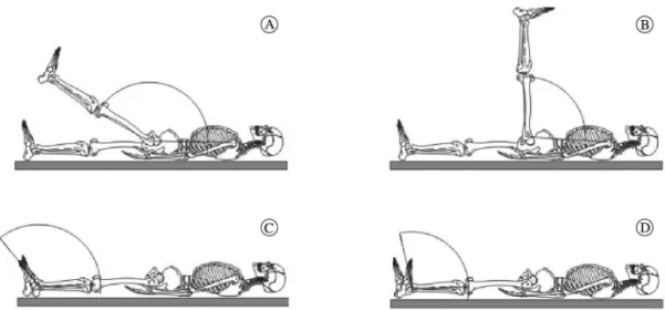
The joint positioning sense assessment was performed with the participants positioned in a supine posture
and had determined their maximum passive lexion
range of motion of the right hip and ankle. The hip and ankle joints were selected because they play a relevant role during the maintenance and recovery of
balance9. The range of motion was determined in the
anterior aspect taking into account the angle between the thigh and the trunk for the hip angle and the foot and the shank for the ankle. In both joints, a fully extended position corresponded to 180°. A goniometer (positioned at the joint center) helped to determine the maximal ranges of movement. Then, the relevant joint was moved during 5s through a “narrow range” (i.e., less than half of the maximum range) or through a “wide range” (i.e., more than half the maximum range) in a random order. These “narrow” and “wide ranges” were chosen as postures that should be replicated while repositioning the segments, i.e., as a target posture. The narrow and wide ranges of the hip were 136.95°±8.61° and 110.10°±7.62° for the elderly and 138.76°±8.68° and 95.67°±10.65° for the young group, respectively. The narrow and wide ranges of the ankle were 143.00°±8.41° and 119.95°±7.28° for the elderly and 140.86°±7.36° and 110.43±6.91° for the young group, respectively. Immediately after determining the target angles in each condition (“narrow” and “wide” ranges), participants were allowed three attempts to reproduce each position. Participants were deprived of visual information during the test. The target position in each trial was photographed to determine the variations in joint positioning. Figure 1 shows a schematic representation of the postures.
The photographs were taken on the right side of the body using a camera (Sony Cyber Shot, 4.1), perpendicularly positioned at approximately 1.2 m from the right sagittal plane and with the focus pointing
to the center of the relevant joint. A set of markers (9.5 mm diameter) were previously placed on the skin, over the following landmarks: (1) acromion; (2) greater trochanter; (3) lateral condyle of the femur;
(4) lateral malleolus; (5) base of the ifth metatarsal.
The discrepancy between the average of three trials and the target position was used to determine the ability to reposition the body segments.
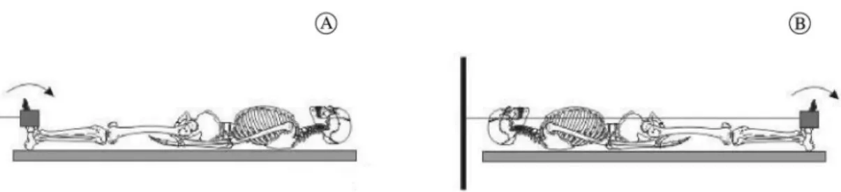
Perceived force level test
The perceived force level test was accomplished by asking the participants to reproduce a percentage of the force determined during a maximal isometric voluntary contraction only around the ankle joint (Figure 2). The maximal isometric voluntary contraction was performed in a supine posture, and participants
were required to perform ankle plantar lexion and dorsilexion at maximum strength. Then, participants
were allowed a period of familiarization with the protocol in which they were demanded to reproduce a target force. A numerical computer display provided visual feedback. They were deemed familiar with the test when variations were less than 10% of the target value.
In the perceived force level test, participants were asked to perform an isometric contraction equivalent to 10% and 20% of the maximal isometric voluntary contraction. The ankle was positioned at approximately 90°, and tests were executed in a random order. Visual feedback was provided once in each test condition, and then two trials without verbal or visual information were performed. Participants signaled as soon as they believed they had reached the desired force level. The discrepancy between each target condition (10% and 20% of maximum isometric voluntary contraction) and the mean of three perceived values of the respective condition were used to indicate the
ability to generate speciic force levels.
The force was determined in the dominant side using a load cell (Kratos, Model IK - 1C, Brazil - 500 Kgf capacity and 0.1 Kg resolution). The load cell was perpendicularly connected to the foot by steel cable attached to a Velcro strap.
Two‑Point discrimination test
The two-point discrimination test was described by Franco et al.10 who conirmed the intra-rater
reproducibility of the test (range from 0.7 to 1.5%). The test was carried out on the sole of the right foot
around the region of the irst metatarsal. A two-point
discriminator device (Touch - Test TM, model NC12776, measures 1 to 25 mm, North Coast Medical, Inc., Ireland) was applied perpendicularly to the sole and the weight of the device was applied simultaneously to both ends. Participants were asked whether one or two ends were in touch with the skin. The distance between the tips was reduced in increments of 1 to 2 mm, until participants were unable to distinguish whether two points were being applied to the skin. Further details about the two-point discrimination test can be found elsewhere10.
Statistics
Initially, data was processed using standard descriptive statistics (mean and standard deviation). Then, the
Shapiro-Wilk test was applied and conirmed data normality. The Levene test conirmed data homogeneity. The data that did not fulill the criteria for normal
distribution were normalized using logarithmic, quadratic or exponential functions. Proprioception was compared between groups using a t-test for independent measures. Bonferroni’s approach was
applied to adjust signiicance level due to multiple
comparison effects. Statistical tests were performed with Statistica software (StatSoft Inc.®, version 7.0), and the level of signiicance was set at p≤0.05.
Figure 2. Schematic representation of the ankle during the perceived force level test. Participants performed maximal isometric voluntary
Results
The sample was composed of 42 participants, who were allocated to two groups: young group (24.62±3.88 years; 64.43±9.53 Kg; 1.69±0.09 m) and elderly group (66.95±5.55 years; 65.34±13.67 Kg;
1.56±0.08 m). The participants were classiied as
active by the IPAQ and were able to understand and perform the tests.
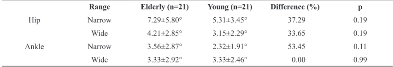
The joint positioning test was not able to distinguish young from the elderly (p>0.05). The absolute errors in repositioning the hip and ankle joints and the percentage differences between the groups are shown in Table 1.
No differences were observed between groups in the level of perceived force test. However, differences were borderline and indicated that there is a trend of a reduced perception of force between groups when the ankle was tested at 20% of maximum voluntary
isometric contraction (p=0.06, dorsilexion and p=0.09, plantar lexion).
Although no differences were found between groups (p>0.05), errors in perceived force increased when higher force levels were required. The perceived force errors and the percentage difference between groups are shown in Table 2.
The elderly group showed a higher threshold in the two-point discrimination test of the sole of the foot when compared to the young group (p=0.05; 13.43±2.62 mm and 6.19±2.09 mm, respectively).
Discussion
The main outcomes of the study revealed no differences between active elderly and young subjects in the joint positioning sense and perceived force level tests. In contrast, the two-point discrimination test showed that young subjects are able to better identify stimulus applied to the sole of the foot.
The joint positioning sense test evaluated the ability of subjects to perceive their body arrangement in order to reproduce a target posture. Passive segment repositioning tests are less complex and may not represent the real conditions performed in daily life actions, where movements should be intentionally controlled to meet the demands and interact with the environment accordingly. Daily actions are controlled based on proprioceptive feedback11 during movement
rather than when the segment is positioned in a particular posture, as in passive tests. Therefore, the active joint positioning sense tests seem to be more
suitable and speciic for functional assessments than
passive tests.
When trying to reproduce a previously established position, the elderly participants showed a slight
(non-signiicant) tendency to present larger errors
than did their young counterparts. Several arguments may explain the similarities between the elderly and young groups. Physical activity has been reported to modulate the functional state of the muscle and
therefore inluence proprioception8. Franco and
Rodacki12 found that differences in proprioception
Table 1. Relative difference between groups and absolute discrepancies between target and intended positions during the joint positioning test performed at narrow and wide hip and ankle ranges of elderly and young subjects.
Range Elderly (n=21) Young (n=21) Difference (%) p
Hip Narrow 7.29±5.80° 5.31±3.45° 37.29 0.19
Wide 4.21±2.85° 3.15±2.29° 33.65 0.19
Ankle Narrow 3.56±2.87° 2.32±1.91° 53.45 0.11
Wide 3.33±2.92° 3.33±2.46° 0.00 0.99
Table 2. Relative difference between groups and absolute discrepancies between target and executed force exertions during the perceived
force level test of the plantar lexor and dorsilexor muscles of elderly and young subjects.
Elderly (N=21) Young (N=21) Difference (%) p
Plantar lexion 10% 1.87±1.42 1.54±1.08 21.43 0.40
20% 2.59±1.85 1.87±1.62 38.50** 0.09
Dorsilexion 10% 1.59±2.24 1.66±1.06 4.22 0.34
20% 2.82±2.44 1.70±1.10 65.88* 0.06
between young and elderly subjects were present only when active young subjects were compared to sedentary elderly subjects. Tsang and Hui-Chan13 also identiied the inluence of physical conditioning
on the ability to identify segment postures around the knee joint while comparing active and sedentary elderly subjects. It may be argued that physical activity demands continuous muscle involvement, which is able to slow down the effects of advancing age on muscle tissue14. Thus, the results of this study
indicate that the perception of joint repositioning may be more associated with the level of physical activity than chronological age itself. Several studies have shown better performance after a training program in tasks that are heavily dependent on proprioceptive information, such as postural control. Improvements in postural control have been reported after a training program designed to stimulate sensory afference when compared to other training programs that involved strength as the primary goal2.
The elderly showed greater dificulties in repositioning
the lower limbs in narrow range postures during hip
lexion and ankle dorsilexion. The increased rigidity
of the tissues surrounding the joints that is typically present in the senescence process may have played a role and reduced the maximal joint range of motion15.
Therefore, positions that required wide ranges may have imposed a greater distention of the surrounding tissues and facilitated afferent sensory feedback. Thus, it may have produced a greater sensory stimulus that resulted in a small joint repositioning error12. This may be true speciically for the ankle joint, where
narrow ranges tend to be near the maximum joint range of motion. Petrella et al.16 reported signiicant
differences in the ability to actively reproduce a given knee angular position between young, active elderly and sedentary elderly subjects and reinforced the arguments that afferent sensory decline occurs and
that physical activity plays a relevant inluence on
proprioceptive functioning.
Petrella et al.16 reported differences when young
and active elderly subjects were compared, however performing the joint positioning sense test while
standing may have inluenced results as balance
requirements are known to occur in the elderly17. Thus,
one may argue that positioning the knee segment in
a deined position in a standing posture may have
included additional balance demands. The supine posture adopted in the present study is likely to be less
susceptible to the inluence of balance requirements
and represent a more realistic approach of the active
joint positioning sense. Therefore, differences in methodological approaches may at least partially explain contrasting results.
The perceived force level test was designed to evaluate the ability to perceive and voluntarily control low force contraction levels of the dorsi- and
plantar-lexor muscles. The elderly presented greater
variability than the young in all perceived force level
test variables, indicating greater dificulty in controlling
the production of a given level of force, regardless of the intensity of muscle contraction18. These indings
are relatively common in the literature using discrete19
or continuous isometric contractions20. The results of the present study are also consistent with the indings
that variability increases during contractions that require low force although most studies have used very small MVIC percentages (ranged from 2 to 10%21,22).
The percentages of 10 and 20% MVIC were selected to represent the more functional demands of daily tasks.
The results indicated that force perception errors were reduced in the elderly group when loads of 20% MVIC were applied in comparison with the young group. It may be argued that very small levels of force (e.g.,
10% MVIC) are unusual and dificult to control for
both groups. The torque observed around the ankle joint is usually greater than 20% of the maximal torque. For instance, Billot et al.23 reported that during simple
tasks such as one-legged standing, the elderly subjects created a torque around the ankle of approximately 45% of their maximum, while the torque observed in young adults during the same task was 15% of the maximum. Hortobágyi et al.24 have also demonstrated
that elderly subjects present larger relative knee joint torques when compared to young subjects while rising from a chair. Thus, subjects may be more familiar with torques within their daily range than with very small torques. In addition, there is evidence that training can remove differences at low %MVCs22. Therefore,
the active status of the subjects may have masked the differences between elderly and young participants.
The elderly group showed lower ability to discriminate two-point pressure in the sole than the young group, although no peripheral sensory disorders were
self-reported or identiied. The sensitivity responses of
the elderly group can be compared to that reported in type II diabetes patients when a similar methodology was applied25. These indings are in agreement with
others that have reported sensory degeneration in the elderly26. Indeed, similar results are also found in other
The afferent signals from cutaneous receptors in the sole provide spatial and temporal information
and inluence balance and stability. Thus, they play a signiicant role when compensatory actions are
required to sustain an erect posture28. Indeed, Toledo
and Barela17 observed that the more degenerated the
proprioceptive system of the elderly subjects was, the greater the oscillations of the center of pressure,
which reinforced the idea that balance is inluenced by
the proprioceptive information of the sole of the foot. Others have reported that the ability to discriminate two points in the sole (i.e. preserved sensory response) is associated with a lower incidence of falls29.
In the present study, the ability to discriminate two points was compared between the elderly and young groups. The results suggest that physical activity may have had a positive effect on proprioception. Santos et al.30 reported increased tactile sensibility
in the sole and reduced anterior-posterior oscillation in diabetic women after a 12-week training program designed to improve proprioception. These results
were not conirmed in the present study, in which
only the physical activity level was considered. It can be argued that maintaining physical activity may not
be suficient to elicit adaptive responses from the sensory system, which may require more speciic or
intense stimuli.
The results must be viewed with caution, as a group of sedentary elderly subjects was not included. In addition, the sample was relatively small and a
larger sample could inluence the results.
Conclusion
The results of the joint positioning sense and perceived force level tests showed similar results between elderly and young subjects. They also suggest that maintaining a physical activity level may not be
suficient to improve the sensory system and elicit
responses to stimuli applied to the sole. Thus, the reduced sensibility during the two-point discrimination test of the soles explains the poorer performance by the elderly group in comparison with the young group, and it was interpreted as the result of the degenerative senescence process. Thus, it can be inferred that
physical activity may inluence the effects of ageing of
joints and muscle proprioceptors, but not the sensory system of the soles of the feet. Intervention studies including physical activity programs are required to determine the effects of proprioception.
References
1. Chou CH, Hwang CL, Wu YT. Effect of exercise on physical function, daily living activities, and quality of life in the frail older adults: a meta-analysis. Arch Phys Med Rehabil. 2012;93(2):237-44. http://dx.doi.org/10.1016/j. apmr.2011.08.042. PMid:22289232.
2. Gauchard GC, Jeandel C, Tessier A, Perrin PP. Beneficial effect of proprioceptive physical activities on balance control in elderly human subjects. Neurosci Lett. 1999;273(2):81-4. http://dx.doi.org/10.1016/S0304-3940(99)00615-1. PMid:10505621.
3. Zheng J, Pan Y, Hua Y, Shen H, Wang X, Zhang Y, et al. Strategic targeted exercise for preventing falls in elderly people. J Int Med Res. 2013;41(2):418-26. http://dx.doi. org/10.1177/0300060513477297. PMid:23569036. 4. Maciel ACC, Guerra RO. Prevalência e fatores associados
ao defícit de equilíbrio em idosos. Rev Bras Cienc Mov. 2005;13(1):37-44.
5. Garcia R, Leme MD, Garcez-Leme LE. Evolution of Brazilian elderly with hip fracture secondary to a fall. Clinics (Sao Paulo). 2006;61(6):539-44. http://dx.doi.org/10.1590/S1807-59322006000600009. PMid:17187090.
6. Alfieri FM. Distribuição da pressão plantar em idosos após intervenção proprioceptiva. Rev Bras Cineantropome Desempenho Hum. 2008;10(2):137-42.
7. Goble DJ, Coxon JP, Wenderoth N, Van Impe A, Swinnen SP. Proprioceptive sensibility in the elderly: degeneration, functional consequences and plastic-adaptive processes. Neurosci Biobehav Rev. 2009;33(3):271-8. http://dx.doi. org/10.1016/j.neubiorev.2008.08.012. PMid:18793668. 8. Xu D, Hong Y, Li J, Chan K. Effect of tai chi exercise on
proprioception of ankle and knee joints in old people. Br J Sports Med. 2004;38(1):50-4. http://dx.doi.org/10.1136/ bjsm.2002.003335. PMid:14751946.
9. Runge CF, Shupert CL, Horak FB, Zajac FE. Ankle and hip postural strategies defined by joint torques. Gait Posture. 1999;10(2):161-70. http://dx.doi.org/10.1016/S0966-6362(99)00032-6. PMid:10502650.
10. Franco PG, Bohrer RCD, Rodacki ALF. Intra-observer reproducibility of the feet soles two-point discrimination test in asymptomatic elderly and young individuals. Rev Bras Fisioter. 2012;16(6):523-7. http://dx.doi.org/10.1590/ S1413-35552012005000062. PMid:23184279.
11. Fatoye F, Palmer S, Macmillan F, Rowe P, van der Linden M. Proprioception and muscle torque deficits in children with hypermobility syndrome. Rheumatology (Oxford). 2008;48(2):152-7. http://dx.doi.org/10.1093/rheumatology/ ken435. PMid:19088133.
12. Franco PG, Rodacki ALF. Percepção de posicionamento articular e do nível de força em sujeitos idosos e jovens. Rev Educ Fis/UEM. 2011; 22(3):327-335. http://dx.doi.
org/10.4025/reveducis.v22i3.10163.
13. Tsang WWN, Hui-Chan CWY. Effects of exercise on joint sense and balance in elderly men: tai chi versus golf. Med Sci Sports Exerc. 2004;36(4):658-67. http://dx.doi. org/10.1249/01.MSS.0000122077.87090.2E. PMid:15064594. 14. Bickel CS, Cross JM, Bamman MM. Exercise dosing to
adults. Med Sci Sports Exerc. 2011;43(7):1177-87. http:// dx.doi.org/10.1249/MSS.0b013e318207c15d. PMid:21131862. 15. Cristopoliski F, Sarraf TA, Dezan VH, Provensi CLG, Rodacki
ALF. Efeito transiente de exercícios de flexibilidade na articulação do quadril sobre a marcha de idosas. Rev Bras Med Esporte. 2008;14(2):139-44. http://dx.doi.org/10.1590/ S1517-86922008000200011.
16. Petrella RJ, Lattanzio PJ, Nelson MG. Effect of age and activity on knee joint proprioception. Am J Phys Med Rehabil. 1997;76(3):235-41. http://dx.doi.org/10.1097/00002060-199705000-00015. PMid:9207711.
17. Toledo DR, Barela JA. Sensory and motor differences between young and older adults: somatosensory contribution to postural control. Rev Bras Fisioter. 2010;14(3):267-75. http://dx.doi.org/10.1590/S1413-35552010000300004. PMid:20730372.
18. Vaillancourt DE, Larsson L, Newell KM. Time-dependent structure in the discharge rate of human motor units. Clin Neurophysiol. 2002;113(8):1325-38. http://dx.doi.org/10.1016/ S1388-2457(02)00167-0. PMid:12140014.
19. Christou EA, Carlton LG. Old adults exhibit greater motor output variability than young adults only during rapid discrete isometric contractions. J Gerontol A Biol Sci Med Sci. 2001;56(12):B524-32. http://dx.doi.org/10.1093/ gerona/56.12.B524. PMid:11723145.
20. Yan JH. Tai chi practice reduces movement force variability for seniors. J Gerontol A Biol Sci Med Sci. 1999;54(12):M629-34. http://dx.doi.org/10.1093/gerona/54.12.M629. PMid:10647969. 21. Galganski ME, Fuglevand AJ, Enoka RM. Reduced control
of motor output in a human hand muscle of elderly subjects during submaximal contractions. J Neurophysiol. 1993;69(6):2108-15. PMid:8350134.
22. Laidlaw DH, Bilodeau M, Enoka RM. Steadiness is reduced and motor unit discharge is more variable in old adults. Muscle Nerve. 2000;23(4):600-12. http://dx.doi.org/10.1002/ (SICI)1097-4598(200004)23:4<600::AID-MUS20>3.0.CO;2-D. PMid:10716772.
23. Billot M, Simoneau EM, Van Hoecke J, Martin A. Age-related relative increases in electromyography activity and torque according to the maximal capacity during upright standing. Eur J Appl Physiol. 2010;109(4):669-80. http:// dx.doi.org/10.1007/s00421-010-1397-7. PMid:20213469.
24. Hortobágyi T, Mizelle C, Beam S, DeVita P. Old adults perform activities of daily living near their maximal capabilities. J Gerontol A Biol Sci Med Sci. 2003;58(5):M453-60. http:// dx.doi.org/10.1093/gerona/58.5.M453. PMid:12730256. 25. Carvalho VF, Ferreira MC, Vieira SAT, Ueda T. Limiar
de sensibilidade cutânea dos pés em pacientes diabéticos através do pressure specified sensory device: uma avaliação da neuropatia. Rev Assoc Med Bras. 2009;55(1):29-34. http://dx.doi.org/10.1590/S0104-42302009000100011. PMid:19360274.
26. Wickremaratchi MM, Llewelyn JG. Effects of ageing on touch. Postgrad Med J. 2006;82(967):301-4. http://dx.doi. org/10.1136/pgmj.2005.039651. PMid:16679466. 27. van Nes SI, Faber CG, Hamers RMTP, Harschnitz O, Bakkers
M, Hermans MC, et al, PeriNomS Study Group. Revising two-point discrimination assessment in normal aging and in patients with polyneuropathies. J Neurol Neurosurg Psychiatry. 2008;79(7):832-4. http://dx.doi.org/10.1136/ jnnp.2007.139220. PMid:18450792.
28. Perry SD, McIlroy WE, Maki BE. The role of plantar cutaneous mechanoreceptors in the control of compensatory stepping reactions evoked by unpredictable, multi-directional perturbation. Brain Res. 2000;877(2):401-6. http://dx.doi. org/10.1016/S0006-8993(00)02712-8. PMid:10986360. 29. Melzer I, Benjuya N, Kaplanski J. Postural stability in the
elderly: a comparison between fallers and non-fallers. Age Ageing. 2004;33(6):602-7. http://dx.doi.org/10.1093/ageing/ afh218. PMid:15501837.
30. Santos AA, Bertato FT, Montebelo MIL. Effect of proprioceptive training among diabetic women. Rev Bras Fisioter. 2008;12(3):183-7. http://dx.doi.org/10.1590/ S1413-35552008000300005.
Correspondence Karini Borges dos Santos Universidade Federal do Paraná Setor de Ciências Biológicas