O
RIGINALA
RTICLE Revista Brasileira de FisioterapiaEvaluation of lumbar concavity using a
radiographic method and kypholordometry
Avaliação da concavidade lombar pelo método radiográfico
e pela cifolordometria
Souza FR1, Ferreira F2, Narciso FV3, Makhoul CMB2, Canto RST4, Barauna MA2
Abstract
Background: Clinical evaluation is the basis for making decisions regarding treatments. When radiographic images cannot be obtained, few resources allow physical therapists to quantitatively evaluate an individual’s condition. One of these is the kypholordometer, a low-cost noninvasive instrument proposed for measuring spinal curvature in the sagittal plane. Objectives: To evaluate the intra- and inter-examiner reliability of the kypholordometer, to investigate its agreement with radiography, and to determine whether there is any correlation between measurements of lumbar curvature using the radiographic method and the kypholordometer. Methods: Twenty healthy individuals of both sexes aged between 21 and 27 years were evaluated. They underwent radiographic examination of the lumbar spine in right lateral view while standing up. The radiographic images were evaluated by a radiologist using Cobb’s method, with T12 and S1 as the reference points. The kypholordometry was carried out in the same position by three evaluators on two occasions, with the same vertebrae as the reference points. A straight line was drawn from T12 to the least prominent vertebra and another from S1 to the same vertebra, thus identifying the degree of lumbar concavity. Results: The results demonstrated that kypholordometry presented excellent levels of reliability (both intra- and inter-examiner), but low agreement with radiography. However, there was a statistically significant positive correlation between the two methods studied (r=0.88). Conclusion: Kypholordometry is a quantitative method with excellent intra- and inter-examiner reliability for evaluating lumbar curvature. It may contribute greatly towards the clinical practice of physical therapists.
Key words: evaluation; spine; radiography; posture.
Resumo
Contextualização: A avaliação clínica é a base para tomada de decisão referente ao tratamento. Quando não é possível obter a radiografia, poucos recursos permitem ao fisioterapeuta avaliar quantitativamente o estado do indivíduo, um deles é o cifolordômetro, um instrumento não invasivo, de baixo custo, proposto para mensuração das curvas da coluna vertebral no plano sagital. Objetivos:Avaliar a confiabilidade intra e interexaminador do cifolordômetro, verificar sua concordância com a radiografia e se há correlação entre a medida da curva lombar pelo método radiográfico e pelo cifolordômetro. Métodos: Foram avaliados 20 indivíduos saudáveis de ambos os sexos, com idade entre 21 e 27 anos. Os voluntários foram submetidos à radiografia da coluna lombar, incidência perfil direito e em ortostatismo. As radiografias foram avaliadas por um radiologista pelo método de Cobb, tendo como pontos de referência T12 e S1. A cifolordometria foi realizada no mesmo posicionamento e por três avaliadores em dois momentos, tendo como referência as mesmas vértebras. Foi traçada uma reta de T12 à vértebra menos proeminente e outra de S1 à mesma, identificando o grau de concavidade lombar.Resultados: Os resultados demonstram que a cifolordometria apresenta níveis excelentes de confiabilidade, tanto inter quanto intraexaminador, baixa concordância com a radiografia, porém há correlação positiva, estatisticamente significativa entre os dois métodos estudados (r=0.88).
Conclusão: A cifolordometria apresentou-se como um método quantitativo, com excelente confiabilidade intra e interexaminador para a avaliação da curvatura lombar, podendo contribuir de sobremaneira para a prática clínica do fisioterapeuta.
Palavras-chave:avaliação; coluna vertebral; radiografia; postura.
Received: 17/12/2007 – Revised: 23/06/2008 – Accepted: 21/11/2008
1 Doctum Institute of Education and Technology, Faculdades Unificadas Doctum de Iúna, Iúna (ES), Brazil 2 Laboratory of Human Motion, Centro Universitário do Triangulo (UNITRI), Uberlândia (MG), Brazil 3 School of Physical Therapy, Faculdade de Patos de Minas (FPM), Patos de Minas (MG), Brazil 4 School of Medicine, Universidade Federal de Uberlândia (UFU), Uberlândia (MG), Brazil
Correspondence to: Fernanda Ferreira, Rua Martinésia, 322 – apto 802, Bairro Aparecida, CEP 38400-606, Uberlândia (MG), Brazil, e-mail: [email protected]
Introduction
Several methods to quantify body posture and spinal cur-vatures have been described in the literature. Among them, the radiography stands out as the “gold standard” for such evaluations. his method is also the most requested by medi-cal professionals1. However, its application is not very
com-mon in the physical therapy clinical practice, either because the equipment is not available to the physical therapist or be-cause not all health plans cover radiographic examinations. he most common procedure for measuring the angles of the spinal curvatures is Cobb’s method, carried out by means of radiographic studies2.
Radiographic examinations can have harmful efects on the body that may lead to somatic diseases such as cancer, leuke-mia, and even cataracts; it can also have genetic consequences, such as trisomy in newborns. In addition, the low-quality of the images often hinders examination analysis, and the procedure has to be repeated, which increases exposure to radiation and its consequences3-5.
In a retrospective study, Levy et al.6 observed 2039
adoles-cents with idiopathic scoliosis diagnosed through radiographic examination and reported that constant, small doses over months and years may take 20 years or more to show harm-ful efects on an individual. he subjects under study were observed over the course of 40 years, and later post-mortem examinations showed that the incidence of cancer was greater when compared to the general population.
To minimize the harmful efects of repeated radiographic examinations over a lifetime while keeping the changes in the spine under control, some techniques have been presented as noninvasive methods, such as: the pantograph, the DeBrunner kyphometer, the biophotogrammetry, the spondylometer, the lexible ruler, and the kypholordometer1,7-12. he
kypholordom-eter was created by Baraúna and patented at the Instituto Na-cional de Patentes Industriais (INPI) under protocol number PI 9905389-67.
he use of kypholordometry to evaluate spinal curvatures in diferent situations has been studied by various researchers. Adorno11, using noninvasive methods such as
biophotogram-metry and the kypholordometer carried out a study on the lumbar concavity of pregnant women and showed that both are precise methods of quantifying the angles of the lumbar concav-ity and those of the thoracic convexconcav-ity. It must be noted that these instruments allow the professional to evaluate the lumbar and thoracic curvatures in order to observe the individual’s response to interventions. Not only are these instruments noninvasive but they are also inexpensive and easy to use.
In another study, Baraúna et al.7 tested the thoracic convexity
of 30 subjects of both sexes aged 13 to 56, using the radiographic
method and kypholordometry. he results showed that the measures obtained through the kypholordometer had a posi-tive correlation with the radiographic examination, conirming the parallel, intra-examiner and inter-examiner reliability. he authors concluded that the kypholordometer is an eicient instrument for thoracic curvature measurement, and it can be used as often as necessary with no harm to the patient.
For the physical therapist, it is important to use nonin-vasive methods of evaluation because they provide plan-ning and follow-up criteria for interventions13. herefore,
the aims of the present study were: to analyze the intra- and interexaminer reliability of the lumbar concavity evaluation using kypholordometry; to verify the agreement between the radiographic method and kypholordometry; and to de-termine whether there is a correlation between the lumbar curvature measured by the radiographic method and by kypholordometry.
Methods
he study was carried out at Centro Universitário do Triân-gulo (Unitri) after approval by the Research Ethics Committee of that institution under protocol number 650217.
Twenty-eight healthy subjects were evaluated, of which 8 were excluded due to the poor quality of the radiographic im-ages. he following exclusion criteria for exclusion were also used: previous surgery or history of fracture of the spine, pelvis, or lower limb, spondylolisthesis, spondylolisis, scoliosis, sixth lumbar vertebra, history of neoplasy, multiparity, pregnancy or suspected pregnancy.
hus, the sample for the study consisted of 20 individu-als, all of them university students, including 7 men and 13 women, aged 21 to 27. he mean height was 1.68±0.10m and the mean weight was 68.26±11.60kg. All subjects were in-formed about the procedures and goals of the study before signing the consent form.
Kypholordometry
Kypholordometry is an evaluation method that uses an appa-ratus which consists of a vertical aluminum pole (39x58mm thick by 197cm tall) that supports 39 horizontal ¼-inch rods (40cm long). hese rods are mobile, unbendable, equidistant and 4cm apart. he vertical pole is ixed on an orthostatic support plat-form lined with adjustable, non-slip material (73x56cm). here is also a level that allows corrections to the support platform, even when the loor is not completely lat. Attached to the pole, there is a lateral support made from acrylic to hold the sheet of paper where the analyzed curve is recorded7.
To evaluate the lumbar curvature using the kypholordom-eter, palpation was necessary to identify spinous processes T12 to S1. hese vertebrae were chosen according to the cri-teria suggested by Stagnara et al.3, Bernhardt and Bridwell13,
Korovessis, Stamatakis and Baikousis14, and Vedantam et
al.15.Next, the subject was positioned on the kypholordometer
with bare feet and trunk (except for the private parts), arms relaxed next to the body and gazing horizontally, as shown in Figure 1. After that, the points projected by the horizontal rods closest to T12 and S1 and the least prominent point be-tween them were marked on the recording paper, as observed in Figure 1. he subject then stepped of the kypholordometer and, after one minute, a second evaluation was carried out by the same examiner, following the same procedures. hus, each subject was evaluated by three diferent examiners, each of whom executed the evaluation and the angle measurements twice. Once the three points had been recorded, a straight line was drawn from the top point to the least prominent point and another from the bottom point to the least prominent point. he angle formed by the intersection of these lines was measured at the vertex using the goniometer; thus identify-ing the degree of lumbar concavity (two-line method).
Radiographic examination
All subjects underwent the lumbar curvature radiographic examination, carried out by the same technician, on the same apparatus and evaluated by the same radiologist. Shea et al.8
showed that errors are less likely to occur when the angle mea-surement by Cobb’s method is taken by a single examiner. he position for the radiographic examination was standardized according to the criteria used by Propst-Proctor and Bleck4,
Bernhardt and Bridwell13, Gelb et al.16 and Leroux et al.17 who
recommend a right lateral radiograph, with a horizontal gaze, extended knees and parallel feet.
To measure the lumbar curve, Cobb’s method was applied to the same vertebrae marked in the kypholordometer. he follow-ing lines were drawn: a parallel line below the T12 and another parallel line above the S1. Perpendicular lines were drawn and their intersection was recorded ( four-line method). he angle value was measured with a goniometer. he radiographic exam-ination was analyzed by a radiologist who did not have access to the data obtained by kypholordometry. hus, there was no mutual knowledge of the recorded values4,7,14,16-20.
Statistical analysis
After descriptive analysis the information was processed in the computer package SPSS for Windows, version 13.0. he measures are presented as means and standard deviations.
After applying the Kolmogorov-Smirnov test, it was observed that the data follow the normal curve. hus, in order to check the reliability of intra- and interexaminer kypholordometry, the Intraclass Correlation Coeicient (ICC) was applied, where an ICC less than 0.4 indicates poor reproducibility; between 0.4 and 0.75, acceptable reproducibility; and greater than 0.75, ex-cellent reproducibility.
To determine a single value for the analyses involving kypholordometry, we calculated the means of the two values for each examiner, and then the overall mean, that is, the mean of the values obtained by each of the three examiners. Next, to analyze the agreement between the kypholordometry and radiography measurements, we used the Bland-Altman method, as described by Rankin and Stokes21. It consists in
Figure 1. Kypholordometer - The subject’s position and the demarcation of the most prominent points, regarding T12 and S1, and the least prominent points of the lumbar curvature.
graphically representing the diferences of the measures in relation to their mean (where d=mean diference between the measures). A reliability interval of 95% was adopted. Pearson’s correlation was applied to verify the existence of correlation between the lumbar curve measures as evaluated by kyphol-ordometry and radiography.
Results
he 20 subjects evaluated had a mean age of 22.7±2.3 years and BMI of 21.2±2.3Kg/m². he mean lumbar concavity meas-ured by kypholordometry was 19±8 degrees, and by radiogra-phy was 70.5±15 degrees. In the case of kypholordometry, the irst examiner obtained a mean of 20±9.4 degrees in the irst measurement and 19.6±8.6 degrees in the second. he second examiner obtained a mean of 19.2±10-degrees in the irst mea-surement and 19±8.7 degrees in the second. he third examiner obtained a mean of 18.3±7.7 degrees in the irst measurement, and 18.5±7.4 degrees in the second measurement.
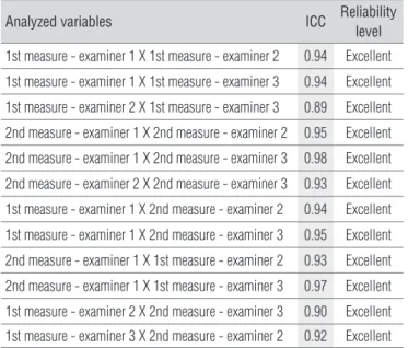
In the reliability evaluation (reproducibility of the method) for all analyses, both intraexaminer (Table 1) and interexam-iner (Table 2), the ICC showed very good coeicients, which shows the excellent reliability of the method of lumbar curva-ture evaluation through kypholordometry.
As can be seen in Figure 2, the Bland-Altman plot21 shows
that there was a great dispersion in relation to the y-axis, in-dicating that there is a low agreement between the kypholor-dometry and the radiography data (d=5.4).
After analyzing the relationship between the lumbar concavity measurements by kypholordometry and the Cobb measurements by radiography, it was possible to detect a sta-tistically signiicant correlation between the methods (r=0.88), as observed in the plot displayed in Figure 3.
Discussion
he results of the present study show that kypholordom-etry, the proposed method of quantitative evaluation of the lumbar curvature, showed high levels of intra- and interexam-iner reliability and reproducibility. his is highly relevant to the physical therapist because it is important to use noninvasive quantitative methods of evaluation that provide criteria for intervention planning and follow-up7,13.
he mean value found in the lumbar curvature evaluation by radiography was 70.5±15.2 degrees. his agrees with the values reported in the literature, which range from 35 and 90 degrees22. However, several studies on the sagittal alignment of
the spine did not deine normal lordosis; on the contrary, they
0 5 10 15 20 25 30 35 40 120
100 80 60 40 20 0
Kypholormetry(º)
Radiography (º)
Figure 3. Relationship between the lumbar curvature measures by kypholordometry and radiography – Positive correlation.
Means of the variables
Difference of the measures (
d
)
25 20 15 10 5 0 -5 -10 -15 -20
20 30 40 50 60 70
Figure 2. Agreement between kypholordometry and radiography measures - Bland-Altman plot.
Analyzed variables ICC Reliability level
1st X 2nd measure - examiner 1 0.98 Excellent
1st X 2nd measure - examiner 2 0.97 Excellent
1st X 2nd measure - examiner 3 0.99 Excellent
Table 1. Intraexaminer reliability for the first and second measurement of each examiner (ICC).
Analyzed variables ICC Reliability
level
1st measure - examiner 1 X 1st measure - examiner 2 0.94 Excellent
1st measure - examiner 1 X 1st measure - examiner 3 0.94 Excellent
1st measure - examiner 2 X 1st measure - examiner 3 0.89 Excellent
2nd measure - examiner 1 X 2nd measure - examiner 2 0.95 Excellent
2nd measure - examiner 1 X 2nd measure - examiner 3 0.98 Excellent
2nd measure - examiner 2 X 2nd measure - examiner 3 0.93 Excellent
1st measure - examiner 1 X 2nd measure - examiner 2 0.94 Excellent
1st measure - examiner 1 X 2nd measure - examiner 3 0.95 Excellent
2nd measure - examiner 1 X 1st measure - examiner 2 0.93 Excellent
2nd measure - examiner 1 X 1st measure - examiner 3 0.97 Excellent
1st measure - examiner 2 X 2nd measure - examiner 3 0.90 Excellent
1st measure - examiner 3 X 2nd measure - examiner 2 0.92 Excellent
Table 2. Interexaminer reliability (ICC).
are more concerned with the frequency distribution of these data in and between the groups being studied. Hence, it is dif-icult to set a standard of normal angles, either by kypholor-dometry or radiography23,24.
he present study also evaluated the reliability of the mea-surements obtained in the lumbar curvature evaluation with the kypholordometer. he results show that the proposed method for lumbar curvature evaluation is reliable when car-ried out by a single examiner at diferent moments or by several examiners. hus, the present work is in line with other studies on kypholordometry, which allows safe monitoring even when conducted by diferent examiners7,11.
he low agreement observed when the Bland-Altman21
agreement test was applied is due to the fact that these results may have been inluenced by the angle evaluation method since Cobb’s angle is drawn along the upper and lower edges of the vertebrae that limit lumbar curvature. After that, perpen-dicular lines are also drawn and their intersection is analyzed. his is the four-line method19, which difers from the angular
evaluation by kypholordometry that uses the two-line method. hese two lines are drawn from the end points of the lum-bar curvature to the less prominent point7; thus, the angle is
deined by the vertex of that intersection. Due to this vertex-based analysis, the angle tends to be smaller, even when using the same vertebrae as a reference, which is easily understood when observing the means of the lumbar concavity evaluations. he mean for kypholordometry was 19±8 degrees, whereas the mean for radiography (Cobb’s angle) was 70.5±15 degrees.
However, there is no reason to discredit the kypholordometry method because, despite the disagreement between the mea-sures obtained by either method, there is a positive correlation between kypholordometry and the radiographic examination in the lumbar curvature evaluation, i.e. the greater the values ob-tained by kypholordometry, the greater the values obob-tained by the radiography, although not necessarily the same for each in-dividual. In a similar study, Baraúna et al.7 detected a signiicant
positive correlation between kypholordometry and radiographic examination in thoracic concavity evaluation. he results also conirmed the parallel reliability of kypholordometry.
Ward and Tidswell25 cite the spondylometer, a similar
ap-paratus to the kypholordometer, although it only monitors the evolution of ankylosing spondylitis. he distances between the most prominent and the least prominent points are measured and recorded on paper. he spondylometer does not include a level and does not allow the measurement of spinal curvature because there is no acrylic lateral support to hold the record-ing paper such as the one used in kypholordometry to draw the evaluated curve and measure the angle. In addition, the spondylometer rods are 5cm apart, whereas the kypholor-dometer rods are positioned 4cm apart, which allows a greater
proximity of the vertebrae to be examined, and therefore more precise angular measurements of the spine7.
Oi et al.26 developed an apparatus called
posture-measur-ing device. It uses a system of wooden rods that move inside aligned metal tubes. he distance between the rods is small but not speciied. he authors showed that this method is very similar to kypholordometry, although it has a serious limitation because its only purpose is visual evaluation and classiication of the individual into four types of proposed postures, making it a subjective method. he authors claim that this method revealed postural deformity in older adults based on compari-sons between radiographic examinations and the spinal out-lines obtained with the posture-measuring device. However, the method does not evaluate that posture quantitatively, but only qualitatively.
After studying the reliability of the lexible ruler for lumbar curvature measurement, Hart and Rose10 stated that
noninva-sive techniques for spinal evaluation in the sagittal plane char-acterize its shape, but may not be as precise as radiographic measurements. However, the radiographic examination may also present angle variations of up to 8 degrees, depending on the focus of the apparatus, time of the day, and radiologist interpretation27.
Harrison et al.28 compared the measures of the lumbar
cur-vature by using Cobb’s two-line method (one line was drawn parallel to and below the T12 body and another line parallel to and above the S1 body) with the four-line method ( four per-pendicular lines are drawn starting from parallel lines). he intersection was recorded and Cobb’s angle was measured. Compared to the four-line method, the two-line method had a smaller absolute diference and a greater correlation coef-icient between examiners. Nevertheless, in clinical practice, there are instances where Cobb’s two-line method cannot be used because the lines do not converge in the radiography it-self, hence the preference for the four-line method, as carried out in the present study.
Radiographic examinations require a specialized team, in-cluding the technician who conducts the procedure, the doctor who reads the examination, and a professional who services the equipment. Investing on radiographic equipment is very costly and increases the cost of the examination for the patient. here is also the need for facilities with suitable internal lining of the walls29,30.
Kypholordometry is easy to carry out, and the data are collected quickly and objectively. It is not necessary to re-cruit a multi-professional team to take the measurements, although there must be at least one trained examiner. he kypholordometer is an inexpensive apparatus, which re-quires little space. Once placed inside the clinic, it can be used whenever necessary to quantify the lumbar concavity
angle in evaluations and to monitor the progress of postural treatments, particularly in cases involving pathologies or pro-visional health conditions that do not allow radiography, such as pregnancy.
he use of kypholordometry ofers immediate access to the results and low costs7. In contrast, it is a method that may result
in variations in the measures collected by the examiners due to palpation, drawing and angle measurement method. hese factors, however, are not restrictive when one considers the beneits the apparatus brings not only to the patient, but also to the physical therapist, who can rely on it as a primary clini-cal evaluation measure. In the event of substantial variations,
the physical therapist can request a radiographic examination however the use of kypholordometry would avoid unnecessary exposure to radiation by the patient and the expenses related to radiography, given that most health plans do not cover ra-diographic examinations requested by the physical therapist.
his study corroborates the eicacy, simplicity and preci-sion of kypholordometry as a method of lumbar curvature eval-uation and leads to the conclusion that there is a correlation between the angular measurements by radiographic examina-tion and by kypholordometry. Furthermore, kypholordometry allows quantitative evaluation of the lumbar curvature with excellent levels of intra- and interexaminer reliability.
1. Singer KP, Jones TJ, Breidahl PD. A comparison of radiographic and computer-assisted measurements of thoracic and thoracolumbar sagittal curvature. Skeletal Radiol. 1990;19(1):21-6.
2. Ferreira DMA, Defino HLA. Estudo clínico da mensuração da gibosidade e suas correlações com medidas radiológicas na escoliose idiopática. I Encontro do Programa de Pós Graduação Interunidades em Bioengenharia da USP; Universidade de São Paulo – USP. Ribeirão Preto, SP: USP; 2001.
3. Stagnara P, De Mauroy JC, Dran G, Gonon GP, Costanzo G, Dimnet J, et al. Reciprocal angulation of vertebral bodies in a sagittal plane: approach to references for the evaluation of kyphosis and lordosis. Spine. 1982;7(4):335-42.
4. Propst-Proctor SL, Bleck EE. Radiographic determination of lordosis and kyphosis in normal and scoliotic children. J.Pediatr Ortop. 1983;3(3):344-6.
5. Juhl JH, Crummy AB, Kuhlman JE. Interpretação radiológica. 7ª ed. Rio de Janeiro: Guanabara Koogan; 2000.
6. Levy AR, Goldberg MS, Mayo NE, Hanley JA, Poitras B. Reducing the lifetime risk of cancer from spinal radiographs among people with adolescent idiopathic scoliosis. Spine. 1996;2(13):1540-8.
7. Baraúna MA, Canto RST, Sanchez HM, Bustamante JC, Ventura-Silva RA, Malusá S. Validade e confiabilidade Intra-indivíduo do cifolordometro na avaliação da convexidade torácica. Rev Bras Fisioter. 2005;9(3): 319-25.
8. Shea KG, Stevens PM, Nelson M, Smith JT, Masters KS, Yandow SA. A comparison of manual versus computer-assisted radiographic measurement: Intraobserver measurement variability for Cobb angles. Spine. 1998;23(5):551-5.
9. Willner S, Johnson B. Thoracic kyphosis and lumbar lordosis during the growth period in children. Acta Paediatr Scand. 1983;72(6):873-8.
10. Hart L, Rose SJ. Reliability of a noninvasive method for measuring the
lumbar curve. J Orthop Sports Phys Ter.1986;8(4):180-4.
11. Adorno MLGR. Avaliação cinesiológica das curvaturas lombar e torácica das gestantes através do cifolordômetro e da fotogrametria computadorizada e sua correlação com a dor lombar [Dissertação de Mestrado]. Uberlândia, MG: Centro Universitário do Triângulo; 2001.
12. Iunes DH, Castro FA, Salgado HS, Moura IC, Oliveira AS, Bevilaqua-Grossi D. Confiabilidade intra e interexaminadores e repetibilidade da avaliação postural pela fotogrametria. Rev Bras Fisioter. 2005;9(3): 327-34.
13. Bernhardt M, Bridwell KH. Segmental analysis of the sagittal plane alignment of the normal thoracic and lumbar spines and thoracolumbar
junction. Spine.1989;14(7):717-21.
14. Korovessis PG, Stamatakis MV, Baikousis AG. Reciprocal angulation of vertebral bodies in the sagittal plane in asymptomatic Greek population. Spine. 1998;23(6):700-5.
15. Vedantam R, Lenke LG, Bridwell KH, Linville DL, Blanke K. The effect of variation in arm position on sagittal spinal alignment. Spine. 2000;25(17):2204-9.
16. Gelb DE, Lenke LG, Bridwel KH, Blanke K, McEnery KW. An analysis of sagittal spinal alignment in 100 asymptomatic middle and older aged volunteers. Spine. 1995;20(12):1351-8.
17. Leroux MA, Zabjek K, Simard G, Badeaux J, Coillard C, Rivard CH. A noninvasive anthropometric technique for measuring kyphosis and lordosis: an application for idiopathic scoliosis. Spine. 2000;25(13):1689-94.
18 Vedantam R, Lenke LG, Keeney JA, Bridwell KH. Comparison of standing sagittal spinal alignment in asymptomatic adolescents and adults. Spine. 1998;23(2):211-5.
19. Vialle R, Levassor N, Rillardon L, Templier A, Skalli W, Guigui P. Radiographic analysis of the sagittal alignment and balance of the spine in assyntomatic subjects. J Bone Joint Surg Am. 2005;87(2): 260-7.
20. Jackson RP, Hales C. Congruent spinopelvic alignment on standing lateral radiographs of adult volunteers. Spine. 2000;25(21):2808-15.
108
21. Rankin G, Stokes M. Reliability of assessment tools in rehabilitation: an illustration of appropriate statistical analyses. Clin Rehabil. 1998;12(3): 187-99.
22. Voutsinas SA, MacEwen GD. Sagittal profiles of the spine. Clin Orthop Relat Res. 1986;(210):235-42.
23. Roussouly P, Gollogly S, Berthonnaud E, Dimnet J. Classification of the normal variation in the sagittal alignment of the human lumbar spine and pelvis in the standing position. Spine. 2005;30(3): 346-53.
24. Christensen HW. Precision and accuracy of an electrogoniometer. J Manipulative Physiol Ther. 1999;22(1):10-4.
25. Ward DJ, Tidswell ME. As Espondiliopatias. In: Downie PA, editor. Neurologia para fisioterapeutas. 4ª ed. São Paulo: Medicina Panamericana do Brasil Ltda; 1987. p. 237-50.
26. Oi N, Tobimatsu Y, Iwaya T, Okada Y, Gushiken S, Kusano S, et al.
Reliability and validity of classification of senile postural deformity in mass examinations. Tohoku J Exp Med. 2004;202(2):205-12.
27. Souchard PE, Ollier M. As escolioses - seu tratamento fisioterapêutico e ortopédico. São Paulo: Realizações; 2001.
28. Harrison DE, Harrison DD, Cailliet R, Janik TJ, Holland B. Radiographic analysis of lumbar lordosis: centroid, Cobb, TRALL, and Harrison posterior tangent methods. Spine. 2001;26(11):235-42.
29. Dutkowsky JP, Shearer D, Schepps B, Orton C, Scola F. Radiation exposure to patients receiving routine scoliosis radiography measured at depth in an anthropomorphic phantom. J Pediatr Orthop. 1990;10(4):532-4.
30. Carman DL, Browne RH, Birch JG. Measurement of scoliosis and kyphosis radiographs: intraobserver and interobserver variation. J Bone Joint Surg Am. 1990;72(3):328-33.