Angiotensin II type 1 and 2 receptors and lymphatic
vessels modulate lung remodeling and fibrosis in
systemic sclerosis and idiopathic pulmonary fibrosis
Edwin Roger Parra, Aline Domingos Pinto Ruppert, Vera Luiza Capelozzi
Faculdade de Medicina da Universidade de Sa˜o Paulo, Department of Pathology, Laboratory of Histomorphometry and Pulmonary Genetics, Sa˜o Paulo/SP, Brazil.
OBJECTIVE: To validate the importance of the angiotensin II receptor isotypes and the lymphatic vessels in systemic sclerosis and idiopathic pulmonary fibrosis.
METHODS:We examined angiotensin II type 1 and 2 receptors and lymphatic vessels in the pulmonary tissues obtained from open lung biopsies of 30 patients with systemic sclerosis and 28 patients with idiopathic pulmonary fibrosis. Their histologic patterns included cellular and fibrotic non-specific interstitial pneumonia for systemic sclerosis and usual interstitial pneumonia for idiopathic pulmonary fibrosis. We used immunohistochemistry and histomorphometry to evaluate the number of cells in the alveolar septae and the vessels stained by these markers. Survival curves were also used.
RESULTS:We found a significantly increased percentage of septal and vessel cells immunostained for the angiotensin type 1 and 2 receptors in the systemic sclerosis and idiopathic pulmonary fibrosis patients compared with the controls. A similar percentage of angiotensin 2 receptor positive vessel cells was observed in fibrotic non-specific interstitial pneumonia and usual interstitial pneumonia. A significantly increased percentage of lymphatic vessels was present in the usual interstitial pneumonia group compared with the non-specific interstitial pneumonia and control groups. A Cox regression analysis showed a high risk of death for the patients with usual interstitial pneumonia and a high percentage of vessel cells immunostained for the angiotensin 2 receptor in the lymphatic vessels.
CONCLUSION:We concluded that angiotensin II receptor expression in the lung parenchyma can potentially control organ remodeling and fibrosis, which suggests that strategies aimed at preventing high angiotensin 2 receptor expression may be used as potential therapeutic target in patients with pulmonary systemic sclerosis and idiopathic pulmonary fibrosis.
KEYWORDS: Systemic Sclerosis; Idiopathic Pulmonary Fibrosis; Immunohistochemistry; Angiotensin; Survival.
Parra ER, Ruppert AD, Capelozzi VL. Angiotensin II type 1 and 2 receptors and lymphatic vessels modulate lung remodeling and fibrosis in systemic sclerosis and idiopathic pulmonary fibrosis. Clinics. 2014;69(1):47-54.
Received for publication onMay 4, 2013;First review completed onJune 15, 2013;Accepted for publication onJuly 18, 2013
E-mail: [email protected]
Tel.: 55 11 3066 7427
& INTRODUCTION
Diffuse parenchymal lung diseases (DPLD), also known as interstitial lung diseases (ILD), encompass a heteroge-neous group of disorders, including idiopathic pulmonary fibrosis (IPF) and systemic sclerosis (SSc). These disorders result from the remodeling of the lung parenchyma by varying inflammation and fibrosis patterns that frequently affect the alveolar septae and vessels and their respective epithelial, endothelial and muscular linings (1). However,
patients with interstitial pneumonia associated with SSc have a better prognosis than patients with idiopathic interstitial pneumonia (2-6) because of their increased response to immunosuppressive therapies and the preva-lence of septal cellular thickening, which is associated with the pattern of nonspecific interstitial pneumonia (NSIP) (7-12). The prognosis is not as good for IPF patients with a pattern of usual interstitial pneumonia (UIP) (12). Thus, there is great interest in developing methods that can identify the mechanisms of parenchymal and vascular remodeling. We must identify these pathogenic mechanisms to avoid destroying the lungs and to provide effective treatment.
Many studies have investigated the parenchymal and vascular remodeling markers in DPLD to identify the mechanisms behind the disease progression and shortened survival rates (13-17). Because epithelial and endothelial cells and fibroblasts represent an abundant and functionally
Copyrightß2014CLINICS– This is an Open Access article distributed under the terms of the Creative Commons Attribution Non-Commercial License (http:// creativecommons.org/licenses/by-nc/3.0/) which permits unrestricted non-commercial use, distribution, and reproduction in any medium, provided the original work is properly cited.
No potential conflict of interest was reported.
important group of cells in lung fibrosis (18), a group of angiotensins that exert biological effects on these cell types have been described (19) and, therefore, may be targeted as potentially useful modulators of organ remodeling and fibrosis (20-22). Of these angiotensins, angiotensin II (ANG II) has shown promise. ANG II is generated by the cleavage of angiotensin I by the angiotensin-converting enzyme (ACE). ANG II binds to and acts via 2 high-affinity receptor isotypes, the angiotensin II type-1 (AGTR-1) and type-2 (AGTR-2) receptors, both of which belong to the 7-transmembrane G protein–coupled receptor family (23,24). In some studies, AGTR-2 has been detected in different adult human cells and in fetal tissue, in which it seems to play a major role during embryonic development (25-27). Other studies have shown that AGTR-2 inhibits or counteracts the AGTR-1 mediated effects by directly interacting with AGTR-1 (28), but there has been uncertainty about the signaling pathway activated by AGTR-2 (29-31). These studies have indicated that the ACE-ANGII-AGTR axis contributes to the fibrotic response in the lung; however, the role that the AGTRs play in developing lung fibrosis in IPF and SSc remains unclear. To validate the importance of the ACE-ANGII-AGTR axis, to explore the quantitative relationship between this factor and patient outcomes and to examine the relationship between AGTRs and other parenchymal and vascular factors, we studied these markers in 30 SSc and 26 IPF patients.
& PATIENTS AND METHODS
Between January 2002 and July 2004, 30 consecutive patients with SSc and ILD (identified using high-resolution computed tomography [HRCT]) and 26 IPF patients under-went open lung biopsies at the Clinical Hospital, University of Sa˜o Paulo Medical School (1,32). The median patient age was 64 years (range, 57-71 years) for IPF (16 men, 10 women) and 45 years (range, 35-53 years) for SSc (all women). All patients fulfilled the diagnostic and subtype criteria for SSc (33,34) and IPF (35,36). Open lung biopsy was performed using formal thoracotomy, thereby avoiding honeycombing areas. The 56 patients provided written informed consent (protocol number 0960/08), and the study was approved by the local ethics committee. The patients’ clinical records were analyzed. The HRCT and pulmonary function test (PFT) were performed up to 3 months before the biopsies. The pulmonary function tests included the forced expiratory volume in 1 s (FEV1), forced vital capacity (FVC), FEV1/FVC ratio 100, total lung capacity (TLC) and residual volume and carbon monoxide transfer factor (DLCO). The TLC, residual volume and residual volume/ TLC percentages were measured using the helium dilution method with a MasterScreen apparatus (Erich Jaeger GmbH, Wu¨rzburg, Bavaria, Germany), and the DLCO and DLCO/alveolar volume were measured using the single breath-holding helium dilution method (37). The lung function measurements are reported as percentages of the predicted values. The cardiac parameters of these patients were normal. The HRCTs were performed using 1.0- or 1.5-mm-thick sections with patients in the supine position and with full inspiration at 10-mm intervals. A specialized chest radiologist and a pneumologist analyzed the images at 3 pre-established levels (i.e., the trachea, carina and pulmon-ary veins) for the presence of any signs of ILD, such as
ground glass, consolidation, reticular opacities, honeycomb-ing and bronchiectasis.
Histological analysis
The pulmonary tissue was fixed in 10% neutral buffered formalin and embedded in paraffin. Thin sections were stained with hematoxylin and eosin. Additional subserial sections from the paraffin blocks were used for the immunohistochemistry. Two pathologists who specialize in lung diseases and were blinded to the clinical aspects of the patients classified the lung specimens according to the new ILD classification consensus (1). The pathologists made their final diagnoses by consensus. UIP, the histologic pattern of IPF, was characterized by a patchy subpleural and paraseptal distribution of the parenchymal injury. Temporal heterogeneity was observed at low magnifica-tion, with areas of normal lung parenchyma alternating with alveolar collapse, interstitial mononuclear infiltrates, septal fibromyxoid tissue (fibroblastic foci) and honeycomb lung. All SSc patients had histologic patterns that were consistent with non-specific interstitial pneumonia, as defined by temporally homogeneous septal inflammatory thickening and interstitial fibrosis. Normal lung tissue, which was obtained from 10 individuals (3 males and 7 females; median age 47 years, range 31 to 60 years) who had died suddenly from non-pulmonary causes, served as the control.
Immunohistochemistry
A standard peroxidase technique was used with Harris’s hematoxylin as the counterstain to identify AGTR-1 and AGTR-2 expression in the alveolar septae and vascular wall cells. D2-40 was used to identify lymphatic vessels in the control, IPF-UIP pattern (normal, collapsed and fibroblastic foci areas) and SSc-NSIP pattern lungs. Biotinylated goat polyclonal antibodies were used. Anti-AGTR-1, anti-AGTR-2 (Santa Cruz Biotechnology, Inc., Santa Cruz, CA, USA) and anti-D2-40 (Signet Laboratory Inc., USA) antibodies were incubated with the tissue sections at 1:50, 1:50 and 1:100 dilutions, respectively. A Max Polymer Novolink amplification kit (Leica, Newcastle Inc., UK) was used for signal amplification, and 3.39-diaminobenzidine tetrachlor-ide (0.25 mg dissolved in 1 mL 0.02% hydrogen peroxtetrachlor-ide) was used as a precipitating substrate for signal detection. The specificity of the primary antibodies was confirmed by the appropriate reagent controls (omitting the primary antibody or substituting non-immune serum for the primary antibody in the staining protocol), which revealed no staining. The positive expression of AGTR-1, AGTR-2 and D2-40 was indicated by brown cytoplasmic staining of the cells.
Histomorphometry
The area fraction occupied by AGTR-1/AGTR-2+cells in the alveolar septae and vascular walls and lymphatic vessels along the peripheral and periaxial interstitium was deter-mined with a point-counting procedure (38). We used a 100-point grid with a known area (187.500mm2 at 4006
of view (as a proportion of the total grid area). Afterwards, the number of positive cells within the septal area was counted. The immunostaining cellularity was determined as the number of positive cells in each field divided by the septal area. The final results were expressed as the mean¡standard deviation (SD) of the lung specimens for each patient in 10 random, non-coincident microscopic fields.
To control for variation in the scoring between our 2 histologists (ERP and VLC), 20% of the stained slides were independently scored by both observers. The coefficient of variance between the cell counts for the 2 observers was,5%.
Statistical analysis
The data are reported as the means¡SD. The statistical analyses were performed using an analysis of variance (ANOVA) followed by the appropriate post hoc tests, including Bonferroni correction and Student’s t-test for comparisons between 2 groups. The survival curves were determined using the Kaplan-Meier method, and the risk of death was estimated using a Cox regression analysis. The statistical program SPSS, Inc. (USA) was used, andp,0.05 was considered to be significant.
& RESULTS
Clinical features
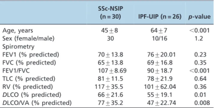
The clinical features of the 56 patients included in this study are shown in Table 1. All studied patients showed a restrictive lung function pattern that was characterized by a decrease in TLC (the mean values were 81% of that predicted for SSc-NSIP and 78% of that predicted for IPF-UIP) and an increased FEV 1/FVC ratio/100 (the mean value was 107% of that predicted for SSc-NSIP vs.90% of that predicted for IPF-UIP). The mean predicted DLCO values were significantly decreased in the IPF-UIP patients (55%) compared with the SSc-NSIP (66%) patients, and they were significantly greater when comparing the DLCO/VA in the IPF-UIP (47%) vs. the SSc-NSIP (77%) patients. No difference was found in the FEV1, FCV, TLC and RV values between the SSc-NSIP and IPF-UIP groups (Table 1).
Angiotensin II type 1 and 2 receptors
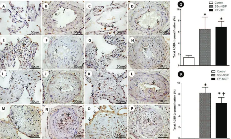
Alveolar septae and vessels from the normal control lungs, NSIP and UIP histologic patterns are shown in Figure 1 (immunostained with the AGTR-1 and AGTR-2 antibodies). Different immunostaining intensity was observed in the epithelial, endothelial, myofibroblast and smooth muscle cells from the alveolar septae and vessels in the SSc-NSIP (cellular SSc-NSIP and fibrotic SSc-NSIP) and IPF-UIP groups compared to the normal control lungs. Table 2 summarizes the morphometric results. A significantly higher percentage of the septal and vessel cells immunostained for AGTR-1/ AGTR-2 in the cellular SSc-NSIP, fibrotic SSc-NSIP and IPF-UIP lungs was observed compared to the controls. In the SSc-NSIP and IPF-UIP lungs, a similar proportion of the septal cells immunostained for AGTR-1 was observed, except for the IPF-UIP lungs, in which the AGTR-2 immunoreactivity represented a significantly reduced proportion of the septal cells compared to the SSc-NSIP lungs. Of equal significance was the increased percentage of the vessel cells that were immunostained for AGTR-1 in the cellular SSc-NSIP and IPF-UIP lungs. Concerning AGTR-2, a similar percentage of the vessel cells was present in the fibrotic SSc-NSIP and IPF-UIP lungs. When we compared the total cellular expression of AGTR-1 in the SSc-NSIP and IPF-UIP groups, a similar proportion was observed; however, both were increased compared to the control group (Figure Q), except for the IPF-UIP lungs, in which the total cellular AGTR-2 expression had a significantly smaller proportion than that of the SSc-NSIP pattern lungs (Figure R).
Distribution of lymphatic vessels
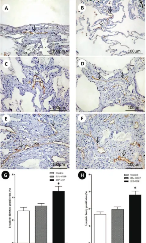
Figure 2 shows the peripheral, interlobular and peri-bronchovascular lymphatic vessels from the normal control lungs and the SSc-NSIP and IPF-UIP histologic patterns, immunostained for D2-40. A different proportion of the lymphatic vessels in the peripheral, interlobular and peribronchovascular interstitium was visualized in the SSc-NSIP (cellular SSc-NSIP and fibrotic SSc-NSIP) and IPF-UIP lungs compared to the control lungs. Table 2 summarizes the morphometric results. In the peripheral, interlobular and peribronchovascular interstitium of the IPF-UIP group, the lymphatic vessels showed a significantly increased area compared to the SSc-NSIP lungs (Figure G). A significantly higher percentage of lymphatic vessels was present in the peripheral, interlobular and periaxial inter-stitium of the IPF-UIP pattern compared to the SSc-NSIP histologic pattern (Figure H).
Pulmonary function tests
A significant inverse association was found between the vascular AGTR-2 and DCLO/VA (R = -0.74,p =0.001) in the SSc-NSIP and (R = -0.78, p =0.001) UIP groups. Equally significant was the negative association between the lymphatic vessel area in SSc-NSIP and the predicted values of DLCO (R = -0.53,p =0.04).
Survival analysis
Nineteen patients died during the 180-month follow-up period (5 SSc-NSIP and 14 IPF-UIP). A preliminary examination of the Kaplan-Meier survival curves showed that in this study, the patients with the SSc-NSIP pattern with percentages of vessel cells immunostained for AGTR-1 that were ,2.49% and AGTR-2 percentages ,7.46% had Table 1 -Clinical data of the patients with systemic
sclerosis and idiopathic pulmonary fibrosis.
SSc-NSIP
(n = 30) IPF-UIP (n = 26) p-value
Age, years 45¡8 64¡7 ,0.001
Sex (female/male) 30 10/16 1.2
Spirometry
FEV1 (% predicted) 70¡13.8 76¡20.01 0.23 FVC (% predicted) 65¡13.8 69¡16.8 0.35
FEV1/FVC 107¡8.69 90¡18.7 ,0.001
TLC (% predicted) 81¡11.5 78¡21.9 0.64 RV (% predicted) 117¡35.5 101¡62.04 0.36 DLCO (% predicted) 66¡21.6 55¡19.1 0.01 DLCO/VA (% predicted) 77¡35.2 47¡22.74 0.008
The data are expressed as the means¡SD or as counts (percentages); p,0.05 was significant.
higher survival rates than the patients with percentages of vessel cells immunostained for AGTR-1 that were .2.49% and AGTR-2 percentages .7.46. The multivariate analyses by the Cox regression model showed statistical significance
(log-likelihood = 59.39,p,0.0001) with a high risk of death for the patients with the IPF-UIP pattern (p =0.01) and a high percentage of vessel cells immunostained for the AGTR-2 and lymphatic vessels (Table 3).
Figure 1 -Cellular expression of the angiotensin II type 1 receptor (AGTR-1) and AGTR-2 divided in septal areas and intrapulmonary vessels from normal control lungs, systemic sclerosis (SSc) and idiopathic pulmonary fibrosis (IPF). In (A) and (B), we observe a diffuse and minimal AGTR-1 (arrows) expression in the alveolar septal areas and in the intrapulmonary vessels of the controls. The AGTR-1 expression shows greater increases in the thickened alveolar septal region from the cellular NSIP (arrows, E) than in the fibrotic NSIP (arrows, I). In the intrapulmonary vessels, the expression of AGTR-1 (arrows) is minimal and similar in both groups (cellular SSc-NSIP, F and fibrotic SSc-SSc-NSIP, J). Increased expression of AGTR-1 (arrows) is observed in the septal areas (M) and intrapulmonary vessels (N) of IPF-UIP. In (C) and (D), we observed AGRT-2 (arrows) expression in the septal areas and intrapulmonary vessels from the controls. In (G) and (K), we observed a similar expression of AGTR-2 (arrows) in the septal areas of cellular and fibrotic SSc-NSIP. Increased expression of AGTR-2 (arrows) is observed in the intrapulmonary vessels of fibrotic SSc-NSIP (L) compared to cellular SSc-NSIP (H). The expression of AGTR-2 (arrows) is more evident in the septal areas of IPF-UIP (O) than in the intrapulmonary vessels (P) of this histologic pattern. Q and R show the total values of AGTR-1 and AGTR-2 and the results analyzed by a one-way ANOVA with the Bonferroni multiple comparisons test for the control, SSc-NSIP and IPF-UIP groups. (*) Significantly higher AGTR-1 expression was observed in the SSc-NSIP and IPF-UIP lungs compared with the control group (p =0.001 andp =0.001, respectively). A similar situation was observed with AGTR-2 expression from the SSc-NSIP and IPF-UIP patterns compared to the control group (p,0.001 andp,0.001, respectively). ({) Significantly higher expression of AGTR-2 in the SSc-NSIP group was observed when compared with the IPF-UIP group (p= 0.04) by Student’s t-test.
Table 2 -Summary of the morphometric resultsa.
Variables Control Cellular SSc-NSIP Fibrotic SSc-NSIP SSc-NSIP UIP
Septal AGTR-1 1.45¡1.02 10.72¡6.20 12.18¡8.00 11.41¡6.97 10.10¡22.23
Vascular AGTR-1 1.06¡1.03 4.36¡5.35 0.51¡1.15 2.53¡4.33 2.13¡1.64
Septal AGTR-2 0.03¡0.02 12.75¡8.05 12.78¡6.96 12.77¡7.36 7.78¡4.31
Vascular AGTR-2 0.01¡0.04 1.26¡1.35 6.91¡4.64 3.83¡4.30 6.06¡4.66
Total AGTR-1 1.25¡0.84 7.54¡4.49 6.34¡3.96 6.97¡4.18 6.26¡10.93
Total AGTR-2 0.02¡0.02 6.51¡3.88 9.85¡5.09 8.18¡4.73 6.38¡3.63
Lymphatic density 1.68¡0.38 2.72¡0.62 2.92¡0.69 2.80¡0.65 3.73¡1.19
Lymphatic area 2.81¡0.90 4.28¡1.32 4.33¡0.64 4.25¡1.08 5.52¡2.24
aThe units of ‘‘% of points’’ indicate the number of points overlying the phenomena of interest divided by the total number of points overlying the septae
& DISCUSSION
Unlike SSc-NSIP, the likely reason that IPF-UIP patients do not respond to immunosuppressive therapies and their clinical course is marked by inexorable deterioration is the imbalance between parenchymal and vascular remodeling. Therefore, it is important to determine whether additional histological details can help us understand the clinical differences between these diseases. After pulmonary injury, parenchymal and vascular remodeling contribute to increased pulmonary epithelial and endothelial permeabil-ity. This early phase is followed by a subacute fibroproli-ferative phase, which may allow repair of the injured lung or may result in progressive obliteration of the interstitial, alveolar and vascular compartments of the lung through a fibroproliferative process that may be established by 24 h after the injury (39).
Recent studies have shown that ANG II contributes to pulmonary fibrosis progression, which is evidenced not only by its potent vasoconstrictor activity but also by its influence on organ remodeling and fibrosis (20,21,22). The AGTR-1 and AGTR-2 binding of ANG II is facilitated by the cleavage of ANG I by the ACE. Both receptors belong to the 7-transmembrane G protein–coupled receptor family (23,24). Whereas most of the ANG II-mediated effects, such as cell growth, inflammation or extracellular matrix synth-esis, have been shown to be mediated via AGTR-1, the function of AGTR-2 has been less clearly investigated (40,41). During the development of fibrosis, AGTR expres-sion undergoes an expresexpres-sion shift in favor of higher AGTR-2 levels, thereby tilting the cellular response to ANG II (42-44). Thus, for all these reasons, we should not be surprised to learn that immunohistochemical staining for AGTRs provides important information about the organ remodeling process in diffuse parenchymal lung diseases, and our results now confirm the pathogenetic importance of AGTRs in IPF and SSc.
We found a significantly greater percentage of septal and vessel cells immunostained for AGTR-1 in SSc-NSIP and IPF-UIP compared with the normal lung. In addition, SSc-NSIP and IPF-UIP had a similar proportion of septal cells immunostained for AGTR-1. We also found an increased percentage of vessel cells immunostained for AGTR-1 in cellular SSc-NSIP and IPF-UIP. Previous reports in the literature have shown the potential role of ANG II in experimental lung fibrosis and in IPF (42-44), while the potency of AGTR-1 in fostering lung fibrosis has been previously demonstrated in multiple in vitro cell culture conditions as well as in vivo animal models (45,42). In experimental animal models of lung fibrosis, using bleo-mycin exposure or radiation, the fibrotic response was
attenuated by the ACE inhibitors and by specific AGTR-1 antagonism (45,46). Studies of the inhibition of the ANG II pathway have demonstrated opposing results. Some studies have shown beneficial results and blocked the fibrosis, principally in bleomycin-induced pulmonary fibrosis (47); others have failed to document a beneficial effect of the ANG II blockade in experimental models of lung fibrosis, indicating that the fibrotic disease process is dependent not only on AGTR-1 activity (48). We found that a small percentage of vessel cells in SSc-NSIP are AGTR-1+. In contrast to the data available for AGTR-1, the function of AGTR-2 is much less well explored. In our study, there was an increased percentage of vessel cells immunostained for AGTR-2 in SSc-NSIP and IPF-UIP, suggesting an important role in the pathogenesis of these disorders. In heart tissue, the AGTR-2 is considered to be a neutralizer, modulating the actions of AGTR-1 (28,29), but many signaling mechan-isms and receptor functions are unclear, particularly the role of AGTR-2 in pulmonary fibrosis.
We also found a significantly higher percentage of lymphatic vessels in the peripheral, interlobular and periaxial interstitium of IPF-UIP compared with the SSc-NSIP histologic pattern. In IPF-UIP, the lymphatic vessels showed a significantly increased area compared with SSc-NSIP. The lymphatic vessels contribute to fibrosis matura-tion and scar formamatura-tion through the drainage of excessive proteins and fluid during fibrosis (49). In the present study, D2-40+ lymphatics were abundant in the peripheral, interlobular and peribronchovascular interstitium. Pre-vious studies performed by our group have shown that the lymphatic vessels play an important role in early remodeling in the development of pulmonary fibrosis (50). The current study clearly demonstrated that the lymphatic vessels were poorly detected in the affected areas of early remodeling, such as cellular SSc-NSIP, compared to the fibrotic SSc-NSIP and IPF-UIP patterns.
Our study has clinical and functional importance. A significant inverse association was found between vascular AGTR-2 and DCLO/VA in the SSc-NSIP and IPF-UIP groups. Of equal significance was the negative association between the lymphatic vessel area in SSc-NSIP and the predicted values of DLCO. To establish the relevance of these findings to disease progression in the patients, the AGTRs were evaluated as a function of survival (controlled for age) in the IPF-UIP and SSc-NSIP histologic patterns. The multivariate analyses using the Cox regression model showed a high risk of death for the patients with the IPF-UIP pattern presenting a high percentage of vessel cells immunostained for AGTR-2 in the lymphatic vessels.
We concluded that the AGTRs and lymphatic vessels in the lung parenchyma offer the potential to control the organ Table 3 -Cox proportional hazards regression to ascertain the individual contribution of the histological patterns (NSIP and UIP) and the morphological factors associated with survival and to compare the adjusted survival between the groups (-2 log-likelihood = 59.93; Chi-squared = 40.10;p,0.001).
b SE Wald test p-value Exp (b) 95.0% CI for Exp (b)
Lower Upper
NSIP pattern -12.70 182.18 5.97 0.94 0.00 0.00 0.60E149
UIP pattern -1.60 0.65 5.97 0.01 0.20 0.56 0.78
AGTR2 vascular 0.26 0.89 9.23 0.002 1.30 1.10 1.55
Lymphatic density -1.20 0.25 6.92 0.008 0.297 0.122 0.73
remodeling and fibrosis involved in SSc-NSIP and IPF-UIP progression, which suggests that strategies aimed at preventing high AGTR synthesis and low lymphangiogen-esis may have a greater impact on SSc and IPF. Further study using a randomized and prospective trial is necessary to finalize this conclusion.
& ACKNOWLEDGMENTS
We are grateful to Sandra de Morais Fernezlian, a biologist from the immunohistochemistry laboratory, for the helpful immunohistochemistry staining results and to Prof. Carlos Roberto Ribeiro de Carvalho and Ronaldo Adib Cairalla, from the Department of Pneumology, for their patient databases. This study was supported by the National Council for Scientific and Technological Development (CNPq) and the Foundation for the Support of Research of the State of Sa˜o Paulo (2008/53022-3 and 2011/06312-9).
& AUTHOR CONTRIBUTIONS
All authors designed the study, analyzed and interpreted the data and wrote the manuscript.
& REFERENCES
1. American Thoracic Society; European Respiratory Society. American Thoracic Society/European Respiratory Society International Mul-tidisciplinary Consensus Classification of the Idiopathic Interstitial Pneumonias. This joint statement of the American Thoracic Society (ATS), and the European Respiratory Society (ERS) was adopted by the ATS board of directors, June 2001 and by the ERS Executive Committee, June 2001. Am J Respir Crit Care Med. 2002;165(2):277-304.
2. Agustı´ C, Xaubet A, Roca J, Agustı´ AG, Rodriguez-Roisin R. Interstitial pulmonary fibrosis with and without associated collagen vascular disease: results of a two year follow up. Thorax 1992;47(12):1035-40, http://dx.doi.org/10.1136/thx.47.12.1035.
3. Douglas WW, Tazelaar HD, Hartman TE, Hartman RP, Decker PA, Schroeder DR, et al. Polymyositis-dermatomyositis-associated interstitial lung disease. Am J Respir Crit Care Med. 2001;164(7):1182-5, http://dx. doi.org/10.1164/ajrccm.164.7.2103110.
4. Flaherty KR, Colby TV, Travis WD, Toews GB, Mumford J, Murray S, et al. Fibroblastic foci in usual interstitial pneumonia: idiopathic versus collagen vascular disease. Am J Respir Crit Care Med. 2003;167(10):1410-5, http://dx.doi.org/10.1164/rccm.200204-373OC.
5. Papiris SA, Vlachoyiannopoulos PG, Maniati MA, Karakostas KX, Constantopoulos SH, Moutsopoulos HH. Idiopathic pulmonary fibrosis and pulmonary fibrosis in diffuse systemic sclerosis: two fibroses with different prognoses. Respiration. 1997;64(1):81-5, http://dx.doi.org/10. 1159/000196648.
6. Wells AU, Cullinan P, Hansell DM, Rubens MB, Black CM, Newman-Taylor AJ, et al. Fibrosing alveolitis associated with systemic sclerosis has a better prognosis than lone cryptogenic fibrosing alveolitis. Am J Respir Crit Care Med. 1994;149(6):1583-90, http://dx.doi.org/10. 1164/ajrccm.149.6.8004317.
7. Cottin V, Thivolet-Be´jui F, Reynaud-Gaubert M, Cadranel J, Delaval P, Ternamian PJ, et al. Interstitial lung disease in amyopathic dermato-myositis, dermatomyositis and polymyositis. Eur Respir J. 2003; 22(2):245-50, http://dx.doi.org/10.1183/09031936.03.00026703. 8. Yamadori I, Fujita J, Bandoh S, Tokuda M, Tanimoto Y, Kataoka M, et al.
Nonspecific interstitial pneumonia as pulmonary involvement of primary Sjo¨gren’s syndrome. Rheumatol Int. 2002;22(3):89-92. 9. Ito I, Nagai S, Kitaichi M, Nicholson AG, Johkoh T, Noma S, et al.
Pulmonary manifestations of primary Sjo¨gren’s syndrome: a clinical, radiologic, and pathologic study. Am J Respir Crit Care Med. 2005;171(6):632-8, http://dx.doi.org/10.1164/rccm.200403-417OC. 10. Kim DS, Yoo B, Lee JS, Kim EK, Lim CM, Lee SD, et al. The major
histopathologic pattern of pulmonary fibrosis in scleroderma is nonspecific interstitial pneumonia. Sarcoidosis Vasc Diffuse Lung Dis. 2002;19(2):121-7.
11. Tansey D, Wells AU, Colby TV, Ip S, Nikolakoupolou A, du Bois RM, et al. Variations in histological patterns of interstitial pneumonia between connective tissue disorders and their relationship to prognosis. Histopathology. 2004;44(6):585-96, http://dx.doi.org/10.1111/j.1365-2559. 2004.01896.x.
12. Park JH, Kim DS, Park IN, Jang SJ, Kitaichi M, Nicholson AG, et al. Prognosis of fibrotic interstitial pneumonia: idiopathic versus collagen vascular disease-related subtypes. Am J Respir Crit Care Med. 2007; 175(7):705-11, http://dx.doi.org/10.1164/rccm.200607-912OC.
13. Parra ER, Silve´rio da Costa LR, Ab’Saber A, Ribeiro de Carvalho CR, Kairalla RA, Fernezlian SM, et al. Nonhomogeneous density of CD34 and VCAM-1 alveolar capillaries in major types of idiopathic inters-titial pneumonia. Lung. 2005;183(5):363-73, http://dx.doi.org/10.1007/ s00408-005-2548-1.
14. Ebina M, Shimizukawa M, Shibata N, Kimura Y, Suzuki T, Endo M, et al. Heterogeneous increase in CD34-positive alveolar capillaries in idio-pathic pulmonary fibrosis. Am J Respir Crit Care Med. 2004; 169(11):1203-8, http://dx.doi.org/10.1164/rccm.200308-1111OC. 15. Yamashita M, Iwama N, Date F, Chiba R, Ebina M, Miki H, et al.
Characterization of lymphangiogenesis in various stages of idiopathic diffuse alveolar damage. Hum Pathol. 2009;40(4):542-51, http://dx.doi. org/10.1016/j.humpath.2008.06.031.
16. Mandal RV, Mark EJ, Kradin RL. Organizing pneumonia and pulmonary lymphatic architecture in diffuse alveolar damage. Hum Pathol. 2008;39(8):1234-8, http://dx.doi.org/10.1016/j.humpath.2008.01.002. 17. Parra ER, Araujo CA, Lombardi JG, Ab’Saber AM, Carvalho CR, Kairalla
RA, et al. Lymphatic fluctuation in the parenchymal remodeling stage of acute interstitial pneumonia, organizing pneumonia, nonspecific inter-stitial pneumonia and idiopathic pulmonary fibrosis. Braz J Med Biol Res. 2012;45(5):466-72.
18. Katzenstein AL, Myers JL. Idiopathic pulmonary fibrosis: clinical relevance of pathologic classification. Am J Respir Crit Care Med. 1998;157(4 Pt 1):1301-15, http://dx.doi.org/10.1164/ajrccm.157.4.970 7039.
19. Dohi M, Hasegawa T, Yamamoto K, Marshall BC. Hepatocyte growth factor attenuates collagen accumulation in a murine model of pulmonary fibrosis. Am J Respir Crit Care Med. 2000;162(6):2302-7, http://dx.doi. org/10.1164/ajrccm.162.6.9908097.
20. Marshall RP, Gohlke P, Chambers RC, Howell DC, Bottoms SE, Unger T, et al. Angiotensin II and the fibroproliferative response to acute lung injury. Am J Physiol Lung Cell Mol Physiol. 2004;286(1):L156-64. 21. Ruiz-Ortega M, Esteban V, Ruperez M, Sanchez-Lopez E, Rodriguez-Vita
J, Carvajal G, et al. Renal and vascular hypertension-induced inflamma-tion: role of angiotensin II. Curr Opin Nephrol Hypertens. 2006;15(2):159-66, http://dx.doi.org/10.1097/01.mnh.0000203190.34643.d4.
22. Schuttert JB, Liu MH, Gliem N, Fiedler GM, Zopf S, Mayer C, et al. Human renal fibroblasts derived from normal and fibrotic kidneys show differences in increase of extracellular matrix synthesis and cell proliferation upon angiotensin II exposure. Pflugers Arch. 2003; 446(3):387-93.
23. De Gasparo M. [at(1) and at(2) angiotensin II receptors: Key features]. Drugs. 2002;62(1):1-10, http://dx.doi.org/10.2165/00003495-200262991-00001.
24. Oro C, Qian H, Thomas WG. Type 1 angiotensin receptor pharmacology: signaling beyond G proteins. Pharmacol Ther. 2007;113(1):210-26, http://dx.doi.org/10.1016/j.pharmthera.2006.10.001.
25. Carey RM. Update on the role of the at2 receptor. Curr Opin Nephrol Hypertens. 2005;14(1):67-71, http://dx.doi.org/10.1097/00041552-20050 1000-00011.
26. Kaschina E, Unger T. Angiotensin at1/at2 receptors: regulation, signalling and function. Blood Press. 2003;12(2):70-88, http://dx.doi. org/10.1080/08037050310001057.
27. Reudelhuber TL. The continuing saga of the at2 receptor: a case of the good, the bad, and the innocuous. Hypertension. 2005;46(6):1261-2, http://dx.doi.org/10.1161/01.HYP.0000193498.07087.83.
28. AbdAlla S, Lother H, Abdel-tawab AM, Quitterer U. The angiotensin II at2 receptor is an at1 receptor antagonist. J Biol Chem. 2001; 276(43):39721-6, http://dx.doi.org/10.1074/jbc.M105253200.
29. D’Amore A, Black MJ, Thomas WG. The angiotensin II type 2 receptor causes constitutive growth of cardiomyocytes and does not anta-gonize angiotensin II type 1 receptor-mediated hypertrophy. Hypertension. 2005;46(6):1347-54, http://dx.doi.org/10.1161/01.HYP. 0000193504.
51489.cf.
30. Miura S, Karnik SS. Ligand-independent signals from angiotensin II type 2 receptor induce apoptosis. EMBO J. 2000;19(15):4026-35, http://dx.doi. org/10.1093/emboj/19.15.4026.
31. Miura S, Karnik SS, Saku K. Constitutively active homo-oligomeric angiotensin II type 2 receptor induces cell signaling independent of receptor conformation and ligand stimulation. J Biol Chem. 2005; 280(18):18237-44.
32. de Souza RB, Borges CT, Capelozzi VL, Parra ER, Jatene FB, Kavakama J, et al. Centrilobular fibrosis: an underrecognized pattern in systemic sclerosis. Respiration. 2009;77(4):389-97, http://dx.doi.org/10.1159/ 000156958.
33. Preliminary criteria for the classification of systemic sclerosis (scler-oderma). Subcommittee for scleroderma criteria of the American Rheumatism Association Diagnostic and Therapeutic Criteria Com-mittee. Arthritis Rheum. 1980;23(5):581-90.
35. American Thoracic Society. Idiopathic pulmonary fibrosis: diagnosis and treatment. International consensus statement. American Thoracic Society (ATS), and the European Respiratory Society (ERS). Am J Respir Crit Care Med. 2000;161(2 Pt 1):646-64.
36. Raghu G, Collard GR, Egan JJ, Martinez FJ, Behr J, Brown KK, et al. An Official ATS/ERS/JRS/ALAT Statement: Idiopathic Pulmonary Fibrosis: Evidence-based Guidelines for Diagnosis and Management. Am J Respir Crit Care Med. 2011;183(6):788-824, http://dx.doi.org/10.1164/rccm. 2009-040GL.
37. Quanjer PH, Tammeling GJ, Cotes JE, Pedersen OF, Peslin R, Yernault JC. Lung volumes and forced ventilatory flows. Report Working Party Standardization of Lung Function Tests, European Community for Steel and Coal. Official Statement of the European Respiratory Society. Eur Respir J Suppl. 1993;16:5-40.
38. Hsia CC, Hyde DM, Ochs M, Weibel ER; ATS/ERS Joint Task Force on Quantitative Assessment of Lung Structure. An Official Research Policy Statement of the American Thoracic Society/European Respiratory Society: Standards for Quantitative Assessment of Lung Structure. Am J Respir Crit Care Med. 2010;181(4):394-418, http://dx.doi.org/10. 1164/rccm.200809-1522ST.
39. Rocco PR, Negri EM, Kurtz PM, Vasconcellos FP, Silva GH, Capelozzi VL, et al. Lung tissue mechanics and extracellular matrix remodeling in acute lung injury. Am J Respir Crit Care Med. 2001;164(6):1067-71, http://dx.doi.org/10.1164/ajrccm.164.6.2007062.
40. Horiuchi M, Mogi M, Iwai M. Signaling crosstalk angiotensin II receptor subtypes and insulin. Endocr J. 2006;53(1):1-5, http://dx.doi.org/10. 1507/endocrj.53.1.
41. Nakashima H, Suzuki H, Ohtsu H, Chao JY, Utsunomiya H, Frank GD, et al. Angiotensin II regulates vascular and endothelial dysfunction: recent topics of angiotensin II type-1 receptor signaling in the vasculature. Curr Vasc Pharmacol. 2006;4(1):67-78, http://dx.doi.org/ 10.2174/157016106775203126.
42. Uhal BD, Kim JK, Li X, Molina-Molina M. Angiotensin-TGF-beta1 crosstalk in human idiopathic pulmonary fibrosis: autocrine mechanisms in myofibroblasts and macrophages. Curr Pharm Des. 2007;13(12):1247-56, http://dx.doi.org/10.2174/138161207780618885.
43. Zhu YC, Zhu YZ, Lu N, Wang MJ, Wang YX, Yao T. Role of angiotensin at1 and at2 receptors in cardiac hypertrophy and cardiac remodeling. Clin Exp Pharmacol Physiol. 2003;30(12):911-8, http://dx.doi.org/10. 1111/j.1440-1681.2003.03942.x.
44. Ruiz-Ortega M, Ruperez M, Esteban V, Rodriguez-Vita J, Sanchez-Lopez E, Carvajal G, et al. Angiotensin II: a key factor in the inflammatory and fibrotic response in kidney diseases. Nephrol Dial Transplant. 2006;21(1):16-20, http://dx.doi.org/10.1093/ndt/gfi265.
45. Molteni A, Wolfe LF, Ward WF, Ts’ao CH, Molteni LB, Veno P, et al. Effect of an angiotensin II receptor blocker and two angiotensin converting enzyme inhibitors on transforming growth factor-beta (TGF-beta) and alpha-actomyosin (alpha SMA), important media-tors of radiation-induced pneumopathy and lung fibrosis. Curr Pharm Des. 2007;13(13):1307-16, http://dx.doi.org/10.2174/13816120778061 8777.
46. Wang R, Ibarra-Sunga O, Verlinski L, Pick R, Uhal BD. Abrogation of bleomycin-induced epithelial apoptosis and lung fibrosis by captopril or by a caspase inhibitor. Am J Physiol Lung Cell Mol Physiol. 2000;279(1):L143-51.
47. Li X, Zhuang J, Rayford H, Zhang H, Shu R, Uhal BD. Attenuation of bleomycin-induced pulmonary fibrosis by intratracheal administration of antisense oligonucleotides against angiotensinogen mRNA. Curr Pharm Des. 2007;13(12):1257-68, http://dx.doi.org/10.2174/138161207 780618867.
48. Keogh KA, Standing J, Kane GC, Terzic A, Limper AH. Angiotensin II antagonism fails to ameliorate bleomycin-induced pulmonary fibrosis in mice. Eur Respir J. 2005;25(4):708-14, http://dx.doi.org/10.1183/ 09031936.05.00090204.
49. Meinecke AK, Nagy N, Lago GD, Kirmse S, Klose R, Schro¨dter K, et al. Aberrant mural cell recruitment to lymphatic vessels and impaired lymphatic drainage in a murine model of pulmonary fibrosis. Blood. 2012;119(24):5931-42, http://dx.doi.org/10.1182/blood-2011-12-396895. 50. Parra ER, Araujo CA, Lombardi JG, Ab’Saber AM, Carvalho CR, Kairalla